. 2023 Nov 17;15(11):2266.
doi: 10.3390/v15112266.

Presence and Persistence of Andes Virus RNA in Human Semen

Affiliations

Presence and Persistence of Andes Virus RNA in Human Semen

Roland Züst et al. Viruses. .

Abstract

When infecting humans, Andes orthohantavirus (ANDV) may cause a severe disease called hantavirus cardiopulmonary syndrome (HCPS). Following non-specific symptoms, the infection may progress to a syndrome of hemorrhagic fever combined with hyper-acute cardiopulmonary failure. The case fatality rate ranges between 25-40%, depending on the outbreak. In this study, we present the follow-up of a male patient who recovered from HCPS six years ago. We demonstrate that the ANDV genome persists within the reproductive tract for at least 71 months. Genome sequence analysis early and late after infection reveals a low number of mutations (two single nucleotide variants and one deletion), suggesting limited replication activity. We can exclude the integration of the viral genome into the host genome, since the treatment of the specimen with RNAse led to a loss of signal. We demonstrate a long-lasting, strong neutralizing antibody response using pseudovirions expressing the ANDV glycoprotein. Taken together, our results show that ANDV has the potential for sexual transmission.

Keywords: Andes virus; neutralizing antibodies; persistence; semen.


Conflict of interest statement

The authors declare no conflict of interest.

Figures

Figure 1
Figure 1
Detection of Andes virus RNA in plasma and semen. RNA levels from plasma (circles) and semen (squares) were assessed on the indicated days post infection using in-house quantitative real-time polymerase chain reaction; viral load on day 1 was from a serum sample, and from day 4 onward was from EDTA blood.
Figure 2
Figure 2
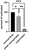
Location of viral material in semen. In total, 1 ml of semen was centrifuged at 600× g for 15 min. The supernatant was transferred into a new tube and the pellet resuspended in 1 ml of PBS. Nucleic acid from 100 μL of input semen, supernatant and resuspended pellet was isolated and analyzed using real-time PCR. Statistical analysis was performed using unpaired the Student’s t-test (***, p < 0.001; **, p < 0.01; *, p < 0.05).
Figure 3
Figure 3
Analysis of viral material. Nucleic acid from semen was isolated and left untreated (black) or subjected to RNase treatment (white). SNV RNA and a plasmid (not sensitive to RNase treatment) containing the target of the SNV RT-qPCR served as controls. After RNase digestion, no signal could be detected in material derived from semen or control RNA (n.d.; not detected).
Figure 4
Figure 4
Neutralization of ANDV using patient sera. The neutralizing antibody titer (reciprocal IC50) against ANDV in serum samples was assessed using a pseudovirion assay. Half maximal inhibitory concentrations were estimated via a model of nonlinear regression fit with settings for log (inhibitor) vs. normalized response curves using GraphPad Prism v9.
Figure 5
Figure 5
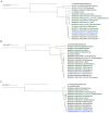
Phylogenetic analysis of ANDV shows a close relationship to previously sequenced ANDV from Chile and Argentina ((A): segment S, (B): segment M, (C): segment L).

References

    1. Maes P., Adkins S., Alkhovsky S.V., Avšič-Županc T., Ballinger M.J., Bente D.A., Beer M., Bergeron É., Blair C.D., Briese T., et al. Taxonomy of the order Bunyavirales: Second update 2018. Arch. Virol. 2019;164:927–941. doi: 10.1007/s00705-018-04127-3. - DOI - PMC - PubMed
    1. Schmaljohn C.S., Dalrymple J.M. Analysis of Hantaan virus RNA:. Evidence for a new genus of bunyaviridae. Virology. 1983;131:482–491. doi: 10.1016/0042-6822(83)90514-7. - DOI - PubMed
    1. Avsic-Zupanc T., Saksida A., Korva M. Hantavirus infections. Clin. Microbiol. Infect. 2019;21S:e6–e16. doi: 10.1111/1469-0691.12291. - DOI - PubMed
    1. Plyusnin A., Sironen T. Evolution of hantaviruses: Co-speciation with reservoir hosts for more than 100 MYR. Virus Res. 2014;187:22–26. doi: 10.1016/j.virusres.2014.01.008. - DOI - PubMed
    1. Bennett S.N., Gu S.H., Kang H.J., Arai S., Yanagihara R. Reconstructing the evolutionary origins and phylogeography of hantaviruses. Trends Microbiol. 2014;22:473–482. doi: 10.1016/j.tim.2014.04.008. - DOI - PMC - PubMed