. 2023 Nov 10;11(11):1707.
doi: 10.3390/vaccines11111707.

Characterization of the Efficacy of a Split Swine Influenza A Virus Nasal Vaccine Formulated with a Nanoparticle/STING Agonist Combination Adjuvant in Conventional Pigs

Affiliations

Characterization of the Efficacy of a Split Swine Influenza A Virus Nasal Vaccine Formulated with a Nanoparticle/STING Agonist Combination Adjuvant in Conventional Pigs

Veerupaxagouda Patil et al. Vaccines (Basel). .

Abstract

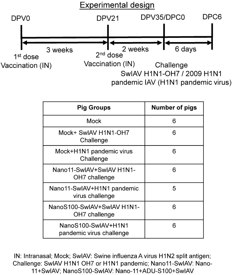
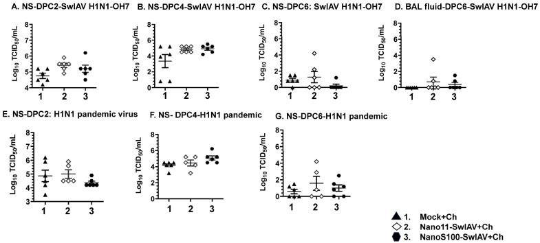
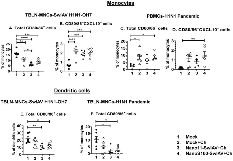
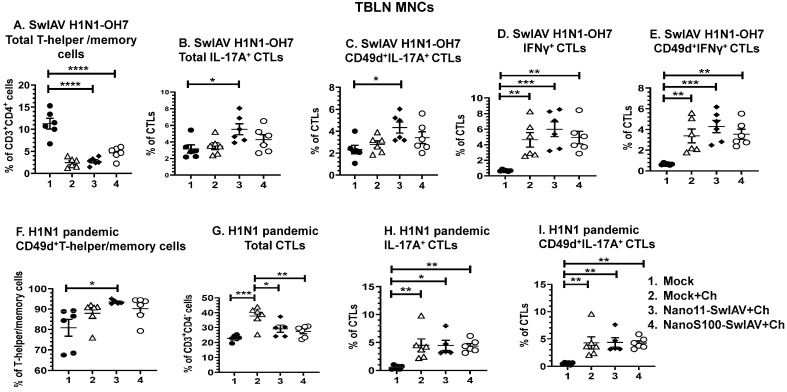
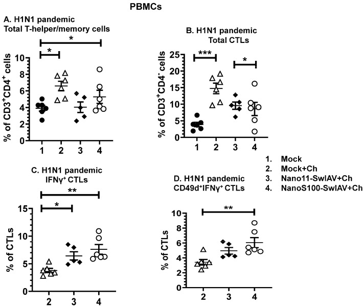
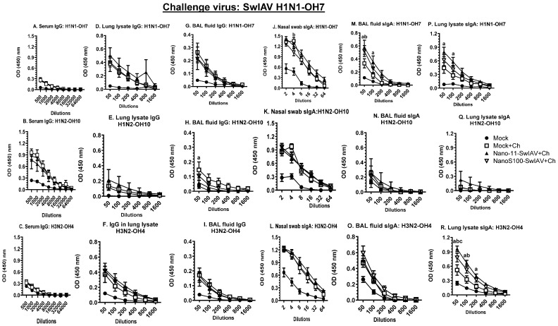
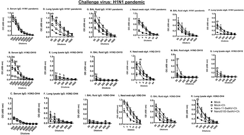
Swine influenza A viruses (SwIAVs) are pathogens of both veterinary and medical significance. Intranasal (IN) vaccination has the potential to reduce flu infection. We investigated the efficacy of split SwIAV H1N2 antigens adsorbed with a plant origin nanoparticle adjuvant [Nano11-SwIAV] or in combination with a STING agonist ADU-S100 [NanoS100-SwIAV]. Conventional pigs were vaccinated via IN and challenged with a heterologous SwIAV H1N1-OH7 or 2009 H1N1 pandemic virus. Immunologically, in NanoS100-SwIAV vaccinates, we observed enhanced frequencies of activated monocytes in the blood of the pandemic virus challenged animals and in tracheobronchial lymph nodes (TBLN) of H1N1-OH7 challenged animals. In both groups of the virus challenged pigs, increased frequencies of IL-17A+ and CD49d+IL-17A+ cytotoxic lymphocytes were observed in Nano11-SwIAV vaccinates in the draining TBLN. Enhanced frequency of CD49d+IFNγ+ CTLs in the TBLN and blood of both the Nano11-based SwIAV vaccinates was observed. Animals vaccinated with both Nano11-based vaccines had upregulated cross-reactive secretory IgA in the lungs and serum IgG against heterologous and heterosubtypic viruses. However, in NanoS100-SwIAV vaccinates, a slight early reduction in the H1N1 pandemic virus and a late reduction in the SwIAV H1N1-OH7 load in the nasal passages were detected. Hence, despite vast genetic differences between the vaccine and both the challenge viruses, IN vaccination with NanoS100-SwIAV induced antigen-specific moderate levels of cross-protective immune responses.

Keywords: ADU-S100; Nano11; cell-mediated immune responses; intranasal vaccination; memory responses; swine; swine influenza A virus.


Conflict of interest statement

The funders had no role in the design of the study; in the collection, analyses, or interpretation of data; in the writing of the manuscript; or in the decision to publish the results.

Figures

Figure 1
Figure 1
Experimental plan to ascertain the NanoS100–SwIAV vaccine efficacy in pigs. Conventional pigs were immunized twice with Nano11–SwIAV or NanoS100–SwIAV split virus vaccine or controls Mock IN and challenged at day 35 post-prime vaccination with SwIAV H1N1-OH7 or H1N1 pandemic virus and euthanized at day 6 post-challenge (DPC6). Nasal swabs, blood, lungs, tracheobronchial lymph nodes [TBLN], and bronchioalveolar [BAL] lavage fluid samples were harvested for the analysis. Five–six pigs were used in each of the experimental groups.
Figure 2
Figure 2
Infectious challenge virus titers in the nasal passage of intranasally (IN) vaccinated pigs. Conventional pigs were immunized twice with Nano11–SwIAV or NanoS100–SwIAV split virus vaccine or controls Mock IN, challenged at day 35 post-prime vaccination with SwIAV H1N1-OH7 or H1N1 pandemic virus, and euthanized at day 6 post-challenge (DPC6). The titers of challenge viruses were characterized by using cell culture method in nasal swab (NS) at (A,E) DPC2; (B,F) DPC4; (C,G) DPC6, and (D) BAL fluid at DPC6. Data indicate the mean value of 5 or 6 pigs ± SEM. Statistical analysis was carried out by one-way ANOVA followed by Tukey’s post-test.
Figure 3
Figure 3
The candidate Nano11-based vaccines modulated the activation of innate myeloid immune cell frequencies in vaccinated pigs. Conventional pigs were immunized twice with Nano11–SwIAV or NanoS100–SwIAV split virus vaccine or controls Mock IN, challenged at day 35 post-prime vaccination with SwIAV H1N1-OH7 or H1N1 pandemic virus, and euthanized at day 6 post-challenge (DPC6). TBLN MNCs and PBMCs were purified at DPC6 and were incubated with SwIAV H1N1-OH7 or H1N1 pandemic in vitro to determine memory responses. The cells were immunolabeled and flow cytometry was employed to investigate the frequencies of dendritic cells (CD3CD172a+SynCAM+) and monocytes (CD3CD172a+SynCAM). In SwIAV H1N1-OH7-challenged pigs, TBLN-MNCs were analyzed: (A) total CD80/86+ monocytes; (B) CD80/86+CXCL10+ monocytes and H1N1 pandemic virus-challenged pigs PBMCs; (C) total CD80/86+ monocytes; and (D) CD80/86+CXCL10+ monocytes. Dendritic cells in (E) SwIAV H1N1-OH7-challenged TBLN-MNCs total CD80/86+ and (F) H1N1 pandemic virus-challenged TBLN MNCs total CD80/86+ cells were characterized. Data represent the mean value of 5–6 pigs ± SEM. Statistical analysis was carried out by one-way ANOVA followed by Tukey’s post-test. Asterisks indicate significant difference between indicated groups (* p < 0.05, ** p < 0.01, *** p < 0.001, **** p < 0.0001).
Figure 4
Figure 4
The candidate Nano11-based vaccines elicited IL-17A+ and IFNγ+ CTL cell responses in TBLN MNCs of vaccinated pigs. Conventional pigs were immunized twice with Nano11–SwIAV or NanoS100–SwIAV split virus vaccine or controls Mock IN, challenged at day 35 post-prime vaccination with SwIAV H1N1-OH7 or H1N1 pandemic virus, and euthanized at day 6 post-challenge (DPC6). TBLN MNCs isolated at DPC6 were restimulated with SwIAV H1N1-OH7 or H1N1 pandemic virus in vitro. The SwIAV H1N1-OH7-restimulated TBLN MNCs were immunolabeled and analyzed by flow cytometry for the frequencies of the following: (A) total T-helper/memory cells [CD3+CD4+CD8α+CD8β]; (B) total IL-17A+CTLs [CD3+CD4-CD8α+CD8β+ IL-17A+]; (C) CD49d+IL-17A+ CTLs [CD3+CD4CD8α+CD8β+IL-17A+CD49d+]; (D) total IFNγ+ CTLs [CD3+CD4CD8α+CD8β+ IFNγ+]; and (E) CD49d+IFNγ+ CTLs. The H1N1 pandemic virus-restimulated TBLN MNCs were immunolabeled and analyzed by flow cytometry for the frequencies of the following: (F) CD49d+T-helper/memory cells [CD3+CD4+CD8α+CD8β]; (G) total CTLs [CD3+CD4CD8α+CD8β+]; (H) total IL-17A+ CTLs [CD3+CD4CD8α+CD8β+IL-17A+]; and (I) CD49d+IL-17A+ CTLs [CD3+CD4CD8α+CD8β+CD49d+IL-17A+]. Data represent the mean value of 5 or 6 pigs ± SEM. Statistical analysis was performed by one-way ANOVA followed by Tukey’s post-test. Asterisks represent significant difference between indicated groups (*p < 0.05, ** p < 0.01, *** p < 0.001, **** p < 0.0001).
Figure 5
Figure 5
NanoS100–SwIAV and Nano11–SwIAV vaccines augmented IFNγ+ CTL frequencies in PBMCs of vaccinated pigs. Conventional pigs were immunized twice with Nano11–SwIAV or NanoS100–SwIAV split virus vaccine or controls Mock IN, challenged at day 35 post-prime vaccination with SwIAV H1N1-OH7 or H1N1 pandemic virus, and euthanized at day 6 post-challenge (DPC6). H1N1 pandemic challenge virus was used for the restimulation of DPC6 isolated PBMCs in vitro. The cells were analyzed for the determination of the frequencies of the following: (A) total T-helper/memory cells [CD3+CD4+CD8α+CD8β]; (B) total CTLs [CD3+CD4CD8α+CD8β+]; (C) IFNγ+ CTLs [CD3+CD4CD8α+CD8β+IFNγ+]; and (D) CD49d+IFNγ+ CTLs [CD3+CD4CD8α+CD8β+IFNγ+CD49d+). Data display the mean value of 5–6 pigs ± SEM. Statistical analysis was conducted by using one-way ANOVA followed by Tukey’s post-test. Asterisks show significant difference between indicated groups (* p < 0.05, ** p < 0.01, *** p < 0.001).
Figure 6
Figure 6
NanoS100–SwIAV and Nano11–SwIAV vaccines upregulated the SwIAV H1N1-OH7-specific sIgA responses in the lungs of vaccinated pigs. Conventional pigs were immunized twice with Nano11–SwIAV or NanoS100–SwIAV split virus vaccine or controls Mock IN and challenged at day 35 post-prime vaccination with SwIAV H1N1-OH7. The vaccinates were subjected to necropsy at day 6 post-challenge (DPC6). Serum, lung lysate, and BAL fluid specimens collected at DPC6 were investigated by ELISA to detect antigen-specific IgG antibody responses against SwIAV (A,D,G) H1N1-OH7; (B,E,H) H1N2-OH10; (C,F,I) H3N2-OH4 and to detect antigen-specific sIgA antibody responses in nasal swab, BAL fluid, and lung lysate samples against SwIAV (J,M,P) H1N1-OH7; (K,N,Q) H1N2-OH10; (L,O,R) H3N2-OH4. Data exhibit the mean value of 5–6 pigs ± SEM. Statistical analysis was conducted by using two-way ANOVA followed by Bonferroni post-test. Letters a, b, and c indicate significance between Mock+Ch versus Nano11–SwIAV+Ch, Mock+Ch versus NanoS100–SwIAV+Ch, and Nano11–SwIAV+Ch versus NanoS100–SwIAV+Ch, respectively, at the indicated sample dilutions.
Figure 7
Figure 7
NanoS100–SwIAV increased the H1N1 pandemic-specific IgG and sIgA responses in both lungs and blood of vaccinated pigs. Conventional pigs were immunized twice with Nano11–SwIAV or NanoS100–SwIAV split virus vaccines or control Mock IN and challenged at day 35 post-prime vaccination with H1N1 pandemic virus. The experimental animals were sacrificed at day 6 post-challenge (DPC6). Serum, lung lysate, and BAL fluid samples harvested at DPC6 were investigated by ELISA to detect IgG antibody responses against SwIAV (A,D,G) H1N1 pandemic; (B,E,H) H1N2-OH10; (C,F,I) H3N2-OH4 and to determine sIgA antibody responses in nasal swab, BAL fluid, and lung lysate samples against SwIAV (J,M,P) H1N1 pandemic; (K,N,Q) H1N2-OH10; (L,O,R) H3N2-OH4. Data show the mean value of 5–6 pigs ± SEM. Statistical analysis was conducted by using two-way ANOVA followed by Bonferroni post-test. Letters a, b, and c display the significance between Mock+Ch versus Nano11–SwIAV+Ch, Mock+Ch versus NanoS100–SwIAV+Ch, and Nano11–SwIAV+Ch versus NanoS100–SwIAV+Ch, respectively, at the indicated sample dilutions.

Similar articles

References

    1. Anderson T.K., Chang J., Arendsee Z.W., Venkatesh D., Souza C.K., Kimble J.B., Lewis N.S., Davis C.T., Vincent A.L. Swine Influenza A Viruses and the Tangled Relationship with Humans. Cold Spring Harb. Perspect. Med. 2021;11:a038737. doi: 10.1101/cshperspect.a038737. - DOI - PMC - PubMed
    1. Ma W. Swine influenza virus: Current status and challenge. Virus Res. 2020;288:198118. doi: 10.1016/j.virusres.2020.198118. - DOI - PMC - PubMed
    1. Hayward A.C., Wang L., Goonetilleke N., Fragaszy E.B., Bermingham A., Copas A., Dukes O., Millett E.R., Nazareth I., Nguyen-Van-Tam J.S., et al. Natural T Cell-mediated Protection against Seasonal and Pandemic Influenza. Results of the Flu Watch Cohort Study. Am. J. Respir. Crit. Care Med. 2015;191:1422–1431. doi: 10.1164/rccm.201411-1988OC. - DOI - PMC - PubMed
    1. Gras S., Kedzierski L., Valkenburg S.A., Laurie K., Liu Y.C., Denholm J.T., Richards M.J., Rimmelzwaan G.F., Kelso A., Doherty P.C., et al. Cross-reactive CD8+ T-cell immunity between the pandemic H1N1-2009 and H1N1-1918 influenza A viruses. Proc. Natl. Acad. Sci. USA. 2010;107:12599–12604. doi: 10.1073/pnas.1007270107. - DOI - PMC - PubMed
    1. Christensen J.P., Doherty P.C., Branum K.C., Riberdy J.M. Profound protection against respiratory challenge with a lethal H7N7 influenza A virus by increasing the magnitude of CD8(+) T-cell memory. J. Virol. 2000;74:11690–11696. doi: 10.1128/JVI.74.24.11690-11696.2000. - DOI - PMC - PubMed