

Increases in Cellular Immune Responses Due to Positive Effect of CVC1302-Induced Lysosomal Escape in Mice
- PMID: 38006050
- PMCID: PMC10675172
- DOI: 10.3390/vaccines11111718
Increases in Cellular Immune Responses Due to Positive Effect of CVC1302-Induced Lysosomal Escape in Mice
Abstract
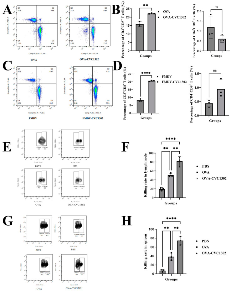
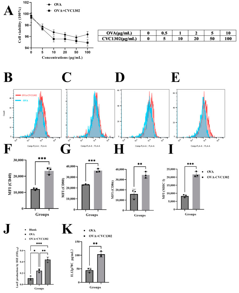
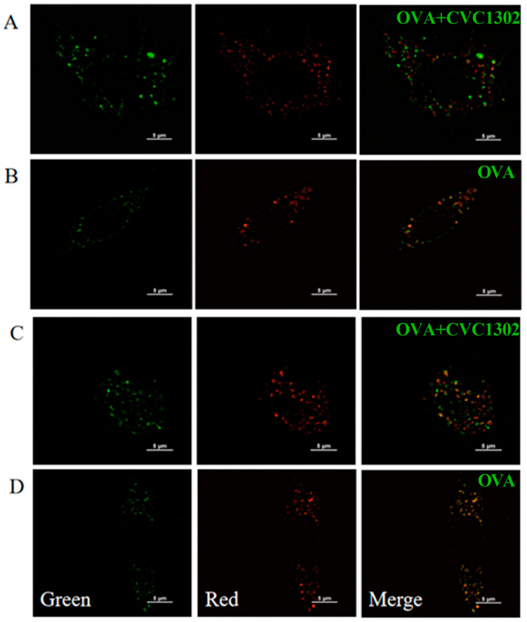
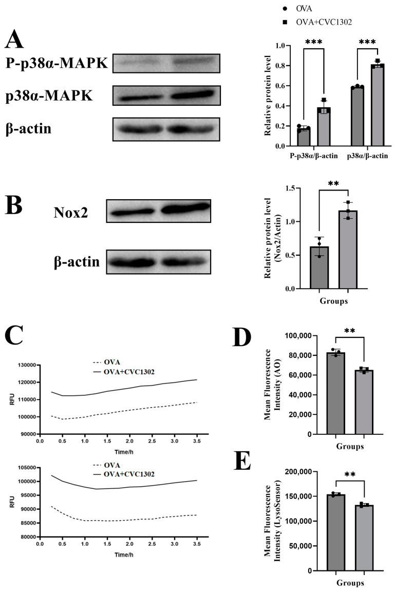
This study found a higher percentage of CD8+ T cells in piglets immunized with a CVC1302-adjuvanted inactivated foot-and-mouth disease virus (FMDV) vaccine. We wondered whether the CVC1302-adjuvanted inactivated FMDV vaccine promoted cellular immunity by promoting the antigen cross-presentation efficiency of ovalbumin (OVA) through dendritic cells (DCs), mainly via cytosolic pathways. This was demonstrated by the enhanced levels of lysosomal escape of OVA in the DCs loaded with OVA and CVC1302. The higher levels of ROS and significantly enhanced elevated lysosomal pH levels in the DCs facilitated the lysosomal escape of OVA. Significantly enhanced CTL activity levels was observed in the mice immunized with OVA-CVC1302. Overall, CVC1302 increased the cross-presentation of exogenous antigens and the cross-priming of CD8+ T cells by alkalizing the lysosomal pH and facilitating the lysosomal escape of antigens. These studies shed new light on the development of immunopotentiators to improve cellular immunity induced by vaccines.
Keywords: cellular immunity; cross-presentation; cytosolic pathways; lysosomal escape.
Conflict of interest statement
The authors declare no conflict of interest.
Figures
Similar articles
-
Identification of mechanisms conferring an enhanced immune response in mice induced by CVC1302-adjuvanted killed serotype O foot-and-mouth virus vaccine.Vaccine. 2019 Oct 8;37(43):6362-6370. doi: 10.1016/j.vaccine.2019.09.014. Epub 2019 Sep 13. Vaccine. 2019. PMID: 31526618
-
Long-term humoral immunity induced by CVC1302-adjuvanted serotype O foot-and-mouth disease inactivated vaccine correlates with promoted T follicular helper cells and thus germinal center responses in mice.Vaccine. 2017 Dec 18;35(51):7088-7094. doi: 10.1016/j.vaccine.2017.10.094. Epub 2017 Nov 9. Vaccine. 2017. PMID: 29129452
-
Pattern-Recognition Receptor Agonist-Containing Immunopotentiator CVC1302 Boosts High-Affinity Long-Lasting Humoral Immunity.Front Immunol. 2021 Nov 18;12:697292. doi: 10.3389/fimmu.2021.697292. eCollection 2021. Front Immunol. 2021. PMID: 34867941 Free PMC article.
-
The immunopotentiator CVC1302 enhances immune efficacy and protective ability of foot-and-mouth disease virus vaccine in pigs.Vaccine. 2018 Dec 18;36(52):7929-7935. doi: 10.1016/j.vaccine.2018.11.012. Epub 2018 Nov 20. Vaccine. 2018. PMID: 30470640
-
Polymer nanoparticles for cross-presentation of exogenous antigens and enhanced cytotoxic T-lymphocyte immune response.Int J Nanomedicine. 2016 Aug 5;11:3753-64. doi: 10.2147/IJN.S110796. eCollection 2016. Int J Nanomedicine. 2016. PMID: 27540289 Free PMC article.
References
-
- Den Brok M.H., Bull C., Wassink M., de Graaf A.M., Wagenaars J.A., Minderman M., Thakur M., Amigorena S., Rijke E.O., Schrier C.C., et al. Saponin-based adjuvants induce cross-presentation in dendritic cells by intracellular lipid body formation. Nat. Commun. 2016;7:13324. doi: 10.1038/ncomms13324. - DOI - PMC - PubMed
-
- Greter M., Helft J., Chow A., Hashimoto D., Mortha A., Agudo-Cantero J., Bogunovic M., Gautier E.L., Miller J., Leboeuf M., et al. GM-CSF controls nonlymphoid tissue dendritic cell homeostasis but is dispensable for the differentiation of inflammatory dendritic cells. Immunity. 2012;36:1031–1046. doi: 10.1016/j.immuni.2012.03.027. - DOI - PMC - PubMed
Grants and funding
LinkOut - more resources
Full Text Sources
Research Materials
