Mimicking Transmural Helical Cardiomyofibre Orientation Using Bouligand-like Pore Structures in Ice-Templated Collagen Scaffolds
- PMID: 38006145
- PMCID: PMC10675392
- DOI: 10.3390/polym15224420
Mimicking Transmural Helical Cardiomyofibre Orientation Using Bouligand-like Pore Structures in Ice-Templated Collagen Scaffolds
Abstract
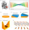
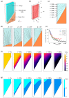
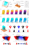
The helical arrangement of cardiac muscle fibres underpins the contractile properties of the heart chamber. Across the heart wall, the helical angle of the aligned fibres changes gradually across the range of 90-180°. It is essential to recreate this structural hierarchy in vitro for developing functional artificial tissue. Ice templating can achieve single-oriented pore alignment via unidirectional ice solidification with a flat base mould design. We hypothesise that the orientation of aligned pores can be controlled simply via base topography, and we propose a scalable base design to recapitulate the transmural fibre orientation. We have utilised finite element simulations for rapid testing of base designs, followed by experimental confirmation of the Bouligand-like orientation. X-ray microtomography of experimental samples showed a gradual shift of 106 ± 10°, with the flexibility to tailor pore size and spatial helical angle distribution for personalised medicine.
Keywords: anisotropic porosity; cardiac tissue engineering; freeze drying; polymer processing.
Conflict of interest statement
The authors declare no conflict of interest. The funders had no role in the design of the study; in the collection, analyses, or interpretation of data; in the writing of the manuscript; or in the decision to publish the results.
Figures


Similar articles
-
Complex architectural control of ice-templated collagen scaffolds using a predictive model.Acta Biomater. 2022 Nov;153:260-272. doi: 10.1016/j.actbio.2022.09.034. Epub 2022 Sep 23. Acta Biomater. 2022. PMID: 36155096
-
Versatile wedge-based system for the construction of unidirectional collagen scaffolds by directional freezing: practical and theoretical considerations.ACS Appl Mater Interfaces. 2015 Apr 29;7(16):8495-505. doi: 10.1021/acsami.5b00169. Epub 2015 Apr 14. ACS Appl Mater Interfaces. 2015. PMID: 25822583
-
Investigation of the intrinsic permeability of ice-templated collagen scaffolds as a function of their structural and mechanical properties.Acta Biomater. 2019 Jan 1;83:189-198. doi: 10.1016/j.actbio.2018.10.034. Epub 2018 Oct 24. Acta Biomater. 2019. PMID: 30366136
-
Ice Templating Soft Matter: Fundamental Principles and Fabrication Approaches to Tailor Pore Structure and Morphology and Their Biomedical Applications.Adv Mater. 2021 Aug;33(34):e2100091. doi: 10.1002/adma.202100091. Epub 2021 Jul 8. Adv Mater. 2021. PMID: 34236118 Review.
-
Aligned Ice Templated Biomaterial Strategies for the Musculoskeletal System.Adv Healthc Mater. 2023 Aug;12(21):e2203205. doi: 10.1002/adhm.202203205. Epub 2023 May 1. Adv Healthc Mater. 2023. PMID: 37058583 Free PMC article. Review.
References
-
- Streeter D.D., Bassett D.L. An Engineering Analysis of Myocardial Fiber Orientation in Pig’s Left Ventricle in Systole. Anat. Rec. 1966;155:503–511. doi: 10.1002/ar.1091550403. - DOI
-
- Nielles-Vallespin S., Khalique Z., Ferreira P.F., de Silva R., Scott A.D., Kilner P., McGill L.A., Giannakidis A., Gatehouse P.D., Ennis D., et al. Assessment of Myocardial Microstructural Dynamics by In Vivo Diffusion Tensor Cardiac Magnetic Resonance. J. Am. Coll. Cardiol. 2017;69:661–676. doi: 10.1016/j.jacc.2016.11.051. - DOI - PMC - PubMed
Grants and funding
LinkOut - more resources
Full Text Sources

