. 2025 Mar 18;124(6):876-890.
doi: 10.1016/j.bpj.2023.11.021. Epub 2023 Nov 23.

Membrane extraction in native lipid nanodiscs reveals dynamic regulation of Cdc42 complexes during cell polarization

Affiliations

Membrane extraction in native lipid nanodiscs reveals dynamic regulation of Cdc42 complexes during cell polarization

Lars N Deutz et al. Biophys J. .

Abstract

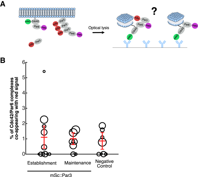
Embryonic development requires the establishment of cell polarity to enable cell fate segregation and tissue morphogenesis. This process is regulated by Par complex proteins, which partition into polarized membrane domains and direct downstream polarized cell behaviors. The kinase aPKC (along with its cofactor Par6) is a key member of this network and can be recruited to the plasma membrane by either the small GTPase Cdc42 or the scaffolding protein Par3. Although in vitro interactions among these proteins are well established, much is still unknown about the complexes they form during development. Here, to enable the study of membrane-associated complexes ex vivo, we used a maleic acid copolymer to rapidly isolate membrane proteins from single C. elegans zygotes into lipid nanodiscs. We show that native lipid nanodisc formation enables detection of endogenous complexes involving Cdc42, which are undetectable when cells are lysed in detergent. We found that Cdc42 interacts more strongly with aPKC/Par6 during polarity maintenance than polarity establishment, two developmental stages that are separated by only a few minutes. We further show that Cdc42 and Par3 do not bind aPKC/Par6 simultaneously, confirming recent in vitro findings in an ex vivo context. Our findings establish a new tool for studying membrane-associated signaling complexes and reveal an unexpected mode of polarity regulation via Cdc42.


Conflict of interest statement

Declaration of interests The authors declare no competing interests.

Figures

Figure 1
Figure 1
Cdc42 is necessary for Par6 recruitment to the plasma membrane during polarity maintenance, but their interaction is undetectable in detergent. (A) Confocal images of C. elegans zygotes carrying endogenously tagged mNG::Cdc42 (top row), mNG::Par3 (middle row), or Par6::mNG (bottom row) during polarity establishment (left) or polarity maintenance (right), with or without cdc-42(RNAi). Scale bars represent 10 μm. (B) Quantification of whole-embryo mNG::Cdc42 fluorescence intensity in control (no RNAi) or cdc-42(RNAi) embryos or in embryos with no mNG tag (N2). Error bars indicate mean ± 95% confidence interval. (C) Ratio of cortex to cytoplasm fluorescence intensity of mNG::Par3, from line scans taken during establishment (measured immediately after pseudocleavage furrow relaxation) or maintenance (measured after nuclear envelope breakdown) in either control (no RNAi) or cdc-42(RNAi) conditions. Each data point is a separate embryo. Error bars indicate mean ± 95% confidence interval. (D) Ratio of cortex to cytoplasm fluorescence for Par6::mNG as described in (C). (E) Schematic of sc-SiMPull where a fluorescently tagged zygote is lysed in a microfluidic device, and TIRF lasers are used to image bait (mNG::Cdc42) and prey (mSc::Halo or Par6::Halo) proteins as Cdc42 binds to a coverslip coated with anti-mNG nanobodies. (F) Fraction of mNG::Cdc42 molecules co-appearing with a noninteracting control protein (mSc::Halo) as a function of time, for experiments performed in detergent conditions (0.1% TX-100). N = 532,212 mNG::Cdc42 bait molecules counted from eight embryos. (G) Fraction of mNG::Cdc42 molecules co-appearing with Par6::HaloTag as a function of time, for experiments performed in detergent conditions (1% TX-100). N = 822,844 mNG::Cdc42 bait molecules counted from eight embryos.
Figure 2
Figure 2
Nanodisc polymers rapidly solubilize membrane proteins from C. elegans embryos. All panels show confocal images of one-cell embryos at the indicated times before and after rapid laser-induced cell lysis in the indicated lysis buffers. Images are representative of three to 10 embryos per condition. Scale bars represent 10 μm. (A and B) mNG::PH membrane marker. (C) Endogenously tagged mNG::Cdc42.
Figure 3
Figure 3
DIBMA polymers rapidly form lipid nanodiscs from C. elegans embryos. (A–C) Electron micrographs of cell lysate extracted from C. elegans embryos. Embryos were lysed in either (A) detergent buffer, (B) 1% DIBMA, or (C) 1% DIBMA with 7.5mM CaCl2 and then extracted from the microfluidic channel, applied to an EM grid, and negative stained. (D) Quantification of average nanodisc diameter measured in either 1% DIBMA or 1% DIBMA with CaCl2. The dotted red line is the mean with error bars indicating the 95% confidence intervals. (E) Electron micrograph of DIBMA buffer on its own, with no embryos, after negative staining.
Figure 4
Figure 4
The interaction of Cdc42 and Par6 is developmentally regulated. (A) Schematic of the sc-SiMPull experiment with embryos being lysed in DIBMA buffer. (B) Image showing mNG::Cdc42/Par6::HaloTag complexes detected after lysis in DIBMA buffer. The image is a maximum-intensity time projection of 15 consecutive difference images used for single-molecule detection. To generate these difference images, a raw image series is binned into 50-frame windows, and an average intensity projection is made for each window. Then, each averaged image is subtracted from the following averaged image, producing a difference image that eliminates background and highlights appearing molecules (6). Arrows indicate single mNG::Cdc42/Par6::HaloTag complexes. Scale bar represents 5 μm. (C) Number of Cdc42/Par6 complexes detected per experiment in the indicated conditions. Each data point represents one embryo, and red lines indicate the medians. (D) Fraction of mNG::Cdc42 molecules co-appearing with a noninteracting control protein (mSc::Halo) as a function of time, for experiments performed in 1% DIBMA conditions. N = 234,416 mNG::Cdc42 bait molecules counted from six embryos. (E and F) Fraction of mNG::Cdc42 molecules co-appearing with Par6::HaloTag as a function of time, for experiments performed in 1% DIBMA. Embryos were staged via brightfield microscopy immediately before lysis, and results from establishment (C) and maintenance phase (D) are shown separately. Curves were fit to single-exponential decay functions, resulting in the indicated estimates for koff. N = 2753 mNG::Cdc42/Par6::HaloTag complexes from 17 embryos (establishment); N = 7746 mNG::Cdc42/Par6::HaloTag complexes from 34 embryos (maintenance). (G) Image showing mNG::Cdc42 molecules associated with lipids (marked by Nile blue staining) after lysis in DIBMA buffer. The image is a maximum-intensity time projection of six consecutive difference images used for single-molecule detection, as in (B). Arrows indicate single mNG::Cdc42 molecules associated with lipids. Scale bar represents 5 μm. (H) Number of mNG::Cdc42 or mNG::HaloTag molecules co-appearing with Nile blue signal in the indicated conditions. Each data point represents one embryo, and the lines indicate the medians. (I–K) Calculation of koff values for the Cdc42/Par6 interaction based on the distribution of single-molecule dwell times. (I) Kaplan-Meier survival curves showing the probability of HaloTag-JF646 signal survival as a function of time since mNG::Cdc42/Par6::HaloTag complex capture. Thin curves show data from individual embryos, and the bold curves show the combined data from all replicates of each condition, obtained by pooling all observed complexes without regard to which embryo they originated from. A difference in survival probability between establishment- and maintenance-phase embryos is already apparent from these raw data, even without correcting for photobleaching. (J) Kaplan-Meier survival curves showing the probability of HaloTag-JF646 signal survival as a function of time since mNG::HaloTag control protein capture. The rate of JF646 signal loss due to photobleaching varies from day to day, due to fluctuations in laser power and system alignment, so these control data were used to calculate kbleach for each experimental session. (K) Calculated koff values obtained by correcting the experimental measurements of kdisappear for photobleaching (see materials and methods). Each gray circle shows the results from a single cell, the size of the circle represents the number of Cdc42/Par6 complexes analyzed, and the error bar shows the Bayesian 95% credible interval on koff. The colored bars represent the maximum probability estimate of koff obtained by pooling all observed complexes from all embryos of a given stage, along with the 95% credible interval calculated from the splitting probability analysis (see materials and methods). Embryos were staged via brightfield microscopy immediately before lysis. N = 3473 mNG::Cdc42/Par6::HaloTag complexes from 18 embryos (establishment); N = 3841 mNG::Cdc42/Par6::HaloTag complexes from 19 embryos (maintenance).
Figure 5
Figure 5
Par6 does not interact simultaneously with Cdc42 and Par3. (A) Schematic of the sc-SiMPull experiment: mNG::Cdc42/Par6::HaloTag complexes were captured and tested for co-appearance with mSc::Par3. (B) Fraction of mNG::Cdc42/Par6::HaloTag complexes co-appearing with mSc::Par3 in embryos lysed in 1% DIBMA conditions. Triple-labeled embryos (mNG::Cdc42; Par6::HaloTag; mSc::Par3) were staged via brightfield microscopy immediately before lysis, and results from establishment- and maintenance-phase embryos are plotted separately. Double-labeled negative control embryos (mNG::Cdc42; Par6::HaloTag) are mixed one-cell stages. N = 1720 mNG::Cdc42/Par6::HaloTag complexes from 11 embryos (establishment); N = 2115 mNG::Cdc42/Par6::HaloTag complexes from nine embryos (maintenance); N = 1793 mNG::Cdc42/Par6::HaloTag complexes from 8 embryos (negative control).

Similar articles

Cited by

References

    1. Goldstein B., Macara I.G. The PAR proteins: fundamental players in animal cell polarization. Dev. Cell. 2007;13:609–622. - PMC - PubMed
    1. Lang C.F., Munro E. The PAR proteins: from molecular circuits to dynamic self-stabilizing cell polarity. Development. 2017;144:3405–3416. - PMC - PubMed
    1. Joberty G., Petersen C., et al. Macara I.G. The cell-polarity protein Par6 links Par3 and atypical protein kinase C to Cdc42. Nat. Cell Biol. 2000;2:531–539. - PubMed
    1. Lin D., Edwards A.S., et al. Pawson T. A mammalian PAR-3-PAR-6 complex implicated in Cdc42/Rac1 and aPKC signalling and cell polarity. Nat. Cell Biol. 2000;2:540–547. - PubMed
    1. Qiu R.G., Abo A., Steven Martin G. A human homolog of the C. elegans polarity determinant Par-6 links Rac and Cdc42 to PKCζ signaling and cell transformation. Curr. Biol. 2000;10:697–707. - PubMed

MeSH terms

LinkOut - more resources