. 2024 Feb 1;147(2):444-457.
doi: 10.1093/brain/awad388.

Nigrostriatal tau pathology in parkinsonism and Parkinson's disease

Affiliations

Nigrostriatal tau pathology in parkinsonism and Parkinson's disease

Yaping Chu et al. Brain. .

Erratum in

Abstract

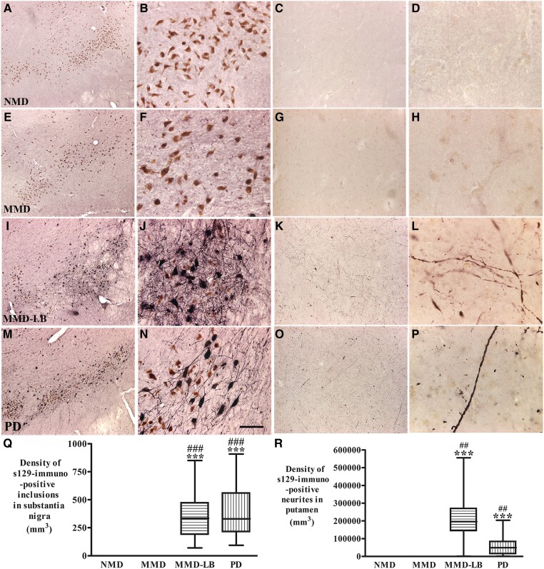
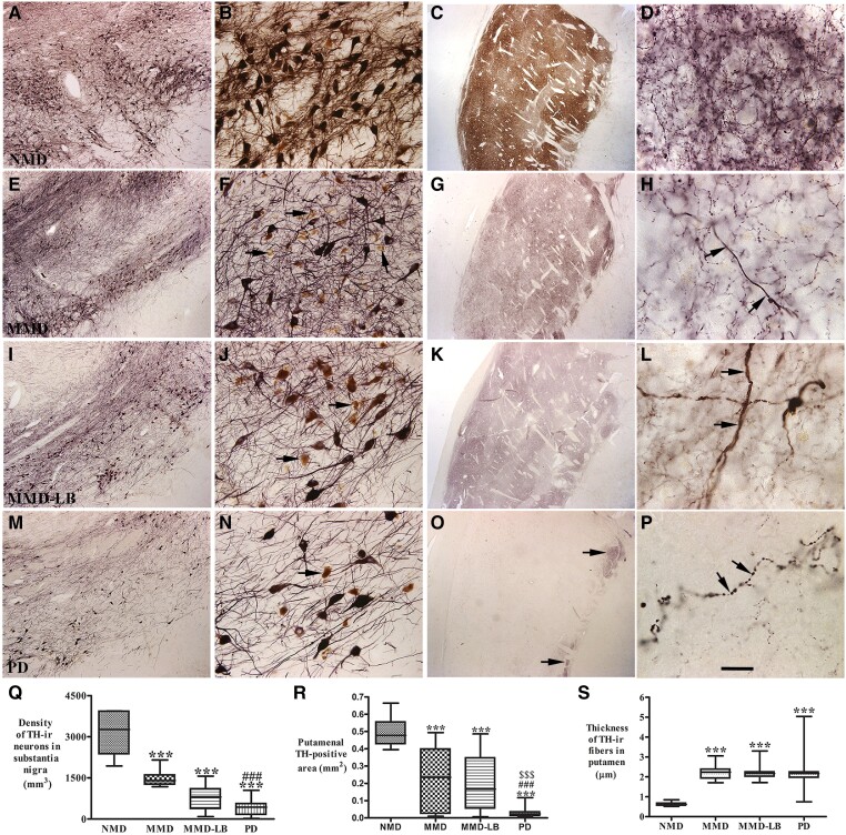
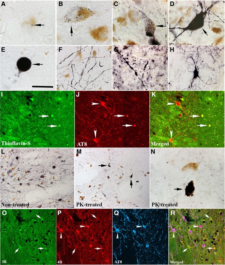
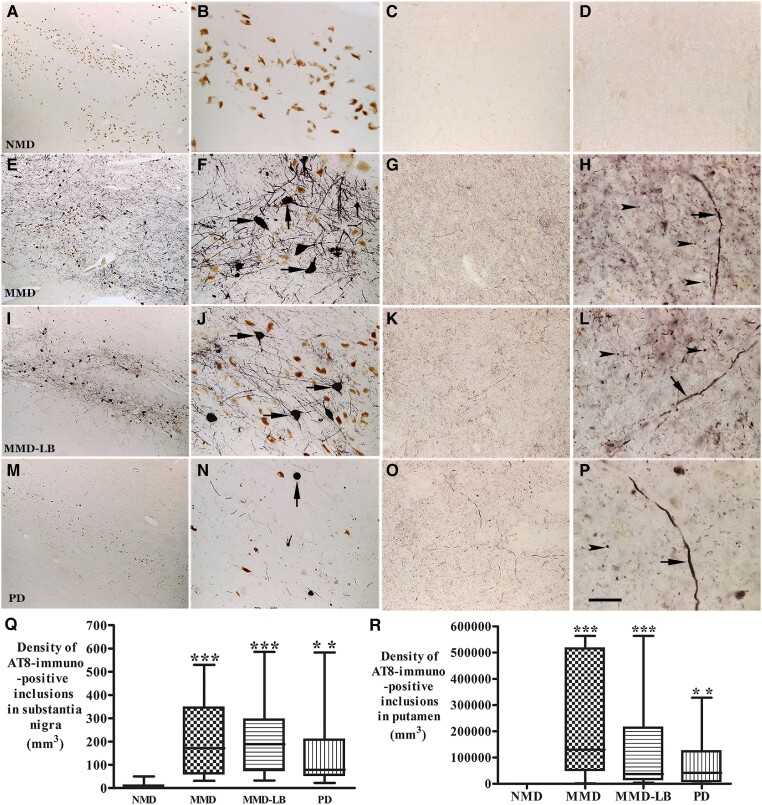
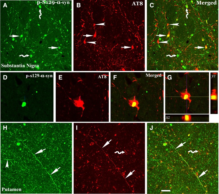
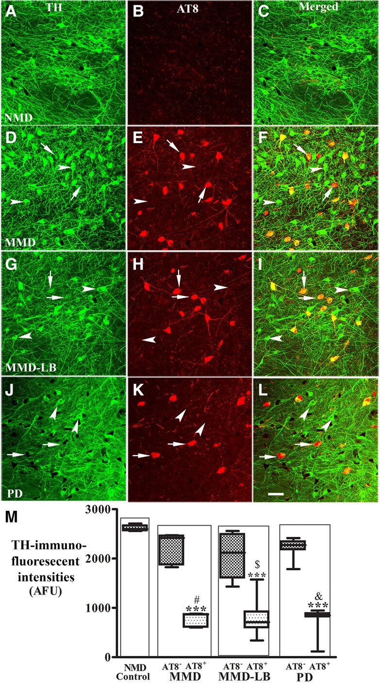
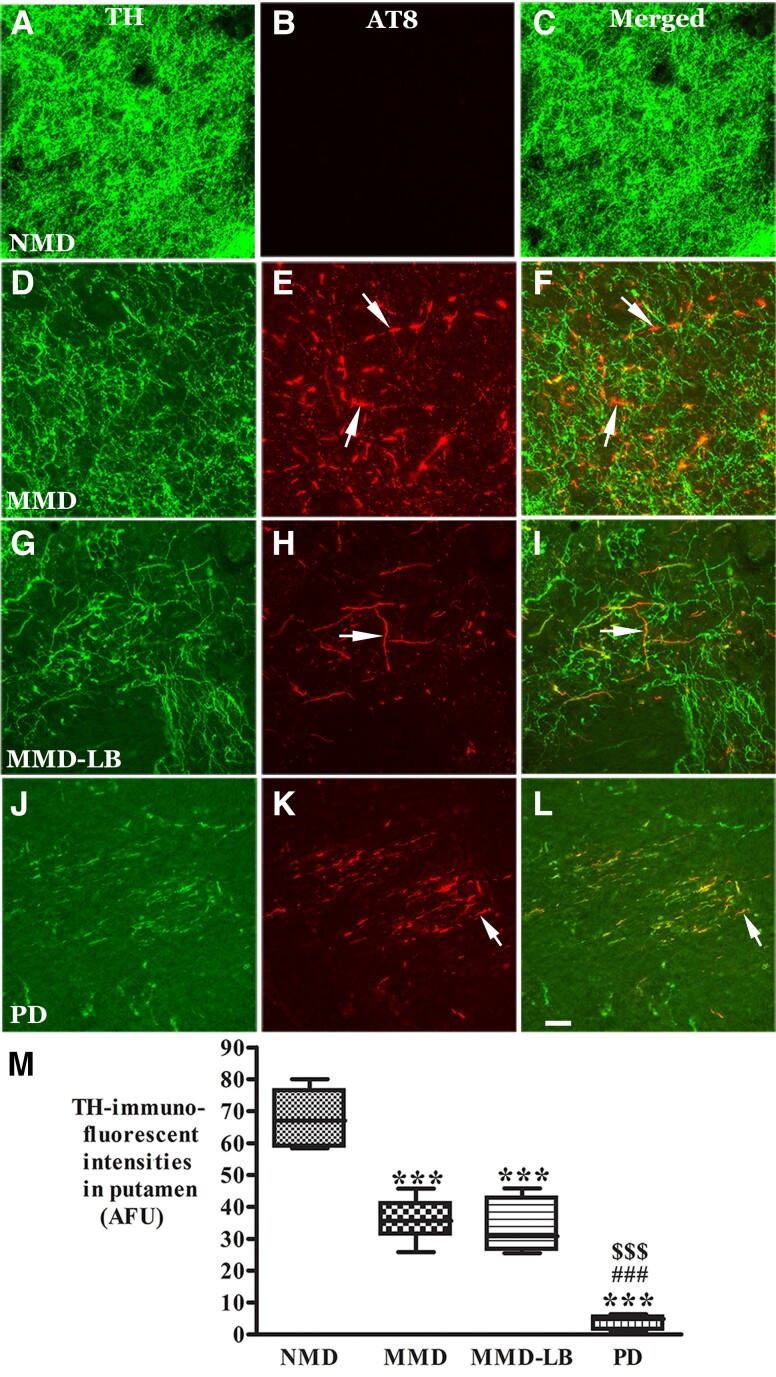
While Parkinson's disease remains clinically defined by cardinal motor symptoms resulting from nigrostriatal degeneration, it is now appreciated that the disease commonly consists of multiple pathologies, but it is unclear where these co-pathologies occur early in disease and whether they are responsible for the nigrostriatal degeneration. For the past number of years, we have been studying a well-characterized cohort of subjects with motor impairment that we have termed mild motor deficits. Motor deficits were determined on a modified and validated Unified Parkinson's Disease Rating Scale III but were insufficient in degree to diagnose Parkinson's disease. However, in our past studies, cases in this cohort had a selection bias, as both a clinical syndrome in between no motor deficits and Parkinson's disease, plus nigral Lewy pathology as defined post-mortem, were required for inclusion. Therefore, in the current study, we only based inclusion on the presence of a clinical phenotype with mild motor impairment insufficient to diagnose Parkinson's disease. Then, we divided this group further based upon whether or not subjects had a synucleinopathy in the nigrostriatal system. Here we demonstrate that loss of nigral dopaminergic neurons, loss of putamenal dopaminergic innervation and loss of the tyrosine hydroxylase-phenotype in the substantia nigra and putamen occur equally in mild motor deficit groups with and without nigral alpha-synuclein aggregates. Indeed, the common feature of these two groups is that both have similar degrees of AT8 positive phosphorylated tau, a pathology not seen in the nigrostriatal system of age-matched controls. These findings were confirmed with early (tau Ser208 phosphorylation) and late (tau Ser396/Ser404 phosphorylation) tau markers. This suggests that the initiation of nigrostriatal dopaminergic neurodegeneration occurs independently of alpha-synuclein aggregation and can be tau mediated.

Keywords: Parkinson’s disease; alpha-synuclein; dopaminergic neurodegeneration; parkinsonism; tau.


Conflict of interest statement

The authors declare no competing interests.

Figures

Figure 1
Figure 1
Qualitative and quantitative evaluation of phosphorylated α-synuclein aggregates in substantia nigra and putamen. Photomicrographs of the mid-substantia nigra (left two columns) and putamen (right two columns) from no motor deficit (NMD; AD), minimal motor deficits (MMD; EH), minimal motor deficits with nigral Lewy body (MMD-LB; IL) and Parkinson’s disease (PD; MP) show phospho-S129 α-syn (p-S129) staining patterns. p-S129 immunoreactivity was undetectable in the subjects with NMD (AD) and MMD (EH). In contrast, p-S129-immunoreactive nigral neurons were observed in subjects with MMD-LB (I and J) and patients with PD (M and N) and p-S129-immunoreactive neurites in putamen were detectable in both subjects with MMD-LB (K and L) and PD (O and P). Scale bar in N = 100 µm in B, F and J, 500 µm in A, E, I and M; 200 µm in C, G, K and O, 20 µm in D, H, L and P. Stereological analyses revealed that no p-S129-positive aggregate in substantia nigra (Q) and putamen (R) was counted in NMD and MMD groups. However, there was a significantly higher densities of p-S129 labelled aggregates in MMD-LB and PD groups. ***P < 0.001 compared with NMD. ##P < 0.01 and ###P < 0.001 compared with MMD.
Figure 2
Figure 2
Qualitative and quantitative evaluation for nigral and putamenal tyrosine hydroxylase expression. Photomicrographs of substantia nigra (left two columns) and putamen (right two columns) from no motor deficit (NMD; AD), minimal motor deficits (MMD; EH), minimal motor deficits with Lewy body pathology (MMD-LB; IL) and Parkinson’s disease (PD; MP) illustrate tyrosine hydroxylase (TH) immunoreactivity. Subject from the NMD group showed intense TH-immunoreactive (TH-ir) somata with extensive local plexus of TH-ir processes (A and B) in substantia nigra and dense TH-ir fibres throughout putamen (C and D). Nigral TH immunoreactivity was reduced in MMD (E and F) and MMD-LB subjects (I and J) compared with NMD (A and B) and some remaining nigral melanized neurons exhibited no detectable TH immunoreactivity (arrows; F and J). Putamenal TH immunoreactivities were reduced in MMD (G and H) and MMD-LB (K and L) and some of remaining TH-ir fibres displayed swollen varicosities (arrows; H and L). PD cases displayed severe reduction of TH immunoreactivity in substantia nigra (M and N) and undetectable TH immunoreactivity in major putamen (O and P), except the ventromedial putamen near globus pallidus (arrows; O) and few remaining TH immunoreactive fibres exhibited swollen segments (arrows; P). Scale bar in P = 20 µm in D, H, L, 2.0 mm in C, G, K and O , 100 µm in B, F, J and N, 500 µm in A, E, I and M. (Q) Stereological analyses revealed that the density of TH-positive neurons was gradually reduced from MMD, MMD-LB to PD relative to the NMD group. Particle analyses (R) demonstrated that the TH-immunoreactive area/mm2 of putamen was accordingly reduced from MMD, MMD-LB to PD relative to the NMD group. (S) The thickness of remaining TH-ir fibres were larger in MMD, MMD-LB and PD relative to NMD. ***P < 0.001 compared with NMD, ###P < 0.001 compared with MMD and $$$P < 0.001 compared with MMD-LB.
Figure 3
Figure 3
Morphologic features of tau aggregation in nigrostriatal system. Photomicrographs of substantia nigra (AF) and putamen (G and H) show the shapes of AT8-immunoreactive (AT8-ir) aggregates. AT8-ir punctate granule was seeded (black, arrow; A) and mixed (black; arrow; B) into neuromelanin (NM; brown). AT8-ir clumpy granules were accumulated within perikarya (arrows; C) or filled with whole neuron including somata and main processes (arrow; D) that neurons displayed NM loss. AT8-ir spherical aggregates, like a Lewy body (arrow; E) with cytoplasm and processes disappear. AT8-ir fragmental threads (F) were distributed in substantia nigra. AT8-ir varicosities and punctuated boutons dispersed into putamen (G). Few AT8-ir putamenal neuron displayed dark stained somata with abundant processes (H). Double-labelling revealed the dense phosphorylated tau accumulations (AT8, red, arrows; J) were thioflavin-S positive (green, arrows; I) but the granules seen with phosphorylated tau were thioflavin negative (arrowheads; IK). Phosphorylated tau immunolabelling (L) observed from the section without proteinase K (PK) treatment was basically eliminated following PK treated section (M and N). However, the dense tau accumulations (arrows; M and N) were resistant to PK treatment. Fluorescent triple-labelling (OR) showed that both 3R (green, arrows; O) and 4R (red, arrows and arrowheads; P) tau isoforms existed in nigral melanized neurons. 4R isoform in same melanized neurons displayed stronger intense staining than 3R and co-localized with phosphorylated tau marker AT8 (blue, arrowheads; PR). Scale bar in E = 20 µm in AH and N, 100 µm in IK, 120 µm in L, M and OR.
Figure 4
Figure 4
Qualitative and quantitative evaluation of tau aggregates in substantia nigra and putamen. Photomicrographs of the mid-substantia nigra (left two columns) and putamen (right two columns) from no motor deficit (NMD; AD), minimal motor deficits (MMD; EH), minimal motor deficits with nigral Lewy body (MMD-LB; IL) and Parkinson’s disease (PD; MP) show AT8-immunoreactive (AT8-ir) patterns. AT8 immunoreactivity was not detected in NMD group (AD). In contrast, numerous AT8-ir neurons were distributed throughout substantia nigra (E and I) and displayed dark somata with abundant processes in subjects with MMD (arrows; F) and MMD-LB (arrows; J). AT8-ir intensities in putamen were higher in MMD (G) and MMD-LB (K) and displayed punctuated dots (arrowheads; H and L) and the same expanded fibre (arrow; H and L). The AT8-ir aggregates with limited processes (arrow; N) in substantia nigra (M) and relative less AT8-ir punctuated dots (arrowhead; O and P) in putamen were observed in PD. Scale bar in P = 20 µm in D, H, L, 100 µm in B, C, F, G, J, K, N and O, 500 µm in A, E, I and M. (Q) Stereological analyses revealed that the density of AT8-ir aggregates in substantia nigra and (R) the density of AT8-ir dots and threads in putamen were significant higher in MMD, MMD-LB and PD relative to NMD group. **P < 0.01 and ***P < 0.001 compared with NMD.
Figure 5
Figure 5
Co-localization analyses of phosphorylated tau (AT8) and αsyn (pS129αsyn) in nigrostriatal system. Confocal microscopic images of substantia nigra (AG) and putamen (HJ) illustrated p-S129-α-syn (green; A, D and H), AT8 (red; B, E and I) and co-localization of AT8 and p-S129 (merged; C, F, G and J). There were three populations of immunofluorescence-labelling aggregates: p-S129-α-syn and AT8 double-labelling (arrows; AC), AT8 single-labelling (arrowhead; B and C) or p-S129-α-syn single-labelling (curved arrow; A and C). The p-S129-α-syn-labelled aggregate (D) deposited within AT8-labelled perikarya (E and F). 3D reconstruction of confocal image further illustrated the co-localization of labelled p-S129-α-syn and AT8 (G): the large panel represents a cross section of the cell layer; the horizontal (yellow) and vertical (pink) lines through them denote the planes of the adjoining xz and yz sections, respectively. Bottom and right: The xz and yz cross sections were obtained from the combined serial optical sections of these cell layers using Olympus Confocal Fluoroview software. The 3D reconstruction analyses revealed that p-S129-α-syn were co-localized with AT8 (yellow). In putamen p-S129-α-syn (arrowhead; H) and AT8-labelled threads and dots (curved arrow; I) were not co-localized, but the longer fibre with AT8 labelling was p-S129-α-syn immunopositive (arrows; HJ). Scale bar in J = 50 μm in HJ, 25μm in DG, 100μm in AC.
Figure 6
Figure 6
Reduction of tyrosine hydroxylase levels in nigral neurons with AT8-immunorepressive aggregates. Confocal microscopic images of substantia nigra from no motor deficit (NMD; AC), minimal motor deficits (MMD; DF), minimal motor deficits with nigral Lewy body (MMD-LB; GI) and Parkinson’s disease (PD; JL) illustrated immunostaining for tyrosine hydroxylase (TH; green; A, D, G and J), AT8 (red; B, E, H and K) and co-localization of TH and AT8 (merged; C, F, I and L). Note that TH immunofluorescent intensity was extensively reduced in the neurons with tau aggregates (arrows; DL) but not in the neurons without tau aggregates (arrowheads; DL). Scale bar in L = 100 µm (applies to all panels). Measurements of immunofluorescent intensities (M) further revealed that TH expression was significantly reduced in the neurons with tau aggregates (AT8+) but not in the neurons without tau aggregate (AT8). ***P < 0.001 related to NMD control, #P < 0.05 related to AT8 immunonegative neurons in MMD, $P < 0.05 related to AT8 immunonegative neurons in MMD-LB and &P < 0.05 related to AT8 immunonegative neurons in PD groups. Data: mean ± standard deviation. AFU = arbitrary fluorescence units.
Figure 7
Figure 7
Reduction of tyrosine hydroxylase levels in putamen with AT8-immunorepressive aggregates. Confocal microscopic images of putamen from no motor deficit (NMD; AC), minimal motor deficits (MMD; DF), minimal motor deficits with nigral Lewy body pathology (MMD-LB; GI) and Parkinson’s disease (PD; JL) illustrated immunostaining for tyrosine hydroxylase (TH; green; A, D, G and J), AT8 (red; B, E, H and K) and co-localization of TH and AT8 (merged; C, F, I and L). Note that TH immunofluorescent intensity was extensively reduced in MMD (D), MMD-LB (G) and PD groups (L) relative to NMD group (A). The AT8-labelled threads and punctuated dots (arrows; E, H and K) were TH immunonegative (arrows; F, I and L). Scale bar in L = 40 µm (applies to all panels). Measurements of TH-immunorepressive (ir) intensity (M) further revealed that TH-ir fibres were significantly reduced in the putamen with tau aggregates in MMD, MMD-LB and PD group. ***P < 0.001 related to NMD group, ###P < 0.001 related to MMD group and $$$P < 0.001 related to MMD-LB group. Data: mean ± standard deviation. AFU = arbitrary fluorescence units.

Comment in

Similar articles

Cited by

References

    1. Dickson DW, Braak H, Duda JE, et al. . Neuropathological assessment of Parkinson’s disease: Refining the diagnostic criteria. Lancet Neurol. 2009;8:1150–1157. - PubMed
    1. Gibb WRG, Lees AJ. A comparison of clinical and pathological features of young- and old-onset Parkinson’s disease. Neurology. 1988;38:1402–1402. - PubMed
    1. Braak H, del Tredici K, Rüb U, de Vos RAI, Jansen Steur ENH, Braak E. Staging of brain pathology related to sporadic Parkinson’s disease. Neurobiol Aging. 2003;24:197–211. - PubMed
    1. Milber JM, Noorigian JV, Morley JF, et al. . Lewy Pathology is not the first sign of degeneration in vulnerable neurons in Parkinson disease. Neurology. 2012;79:2307–2314. - PMC - PubMed
    1. Kalia LV, Lang AE, Hazrati LN, et al. . Clinical correlations with Lewy body pathology in LRRK2-related Parkinson disease. JAMA Neurol. 2015;72:100–105. - PMC - PubMed

Publication types