. 2024 Jun;14(6):1567-1581.
doi: 10.1007/s13346-023-01472-y. Epub 2023 Nov 25.

3D printing injectable microbeads using a composite liposomal ink for local treatment of peritoneal diseases

Affiliations

3D printing injectable microbeads using a composite liposomal ink for local treatment of peritoneal diseases

Remo Eugster et al. Drug Deliv Transl Res. 2024 Jun.

Abstract

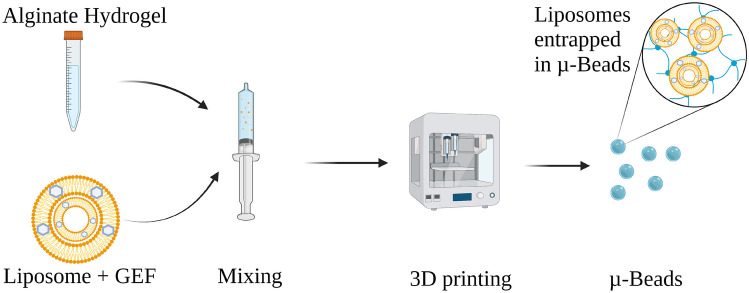
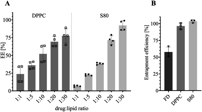
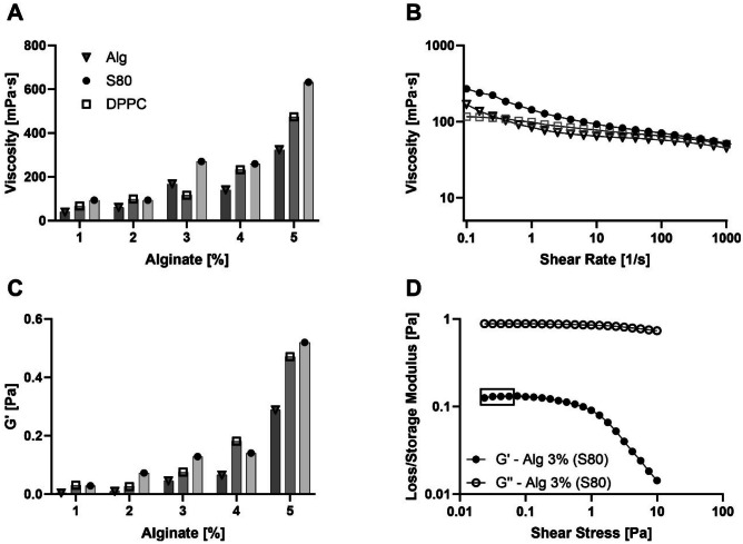
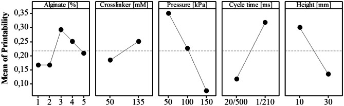
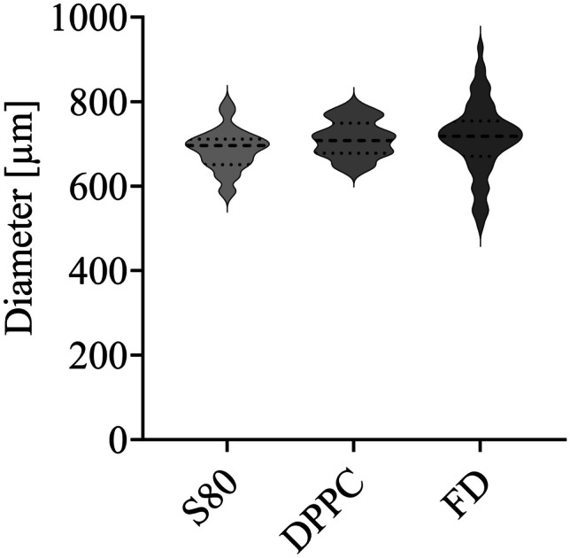
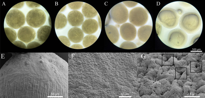
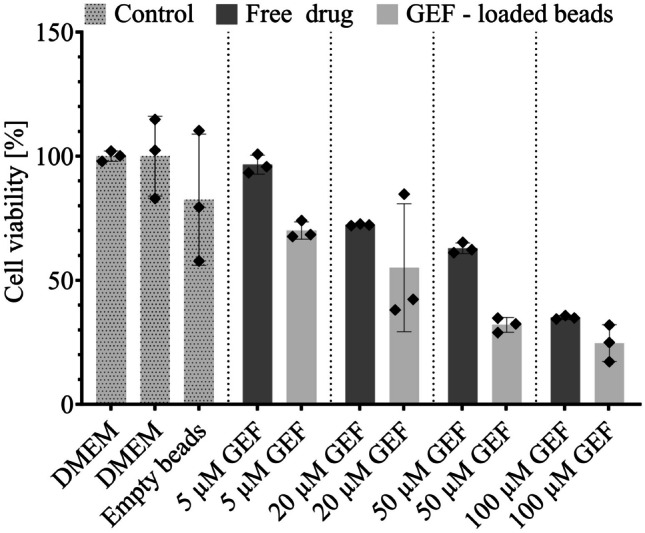
The peritoneal cavity offers an attractive administration route for challenging-to-treat diseases, such as peritoneal carcinomatosis, post-surgical adhesions, and peritoneal fibrosis. Achieving a uniform and prolonged drug distribution throughout the entire peritoneal space, though, is difficult due to high clearance rates, among others. To address such an unmet clinical need, alternative drug delivery approaches providing sustained drug release, reduced clearance rates, and a patient-centric strategy are required. Here, we describe the development of a 3D-printed composite platform for the sustained release of the tyrosine kinase inhibitor gefitinib (GEF), a small molecule drug with therapeutic applications for peritoneal metastasis and post-surgical adhesions. We present a robust method for the production of biodegradable liposome-loaded hydrogel microbeads that can overcome the pharmacokinetic limitations of small molecules with fast clearance rates, a current bottleneck for the intraperitoneal (IP) administration of these therapeutics. By means of an electromagnetic droplet printhead, we 3D printed microbeads employing an alginate-based ink loaded with GEF-containing multilamellar vesicles (MLVs). The sustained release of GEF from microbeads was demonstrated. In vitro studies on an immortalized human hepatic cancer cell line (Huh-7) proved concentration-dependent cell death. These findings demonstrate the potential of 3D-printed alginate microbeads containing liposomes for delivering small drug compounds into the peritoneum, overcoming previous limitations of IP drug delivery.

Keywords: 3D printing; Drop-on-demand manufacturing; Hydrogel microbeads; Liposomes; Peritoneal drug delivery; Sustained drug release.


Conflict of interest statement

No private study sponsors had any involvement in the study design, data collection, or interpretation of data presented in this manuscript. P.L. declares the following competing interests: she has consulted and received research grants from Lipoid, Sanofi-Aventis Deutschland and DSM Nutritional Products Ltd. R.E., A.G., S.A. and A.S. declare no competing interests.

Figures

Fig. 1
Fig. 1
Preparation of microbeads by mixing alginate hydrogel with liposomes encapsulating GEF and 3D printing the formulation
Fig. 2
Fig. 2
Encapsulation and entrapment efficiency. A Encapsulation efficiency (EE%) of GEF in DPPC or S80 MLVs with different drug-to-lipid ratios (mean ± SD, n = 4). B Entrapment efficiency of GEF-loaded beads (mean ± SD, n = 3)
Fig. 3
Fig. 3
Rheological properties of liposomal ink inks. A Viscosity at rest for inks containing alginate spanning from 1 to 5% and either liposomes (S80 or DPPC; 15 mM) or hydration buffer. B Flow curve of inks used in the study (15 mM S80; 3% Alg or 15 mM DPPC; 3% Alg or 3% Alg). C G’ for assessed inks as for viscosity at rest. D Loss/storage modulus of DP ink used for in vitro study (15 mM S80; 3% Alg). Rectangle highlights values used for determining G’
Fig. 4
Fig. 4
Main effects plot for printability in a multifactorial experimental design (120 runs). This plot illustrates the impact of the considered factors on the printability response. Higher response values denote a more desirable printability for each factor level. The slope of the lines connecting the factor levels serves as an indicator of the significance of their influence on the printability response
Fig. 5
Fig. 5
Violin plot depicting the distribution of microbead diameters across three non-consecutive batches per formulation (median ± quartile, n = 60). Considering intra- and inter-batch variation, this representation serves as an indicator of the robustness in the production of liposome-laden microbeads. While the liposome-laden microbeads manifest a similar distribution, the microbeads containing FD result in a larger process variation
Fig. 6
Fig. 6
Images of microbeads after 3D printing. A S80 microbeads (15 mM S80; 3% Alg; 0.5 mM GEF). B S80 microbeads (15 mM S80; 3% Alg 0.5 mM GEF) after injection through an 18G needle. C DPPC microbeads (15 mM DPPC; 3% Alg; 0.5 mM GEF). D FD microbeads (3% Alg; 0.5 mM GEF). SEM images (E, F, G) of dried microbead (15 mM S80; 3% Alg; 0.5 mM GEF). E Surface image of microbeads at a macroscale. F Microscale of surface area shows homogeneous distribution of pores over surface area. G Pores with a diameter of ~ 100 nm on the surface indicated by rectangles
Fig. 7
Fig. 7
Cumulative drug release from 3D-printed microbeads (15 mM lipid; 3% Alg; 0.5 mM GEF) in PSF (mean ± SD, n ≥ 3). Total of 100% drug was retrieved with extraction following release experiment
Fig. 8
Fig. 8
Cell viability of different GEF concentration on Huh-7 cells (mean ± SD, n = 3) normalized to DMEM. GEF was either administered as free drug in presence of 0.5% V/V DMSO (dark grey) or encapsulated in MLVs entrapped in microbeads (light grey), where no DMSO had to be added. Cell viability in DMEM and exposed to empty beads (light grey dotted) was measured as control. For experiments in DMEM, the solubility did not allow the testing under sink conditions

Similar articles

Cited by

References

    1. Roy P, Mignet N, Pocard M, Boudy V. Drug delivery systems to prevent peritoneal metastasis after surgery of digestives or ovarian carcinoma: a review. Int J Pharm. 2021;592:120–141. doi: 10.1016/j.ijpharm.2020.120041. - DOI - PubMed
    1. Mohamed F, Sugarbaker PH. Carrier solutions for intraperitoneal chemotherapy. Surg Oncol Clin N Am. 2003;12:813–824. doi: 10.1016/S1055-3207(03)00039-5. - DOI - PubMed
    1. Braet H, Fransen P-P, Mariën R, Lollo G, Ceelen W, Vervaet C, Balcaen L, Vanhaecke F, Vanhove C, van der Vegte S, Gasthuys E, Vermeulen A, Dankers PYW, De Smedt SC, Remaut K. CO2-driven nebulization of pH-sensitive supramolecular polymers for intraperitoneal hydrogel formation and the treatment of peritoneal metastasis. ACS Appl Mater Interfaces. 2023;15:49022–49034. doi: 10.1021/acsami.3c11274. - DOI - PubMed
    1. Mirahmadi N, Babaei MH, Vali AM, Dadashzadeh S. Effect of liposome size on peritoneal retention and organ distribution after intraperitoneal injection in mice. Int J Pharm. 2010;383:7–13. doi: 10.1016/J.IJPHARM.2009.08.034. - DOI - PubMed
    1. Goodman MD, McPartland S, Detelich D, Saif MW. Chemotherapy for intraperitoneal use: a review of hyperthermic intraperitoneal chemotherapy and early post-operative intraperitoneal chemotherapy. J Gastrointest Oncol. 2016;7:45–57. doi: 10.3978/j.issn.2078-6891.2015.111. - DOI - PMC - PubMed

Publication types

MeSH terms