Review
. 2023 Dec:68:102967.
doi: 10.1016/j.redox.2023.102967. Epub 2023 Nov 18.

Oxidative stress in the eye and its role in the pathophysiology of ocular diseases

Affiliations
Review

Oxidative stress in the eye and its role in the pathophysiology of ocular diseases

Elsa Wilma Böhm et al. Redox Biol. 2023 Dec.

Abstract

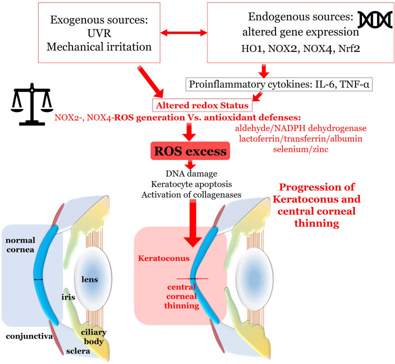
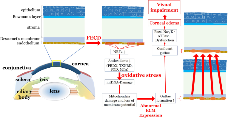
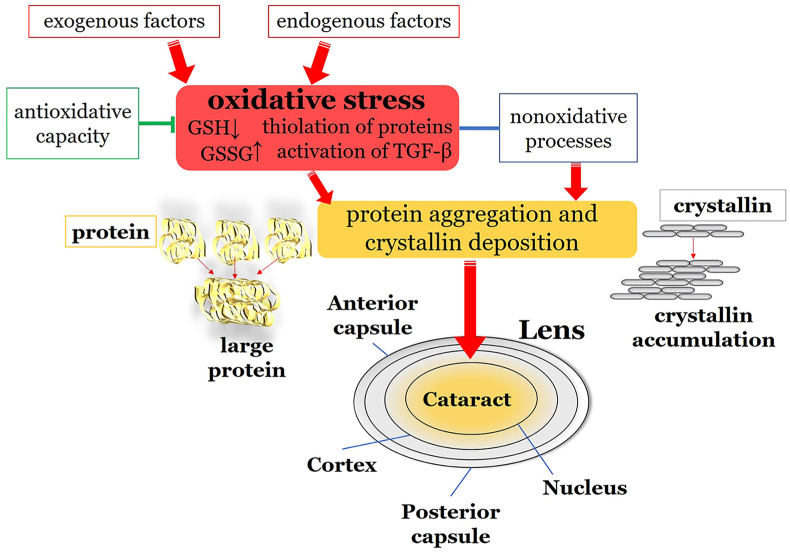
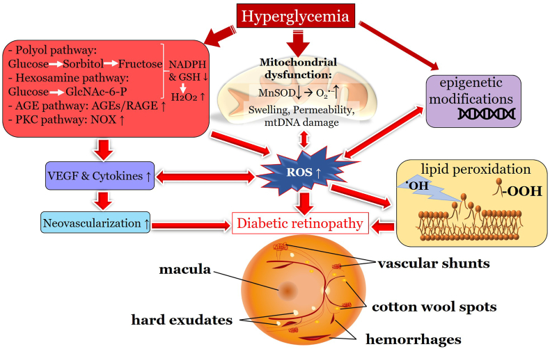
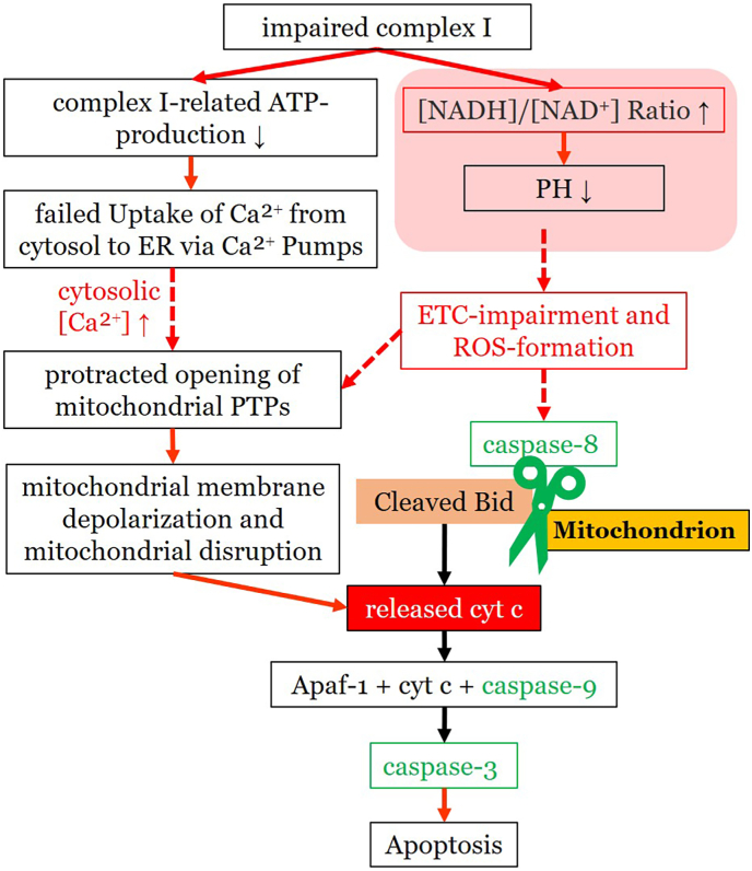
Oxidative stress occurs through an imbalance between the generation of reactive oxygen species (ROS) and the antioxidant defense mechanisms of cells. The eye is particularly exposed to oxidative stress because of its permanent exposure to light and due to several structures having high metabolic activities. The anterior part of the eye is highly exposed to ultraviolet (UV) radiation and possesses a complex antioxidant defense system to protect the retina from UV radiation. The posterior part of the eye exhibits high metabolic rates and oxygen consumption leading subsequently to a high production rate of ROS. Furthermore, inflammation, aging, genetic factors, and environmental pollution, are all elements promoting ROS generation and impairing antioxidant defense mechanisms and thereby representing risk factors leading to oxidative stress. An abnormal redox status was shown to be involved in the pathophysiology of various ocular diseases in the anterior and posterior segment of the eye. In this review, we aim to summarize the mechanisms of oxidative stress in ocular diseases to provide an updated understanding on the pathogenesis of common diseases affecting the ocular surface, the lens, the retina, and the optic nerve. Moreover, we discuss potential therapeutic approaches aimed at reducing oxidative stress in this context.

Keywords: Antioxidants; Eye; Ocular diseases; Oxidative stress; Pathophysiology; Reactive oxygen species.


Conflict of interest statement

Declaration of competing interest The authors declare that they do not have financial or other conflicts of interest. All authors made substantial contributions to the conception and design of the article, drafting or revising the article for critically important intellectual content and approved the final version of the article.

Figures

Fig. 1
Fig. 1
Structure of the ocular tear film and conjunctiva. The ocular tear film is composed of three different layers. The lipid layer is produced by the Meibomian glands located in the eyelids and is separated into a non-polar lipid sublayer and a polar lipid sublayer. The aqueous layer is produced by the lacrimal gland and accessory lacrimal glands and contains various proteins and electrolytes, including growth factors, cytokines, and the antioxidant defense system of the ocular tear film. The mucin layer is the innermost layer secreted by conjunctival goblet cells. The ocular conjunctiva consists of the conjunctival epithelium, a non-keratinized stratified squamous epithelium, and the substantia propria with vessels, connective tissue and lymphocytes. AA: ascorbic acid; GSH: glutathione; SOD: superoxide dismutase.
Fig. 2
Fig. 2
Superficial punctate keratitis in a patient with dry eye disease. This slit lamp photograph of the anterior eye segment stained with sodium fluorescein-containing drops reveals fluorescein-positive yellow spots on the corneal surface, which represent epithelial defects often seen in patients with dry eye disease. Unpublished image. Department of Ophthalmology, University Medical Center Mainz. (For interpretation of the references to colour in this figure legend, the reader is referred to the Web version of this article.)
Fig. 3
Fig. 3
Role of oxidative stress in the pathogenesis of dry eye disease. Environmental factors, such as air pollution, particulate matter, gaseous substances or UVR as well as aging lead to increased inflammation with consequent imbalance between ROS generation and the antioxidant defense. Additionally, hyperosmolarity and instability of the tear film further aggravates inflammation and oxidative stress contributing to the vicious circle of dry eye disease. DED: dry eye disease; UVR: ultraviolet radiation; IL-1: interleukin 1; IL-6: interleukin 6; TNF-α: tumor necrosis factor α; O2•−: superoxide; H2O2: hydroxyl peroxide; Nrf2: nuclear factor erythroid 2-related factor 2; SOD: superoxide dismutase; CAT: catalase.
Fig. 4
Fig. 4
Pterygium in a 30-year-old man. The photograph of the anterior segment of the right eye shows the typical appearance of a pterygium with a fleshy, triangular-shaped mass of conjunctival tissue growing from the nasal side onto the cornea. Unpublished image. Department of Ophthalmology, University Medical Center Mainz.
Fig. 5
Fig. 5
Sources and role of oxidative stress in keratoconus. Exogenous sources of oxidative stress include UV radiation and mechanical irritation. Moreover, altered gene expression of ROS-related genes, such as HO1, NOX2, NOX4 and NRF2, with consequently elevated expression of proinflammatory cytokines, such as IL-6, TNF-α and MMP, are found. By increased levels of ROS, protein damage and keratocyte death as well as changes in the extracellular matrix occur causing progressive thinning of the central cornea. UVR: ultraviolet radiation; HO1: heme oxygenase-1; NOX: nicotinamide adenine dinucleotide phosphate oxidase; Nrf2: nuclear factor erythroid 2-related factor 2; IL-6: interleukin 6; TNF-α: tumor necrosis factor α; ROS: reactive oxygen species; NADPH: reduced form of nicotinamide adenine dinucleotide.
Fig. 6
Fig. 6
Oxidative stress related pathways in the pathogenesis of FECD. In patients with FECD, downregulation of Nrf2, which is in turn responsible for the expression of numerous antioxidants, leads to increased oxidative stress. Damaged mitochondrial DNA causes mitochondrial dysfunction with loss of membrane potential. This leads to increased formation of guttae. Mitochondrial loss further leads to dysfunction of Na+/K+-ATPase. Consecutive loss of the osmotic gradient between the anterior chamber and the corneal stroma causes water influx and stromal edema with decrease of visual acuity. Nrf2: nuclear factor erythroid 2-related factor 2; PRDX: peroxiredoxin; TXNRD: thioredoxin reductase; SOD: superoxide dismuatse; MT3: metallothionein-3; ECM: extracellular matrix; FECD: Fuchs endothelial corneal dystrophy.
Fig. 7
Fig. 7
Cataract in an 83-year-old woman. The photograph of the anterior segment of the right eye shows the typical clinical appearance of cataract with corticonuclear lens opacification. Unpublished images. Department of Ophthalmology, University Medical Center Mainz.
Fig. 8
Fig. 8
Role of oxidative stress in cataract formation. Exogenous factors, such as UV radiation, and endogenous factors, such as aging cause increased oxidative stress. This leads to crystallin deposition, ultimately resulting in the formation of large protein conglomerates causing the typical opacity of cataract and leading to loss of visual acuity. GSH: glutathione; GSSG: oxidized glutathione; TGF-β: transforming growth factor-beta.
Fig. 9
Fig. 9
Non-neovascular age-related macular degeneration in an 80-year-old woman. Optical coherence tomography and fundus photograph of a patient with nonvascular (dry) age related macular degeneration are shown. The green arrows point to drusen, that appear as focal yellow extracellular deposits within and around the macula. Unpublished image. Department of Ophthalmology, University Medical Center Mainz. (For interpretation of the references to colour in this figure legend, the reader is referred to the Web version of this article.)
Fig. 10
Fig. 10
Non-proliferative diabetic retinopathy in a 70-year old woman. Fundus photographs of both eyes and optical coherence tomography of left eye in a patient with severe non-proliferative diabetic retinopathy are shown. The green arrows point to intraretinal hemorrhages and hard exsudates around the macula. Optical coherence tomography demonstrates diabetic macular edema. Unpublished image. Department of Ophthalmology, University Medical Center Mainz. (For interpretation of the references to colour in this figure legend, the reader is referred to the Web version of this article.)
Fig. 11
Fig. 11
Source and role of oxidative stress in diabetic retinopathy. Hyperglycemia and epigenetic modifications induce mitochondrial dysfunction and metabolic pathways, such as polyol, hexosamine, PKC pathways and AGEs leading elevated levels of ROS. Elevated levels of ROS cause lipid peroxidation and neovascularization through elevated VEGF and cytokine levels, finally terminating in diabetic retinopathy. AGEs: Advanced Glycation End Products; PKC: Protein Kinase C; VEGF: vascular endothelial growth factor; ROS: reactive oxygen species; NADPH: reduced form of nicotinamide adenine dinucleotide; GSH: glutathione; H2O2: hydroxyl peroxide; GlcNAc-6-P: N-acetlyglucosamine 6-phosphate; MnSOD: mitochondrial superoxide dismutase; O2•−: superoxide.
Fig. 12
Fig. 12
Branch retinal vein occlusion in a 62-year-old patient. Fundus photograph of a left eye with retinal vein occlusion in the inferior branch of central artery is demonstrated. Retinal occlusion leads to dilatation and tortuosity of retinal veins. Retinal hemorrhage as well as macular edema are demonstrated. Unpublished image. Department of Ophthalmology, University Medical Center Mainz.
Fig. 13
Fig. 13
Retinitis pigmentosa in a 42-year-old patient. Fundus photograph und perimetry of both eyes with retinitis pigmentosa are demonstrated. Typical clinical signs, such as bone spicule-shaped pigment deposits, retinal atrophy, attenuation of retinal vessels, and a pale appearance of the optic nerve head can be detected. Perimetry shows typical concentric visual field loss with consequent tunnel vision. Unpublished image. Department of Ophthalmology, University Medical Center Mainz.
Fig. 14
Fig. 14
Model representing the trabecular outflow of aqueous humor to the Schlemm’s canal. The aqueous humor first flows through the large intercellular spaces of the uveal trabecular layer. Then, it passes via the intercellular as well as via transcellular route through the trabecular lamellae of the corneoscleral stratum. Subsequently, the aqueous humor reaches the juxtacanalicular trabecular layer, characterized by densely compacted extracellular matrix and reduced intercellular spaces. Finally, through the passage of this last layer, the aqueous humor arrives at the Schlemm’s canal. TM: trabecular meshwork.
Fig. 15
Fig. 15
ROS sources and ROS-induced damage in glaucoma. Studies reported decreased systemic antioxidative parameters such as TAC or TAS and elevated redox biomarkers in patients with glaucoma, evidencing the role of the oxidative stress in this disorder. Possible pathophysiological initiators, which drive to an altered redox status, are suggested to be a loss of integrity and function of the trabecular meshwork, with subsequent elevation of IOP, and abnormal perfusion of the retina and optic nerve, causing hypoxia. These conditions trigger ROS-sources, such as NOX2, leading to oxidative stress. The ROS excess in turn exacerbates hypoxia and raised IOP, promoting the glial activation, TMC and RGC death and ONH remodeling. TAS: total antioxidant status; BAP: biological antioxidant potential; TRAP: total reactive antioxidant potential; TAC: total antioxidant capacity; MDA: malonyldialdehyde; 8-OHdG: 8'-hydroxy-2'-deoxyguanosine; ROS: reactive oxygen species; IOP: intraocular pressure; HIF-1α: hypoxia inducible factor 1α; NOX2: nicotinamide adenine dinucleotide phosphate oxidase 2; XO: xanthine oxidase; COX: cyclooxigenase; ER: endoplasmic reticulum; TNF-α: tumor necrosis factor α; NF-kB: nuclear factor ‘kappa-light-chain-enhancer’ of activated B-cells; TMC: trabecular meshwork cell; RGC: retinal ganglion cell; ONH: optic nerve head.
Fig. 16
Fig. 16
Proposed model of interrelation between mitochondria and endoplasmic reticulum in retinal ganglion cells and trabecular meshwork cells in glaucoma. In the context of glaucoma, TMCs and RGCs establish an interplay between the ER and mitochondrial stress. Under conditions of oxidative stress, the ER becomes oversaturated of misfolded proteins. Consequently, through activation and chronic stimulation of the unfolded protein response, some specific pathways, such as the PERK/ATF-4/CHOP pathway, lead to apoptosis, inflammation, and ROS generation. In addition, calcium ions are released by the stressed ER, and are indirectly gained by the mitochondria, therefore inducing a ROS overproduction and downstream an amplification of inflammation and apoptosis. IOP: intraocular pressure; ER: endoplasmic reticulum; TM: trabecular meshwork; NF-kB: nuclear factor ‘kappa-light-chain-enhancer’ of activated B-cells; ROS: reactive oxygen species; ERAD: ER-associated degradation; eIF2α: eukaryotic initiation factor 2α; PERK: protein kinase RNA-like ER-kinase; IRE-1: inositol-requiring protein 1; ATF: activating transcription factor; CHOP: CCAAT-enhancer-binding protein homologous protein; sXBP1: spliced X-box binding protein-1; TNFR2: tumor necrosis factor receptor 2; ASK-1: apoptosis signal-regulating kinase 1; JNK: Janus kinase; ERK: extracellular-signal-regulated kinase; NOX: nicotinamide adenine dinucleotide phosphate oxidase; ERO1: ER-oxidoreductin 1; RyR: Ryanodine receptor; IP3R: Inositol 1,4,5-trisphosphate receptor.
Fig. 17
Fig. 17
Model representing the LHON pathogenesis and the apoptotic cascade via cytochrome c. In LHON, a defected complex I, a main component of the mitochondrial ETC, causes an increased NADH/NAD⁺ ratio and subsequently a decreased PH, driving to an impairment of the oxidative phosphorylation and to ROS overproduction, which ultimately activates caspase-dependent apoptosis. Additionally, the complex I-related ATP production is reduced, therefore driving to a decreased activity of the ATP-dependent calcium pumps of endoplasmic reticulum and consequently to increased cytosolic concentrations of calcium ions. Hereby, a prolonged opening of the mitochondrial PTPs guides to a membrane depolarization and to a mitochondrial disruption, which finally amplifies the cytochrome c release and the caspase-dependent apoptosis. ROS: reactive oxygen species; NAD: nicotinamide adenine dinucleotide; NADH: reduced form of nicotinamide adenine dinucleotide; ATP: adenosine triphosphate; Bid: BH3 interacting-domain death agonist; cyt c: cytochrome c; Apaf-1: apoptotic protease-activating factor 1; PTP: permeability transition pore; LHON: Leber's hereditery optic neuropathy.

Similar articles

Cited by

References

    1. Wang J., Li M., Geng Z., Khattak S., Ji X., Wu D., et al. Role of oxidative stress in retinal disease and the early intervention strategies: a review. Oxid. Med. Cell. Longev. 2022;2022 doi: 10.1155/2022/7836828. - DOI - PMC - PubMed
    1. Lushchak V.I. Classification of oxidative stress based on its intensity. Excli j. 2014;13:922–937. - PMC - PubMed
    1. Ruan Y., Jiang S., Musayeva A., Gericke A. Oxidative stress and vascular dysfunction in the retina: therapeutic strategies. Antioxidants (Basel) 2020;9(8) doi: 10.3390/antiox9080761. - DOI - PMC - PubMed
    1. Taurone S., Ralli M., Artico M., Madia V.N., Scarpa S., Nottola S.A., et al. Oxidative stress and visual system: a review. Excli j. 2022;21:544–553. doi: 10.17179/excli2022-4663. - DOI - PMC - PubMed
    1. Weng M., Xie X., Liu C., Lim K.L., Zhang C.W., Li L. The sources of reactive oxygen species and its possible role in the pathogenesis of Parkinson’s disease. Parkinsons Dis. 2018;2018 doi: 10.1155/2018/9163040. - DOI - PMC - PubMed