. 2023 Nov 24;11(11):e006985.
doi: 10.1136/jitc-2023-006985.

Framework humanization optimizes potency of anti-CD72 nanobody CAR-T cells for B-cell malignancies

Affiliations

Framework humanization optimizes potency of anti-CD72 nanobody CAR-T cells for B-cell malignancies

William C Temple et al. J Immunother Cancer. .

Abstract

Background: Approximately 50% of patients who receive anti-CD19 CAR-T cells relapse, and new immunotherapeutic targets are urgently needed. We recently described CD72 as a promising target in B-cell malignancies and developed nanobody-based CAR-T cells (nanoCARs) against it. This cellular therapy design is understudied compared with scFv-based CAR-T cells, but has recently become of significant interest given the first regulatory approval of a nanoCAR in multiple myeloma.

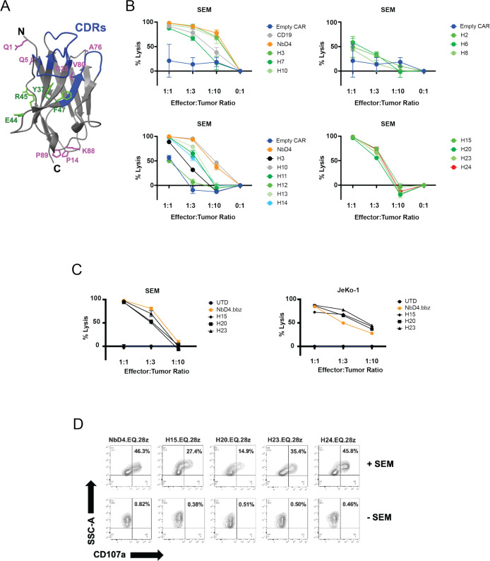
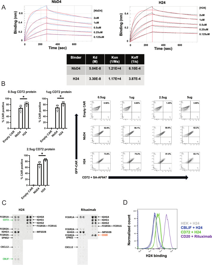
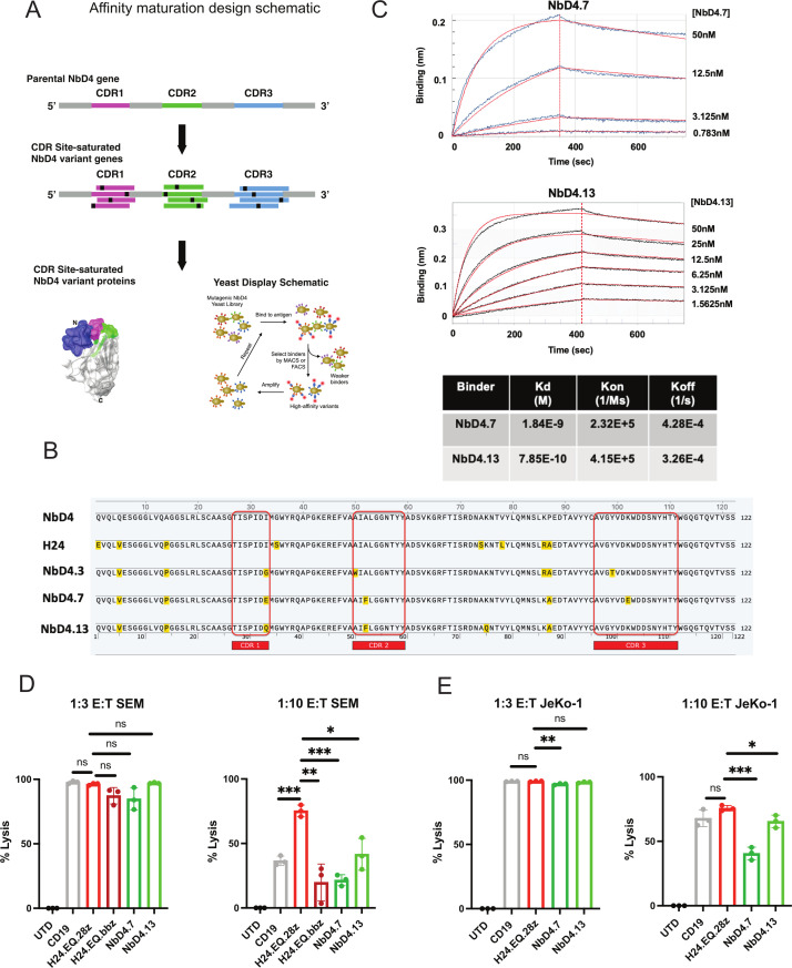
Methods: We humanized our previous nanobody framework regions, derived from llama, to generate a series of humanized anti-CD72 nanobodies. These nanobody binders were inserted into second-generation CD72 CAR-T cells and were evaluated against preclinical models of B cell acute lymphoblastic leukemia and B cell non-Hodgkin's lymphoma in vitro and in vivo. Humanized CD72 nanoCARs were compared with parental ("NbD4") CD72 nanoCARs and the clinically approved CD19-directed CAR-T construct tisangenlecleucel. RNA-sequencing, flow cytometry, and cytokine secretion profiling were used to determine differences between the different CAR constructs. We then used affinity maturation on the parental NbD4 construct to generate high affinity binders against CD72 to test if higher affinity to CD72 improved antitumor potency.

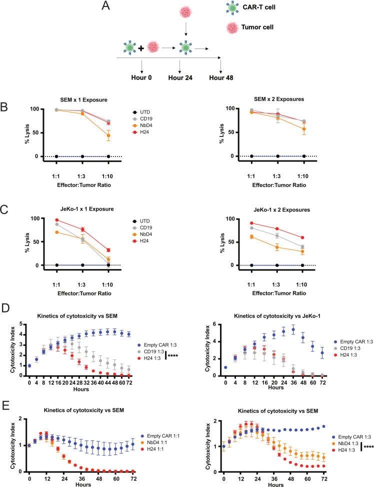
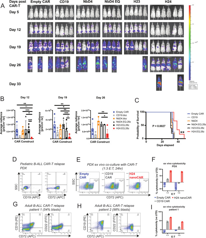
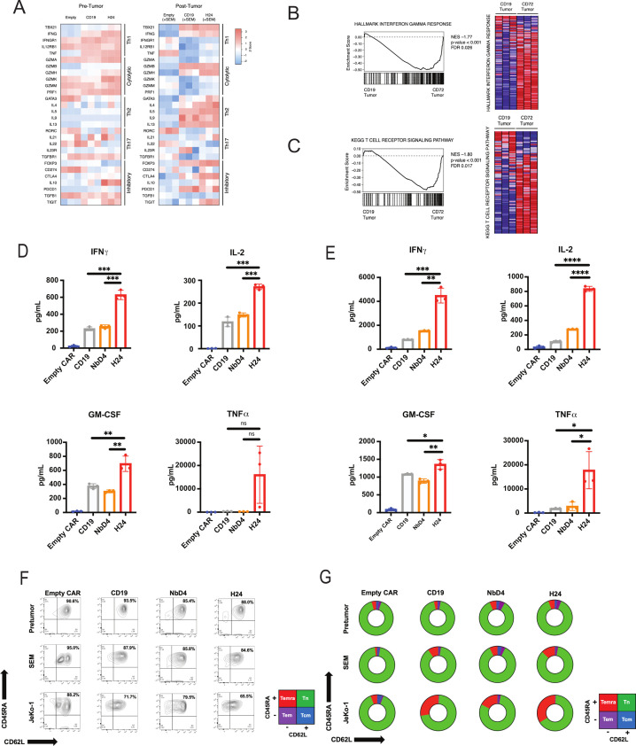
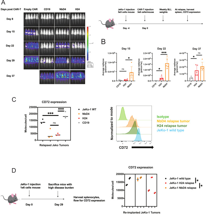
Results: Toward clinical translation, here we humanize our previous nanobody framework regions, derived from llama, and surprisingly discover a clone ("H24") with enhanced potency against B-cell tumors, including patient-derived samples after CD19 CAR-T relapse. Potentially underpinning improved potency, H24 has moderately higher binding affinity to CD72 compared with a fully llama framework. However, further affinity maturation (KD<1 nM) did not lead to improvement in cytotoxicity. After treatment with H24 nanoCARs, in vivo relapse was accompanied by CD72 antigen downregulation which was partially reversible. The H24 nanobody clone was found to have no off-target binding and is therefore designated as a true clinical candidate.

Conclusion: This work supports translation of H24 CD72 nanoCARs for refractory B-cell malignancies, reveals potential mechanisms of resistance, and unexpectedly demonstrates that nanoCAR potency can be improved by framework alterations alone. These findings may have implications for future engineering of nanobody-based cellular therapies.

Keywords: Cell Engineering; Hematologic Neoplasms; Immunotherapy; Receptors, Chimeric Antigen; Translational Medical Research.


Conflict of interest statement

Competing interests: APW and MAN have filed intellectual property claims relevant to the nanobody sequences described here. MAN is an employee and equity shareholder of Cartography Biosciences. APW has received research funding from Genentech/Roche. The other authors declare no relevant conflicts of interest.

Figures

Figure 1
Figure 1
Humanized CD72 nanoCARs have potent anti-leukemia and lymphoma efficacy in vitro. (A) Structural model of the H24 nanobody, in which the framework amino acid substitutions that differentiate NbD4 from H24 are shown in purple. Green amino acids cannot be mutated to human without disrupting obligate monomer nature of nanobody and thus remain as llama-derived sequence in H24. Blue regions represent complementary determining regions (CDRs). Protein structural model created with RaptorX software. (B) In vitro cytotoxicity assays of various humanized anti-CD72 nanoCARs against KMT2A-rearranged B-ALL (SEM). Data are normalized to untransduced T cells (UTD). n=3 technical replicates per data point. (C) In vitro cytotoxicity assays of various humanized anti-CD72 nanoCARs against mantle cell lymphoma (JeKo-1). Data are normalized to untransduced T cells. n=3 technical replicates. (D) Humanized anti-CD72 nanoCARs were co-cultured in the presence or absence of SEM tumor cells for 24 hours, and then flow cytometry was used to determine degranulation by CD107a expression (n=1). B-ALL, B cell acute lymphoblastic leukemia.
Figure 2
Figure 2
H24 nanoCARs have enhanced antitumor potency versus NbD4 nanoCARs and are non-inferior to CD19 CAR-T cells. (A) Experimental design for the tumor rechallenge experiments. (B) In vitro 24-hour cytotoxicity assays comparing H24, NbD4, and CD19 CAR-T cells against SEM and JeKo-1. Data are normalized to untransduced T cells (UTD). n=6 technical replicates. (C) CAR-T cells from figure 2B were exposed again to SEM tumor at 24 hours at noted E:T ratio. n=6 technical replicates. (D) Empty CAR, H24 nanoCARs, and CD19 CAR-T cells were cocultured with SEM or JeKo-1 tumor cells at 1:3 E:T ratio for 72 hours; data obtained using Incucyte live-cell imaging (n=6). (E) Empty CAR, H24 nanoCARs, and CD19 CAR-T cells were co-cultured with SEM tumor cells at either a 1:1 or 1:3 E:T ratio for 72 hours using Incucyte. n=6 technical replicates. Data in figure 2D and figure 2E are generated using two-way ANOVA for multiple comparisons. ****p<0.0001. ANOVA, analysis of variance.
Figure 3
Figure 3
H24 nanoCARs prolong survival in mice implanted with KMT2Ar B-ALL tumor and eliminate pediatric and adult B-ALL tumors relapsed after CD19 CAR-T cell therapy. (A) NSG mice were injected with 1e6 luciferase-labeled SEM B-ALL cells on day −7, and on day 0 mice were treated with a low-dose in vivo CAR stress test with 1.5e6 CAR-T cells per mouse; CAR design as noted per arm. n=4–6 mice per arm. (B) Tumor burden assessed weekly via BLI; quantified BLI intensity shown. (C) Kaplan-Meier curves of overall survival. (D) Flow cytometry for CD19 or CD72 was performed on B-ALL PDX splenocytes, derived from a pediatric patient relapsed after CD19 CAR-T cell therapy (representative of n=3). (E) H24, CD19, or empty CAR-T cells were cocultured ex vivo with PDX splenocytes for 24 hours. Flow cytometry was performed against CD72 to assess tumor cytotoxicity (n=3). (F) Ex vivo cytotoxicity for the various CAR-T cell constructs versus PDX sample, assessed by flow cytometry; percent cytotoxicity normalized to untransduced T cells (n=3). (G, H) Flow cytometry for CD19, CD72, and CD22 was performed on two adult B-ALL patient samples relapsed after CD19 CAR-T cell therapy. (I) Ex vivo cytotoxicity versus adult patient sample #1, performed as in (E) (n=3). Insufficient sample was available for sample #2 to perform cytotoxicity assay. Data in figure 3B are generated using an unpaired two-tailed t-test. Data in figure 3C generated using the log-rank (Mantel-Cox) test. ns=*p<0.05, **p<0.01, ***p<0.001. B-ALL, B cell acute lymphoblastic leukemia; ns not significant.
Figure 4
Figure 4
H24 nanoCARs exhibit a unique transcriptional profile compared with CD19 CAR-T cells after exposure to SEM. Bulk RNA-sequencing performed in triplicate on empty CAR, CD19, and H24 CAR-T cells before and after 24-hour exposure to SEM tumor cells. (A) Heat map of genes relevant for T cell differentiation (classified by reference 30) that are differentially expressed between empty CAR, CD19 CAR-T cells, and H24 nanoCARs, either before or after 24 hours SEM exposure (1:1 E:T). (B, C) GSEA plot and heat map of “hallmark interferon gamma response genes” and “KEGG T cell receptor signaling pathway” between CD19 CAR-T cells and H24 nanoCARs after exposure to SEM. (D) Multiplexed cytokine profiling from culture supernatant after 24-hour SEM exposure (n=3 biological replicates). (E) Multiplexed cytokine profiling from culture supernatant after 24-hour Jeko exposure (n=3 biological replicates). (F) Memory marker profiling for CD45RA and CD62L before tumor exposure, after 24-hour exposure to SEM tumor cells, and after 24-hour exposure to JeKo-1 tumor cells. Representative of n=2 technical replicates. (G) Graphical representation of data from figure 4G, in which the number of naïve T cells (CD45RA+/CD62L+), central memory T cells (Tcm: CD45RA-/CD62L+), T-effector memory RA (TEMRA) cells (CD45RA+/CD62L−), and effector memory T cells (Tem: CD45RA-/CD62L−) are shown from the different CAR-T cell constructs in figure 4F. RNA-seq data are publicly deposited to the Gene Expression Omnibus (GEO) repository with accession number: GSE218791. Data in figure 4D and figure 4E are generated using an unpaired two-tailed t-test. ns=*p<0.05, **p<0.01, ***p<0.001, ****p<0.0001. E:T, effector:tumor; ns, not significant.
Figure 5
Figure 5
H24 nanoCARs have in vivo lymphoma efficacy and lead to persistent CD72 antigen loss. (A) NSG mice were injected with 5e5 firefly-luciferase labeled JeKo-1 mantle cell lymphoma cells on day −4, and on day 0 mice were treated with 3e6 CAR-T cells per mouse (n=5/arm, as shown). BLI was performed on day −1 to randomize the mice into different treatment arms to ensure the disease burden was equal across all CAR constructs. (B) Tumor burden was assessed weekly via BLI, quantified BLI images on each day after CAR-T cell injection are shown. (C) At sacrifice, quantitative flow cytometry for CD72 expression was performed on relapsed Jeko tumors from n=3 mice/arm. (D) Relapsed JeKo-1 tumors were reimplanted into untreated NSG mice in the absence of anti-CD72 immune pressure. 29 days after reimplantation, quantitative flow cytometry was performed on relapsed Jeko tumors for CD72 expression. Data in figure 5B, figure 5C, and figure 5D are generated using an unpaired two-tailed t-test. ns=*p<0.05, ***p<0.001. BLI, biolayer interferometry.
Figure 6
Figure 6
H24 has higher binding affinity to CD72 than NbD4 and is highly specific for CD72. (A) Representative biolayer interferometry plots demonstrating H24 and NbD4 nanobody binding to the biotinylated extracellular domain of CD72. Raw data in black; best fit data from Octet Forte software in red. Data are representative of two technical replicates. Best-fit kon, koff, and calculated KD are listed. (B) Flow cytometry with recombinant biotinylated CD72 (secondary: streptavidin AF647 conjugate) supports higher affinity of H24 versus NbD4. CAR expression was normalized between all CAR-T cells based on intracellular GFP expression. Bar graphs represent the mean±SD for percentage of T cells that are positive for recombinant CD72 binding based on shown gating. Data are representative of two separate experiments from two different T cell donors. (C) HEK cells were engineered to overexpress each of the antigens listed per Retrogenix protocols, and then cells were fixed and evaluated for binding to H24 binder. Possible interactions indicating binding to H24 are shown in green, and non-specific screen hits found with both H24 and rituximab biosimilar in black. (D) HEK cells overexpressing CBLIF, CD72, or wild-type HEK (negative control) were generated per Retrogenix protocols and stained with H24 nanobody-Fc fusion. As a positive control, HEK overexpressing CD20 was stained with rituximab biosimilar. AF647 anti-human IgG Fc detection antibody was used as the secondary antibody for both the H24 and rituximab conditions. H24 binding to CBLIF-expressing cells was considered to be artifactual based on internal validation and assay standards at Retrogenix, given substantial overlap with HEK-alone staining. Data in figure 6B are generated using an unpaired two-tailed t-test. *p<0.05. CBLIF, cobalamin binding intrinsic factor.
Figure 7
Figure 7
High affinity nanobodies generated by affinity maturation do not improve CAR-T cell efficacy. (A) Schematic for the design of the affinity matured nanobody binders against CD72. (B) Shown are the amino acid sequence alignments of parental NbD4, H24, NbD4.3, NbD4.7, and NbD4.13. Amino acids in red are mutations that are in either the CDRs or the framework regions that are different than parental NbD4. (C) Representative biolayer interferometry plots demonstrating NbD4.7 and NbD4.13 binding to the biotinylated extracellular domain of CD72. Raw data is in blue for NbD4.7 and black for NbD4.13, while best fit data is in red. Data are representative of two independent experiments. (D) In vitro 24 hours cytotoxicity assay against SEM comparing affinity matured nanoCARs NbD4.7 and NbD4.13 to H24.EQ.28z at E:T 1:3 and 1:10, cocultured for 24 hours. Data normalized to untransduced T cells (UTD) co-cultured with SEM at the indicated E:T ratios. n=3 technical replicates per data point. (E) In vitro 24 hours cytotoxicity assay against JeKo-1 comparing affinity matured nanoCARs NbD4.7 and NbD4.13, both with EQ.28z backbone, to H24.EQ.28z at various E:T ratios (1:3, 1:10). Data normalized to untransduced T cells (UTD) cocultured with JeKo-1 at the indicated E:T ratios. n=3 technical replicates per data point. Data in figure 7D and figure 7E are generated using an unpaired two-tailed t-test. ns=*p<0.05, **p<0.01, ***p<0.001. E:T, effector:tumor.

References

    1. Maude SL, Frey N, Shaw PA, et al. . Chimeric antigen receptor T cells for sustained remissions in leukemia. N Engl J Med 2014;371:1507–17. 10.1056/NEJMoa1407222 - DOI - PMC - PubMed
    1. Maude SL, Laetsch TW, Buechner J, et al. . Tisagenlecleucel in children and young adults with B-cell Lymphoblastic leukemia. N Engl J Med 2018;378:439–48. 10.1056/NEJMoa1709866 - DOI - PMC - PubMed
    1. Majzner RG, Mackall CL. Clinical lessons learned from the first leg of the CAR T cell journey. Nat Med 2019;25:1341–55. 10.1038/s41591-019-0564-6 - DOI - PubMed
    1. Schultz LM, Eaton A, Baggott C, et al. . Outcomes after Nonresponse and relapse post-Tisagenlecleucel in children, adolescents, and young adults with B-cell acute Lymphoblastic leukemia. JCO 2023;41:354–63. 10.1200/JCO.22.01076 - DOI - PMC - PubMed
    1. Wudhikarn K, Flynn JR, Rivière I, et al. . Interventions and outcomes of adult patients with B-ALL progressing after Cd19 Chimeric antigen receptor T-cell therapy. Blood 2021;138:531–43. 10.1182/blood.2020009515 - DOI - PMC - PubMed

Publication types

Substances