. 2023 Nov 24;11(11):e007515.
doi: 10.1136/jitc-2023-007515.

Chimeric antigen receptor T cells to target CD79b in B-cell lymphomas

Affiliations

Chimeric antigen receptor T cells to target CD79b in B-cell lymphomas

Fuliang Chu et al. J Immunother Cancer. .

Abstract

Background: Chimeric antigen receptor (CAR) T cells targeting CD19 mediate potent and durable effects in B-cell malignancies. However, antigen loss or downregulation is a frequent cause of resistance. Here, we report development of a novel CAR T-cell therapy product to target CD79b, a pan B-cell antigen, widely expressed in most B-cell lymphomas.

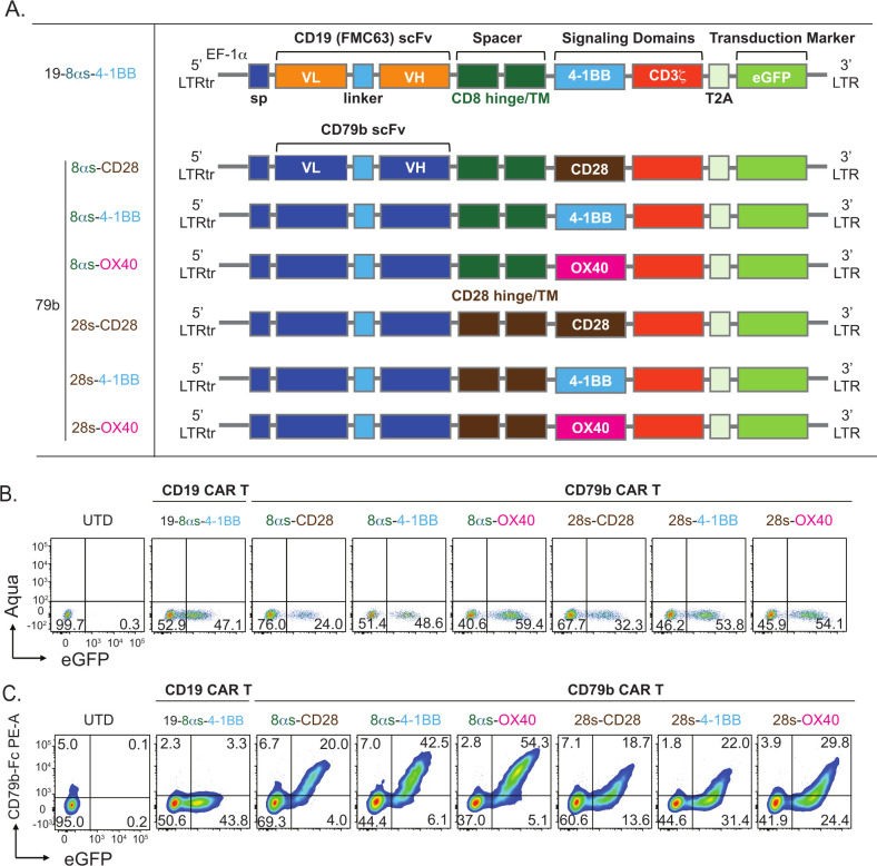
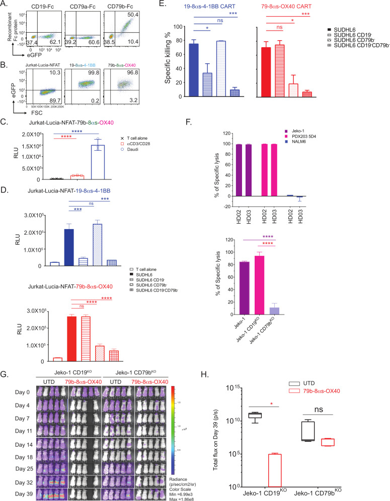
Methods: We generated a novel anti-CD79b monoclonal antibody by hybridoma method. The specificity of the antibody was determined by testing against isogenic cell lines with human CD79b knock-in or knock-out. A single-chain variable fragment derived from the monoclonal antibody was used to make a panel of CD79b-targeting CAR molecules containing various hinge, transmembrane, and co-stimulatory domains. These were lentivirally transduced into primary T cells and tested for antitumor activity in in vitro and in vivo B-cell lymphoma models.

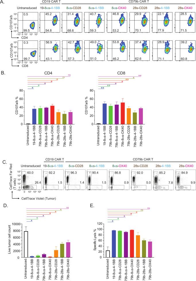
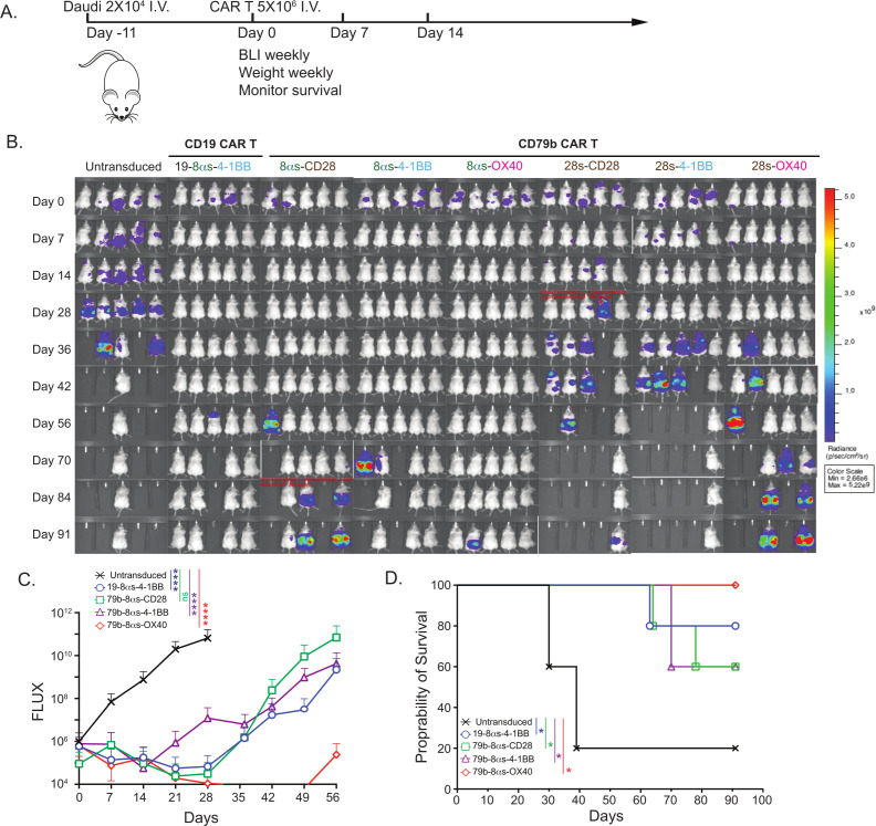
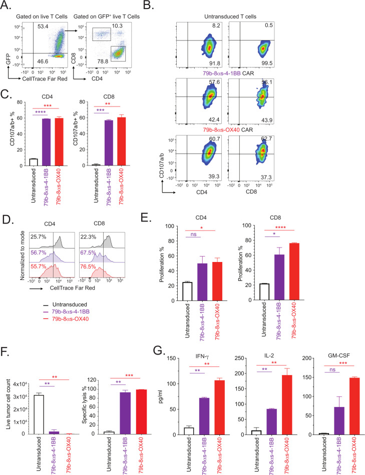
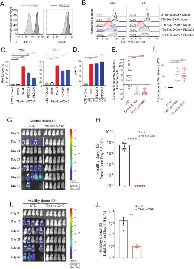
Results: We found that the novel anti-CD79b monoclonal antibody was highly specific and bound only to human CD79b and no other cell surface protein. In testing the various CD79b-targeting CAR molecules, superior antitumor efficacy in vitro and in vivo was found for a CAR consisting CD8α hinge and transmembrane domains, an OX40 co-stimulatory domain, and a CD3ζ signaling domain. This CD79b CAR specifically recognized human CD79b-expressing lymphoma cell lines but not CD79b knock-out cell lines. CD79b CAR T cells, generated from T cells from either healthy donors or patients with lymphoma, proliferated, produced cytokines, degranulated, and exhibited robust cytotoxic activity in vitro against CD19+ and CD19- lymphoma cell lines and patient-derived lymphoma tumors relapsing after prior CD19 CAR T-cell therapy. Furthermore, CD79b CAR T cells were highly efficient at eradicating pre-established lymphoma tumors in vivo in three aggressive lymphoma xenograft models, including two cell line-derived xenografts and one patient-derived xenograft. Notably, these CAR T cells did not demonstrate any significant tonic signaling activity or markers of exhaustion.

Conclusion: Our results indicated that this novel CD79b CAR T-cell therapy product has robust antitumor activity against B-cell lymphomas. These results supported initiation of a phase 1 clinical trial to evaluate this product in patients with relapsed or refractory B-cell lymphomas.

Keywords: B-Lymphocytes; Immunotherapy; Immunotherapy, Adoptive; Receptors, Chimeric Antigen; T-Lymphocytes.


Conflict of interest statement

Competing interests: FC has intellectual property related to cell therapy. JC has intellectual property related to cell therapy. JL has intellectual property related to cell therapy. FV receives research support from Allogene and Geron corporation. SSN received research support from Kite/Gilead, BMS, Allogene, Precision Biosciences, Adicet Bio, and Sana Biotechnology; served as Advisory Board Member/Consultant for Kite/Gilead, Merck, Sellas Life Sciences, Athenex, Allogene, Incyte, Adicet Bio, BMS, Bluebird Bio, Fosun Kite, Sana Biotechnology, Caribou, Astellas Pharma, Morphosys, Janssen, Chimagen, ImmunoACT, Orna Therapeutics, Takeda, and Synthekine; has stock options from Longbow Immunotherapy; and has intellectual property related to cell therapy.

Figures

Figure 1
Figure 1
Schematic of different CD79b CAR constructs and transduction efficiency in primary T cells. (A) Schematic design of CAR constructs to target CD19 or CD79b. (B and C) CAR constructs shown in panel A were transduced lentivirally into primary human T cells and transduction efficiency was determined by flow cytometry by assessing eGFP expression and CAR expression is shown by demonstrating binding to fluorescently labeled recombinant human CD79b-Fc protein. Data is representative of CAR T cells generated from two different donors. CAR, chimeric antigen receptor; eGFP, enhanced green fluorescent protein; LTR, long terminal repeat; LTRtr, LTR truncated; scFv, single chain variable fragment; sp, signal peptide; VL, variable light chain; VH, variable heavy chain; TM, transmembrane domain.
Figure 2
Figure 2
CD79b CAR T cells proliferated and produced cytokines in response to lymphoma cells in vitro. CAR T cells targeting CD19 or CD79b generated from healthy donor T cells were co-cultured with Daudi Burkitt lymphoma tumor cells at an effector:target ratio of 1:1. Untransduced T cells were used as controls. (A) Representative flow cytometric plots illustrating proliferation of CD4+ and CD8+ CAR T cells after 96 hours of co-culture. (B) Summary results showing proliferation from replicate wells (N=2). (C) Cytokine production by CAR T cells in response to Daudi lymphoma cells in replicate wells (N=2) after 24 hours of co-culture. Data represents mean±SD and is representative of at least three separate experiments from three different donors. ***p<0.001; **p<0.01; *p<0.05; (ns) p>0.05. CAR, chimeric antigen receptor; IFN, interferon; IL, interleukin; GM-CSF, granulocyte-macrophage colony-stimulating factor.
Figure 3
Figure 3
CD79b CAR T cells degranulated and exhibited cytotoxic activity against lymphoma cells in vitro. CAR T cells targeting CD19 or CD79b were generated from healthy donor T cells and co-cultured with Daudi Burkitt lymphoma tumor cells at an effector:target ratio of 1:1. Untransduced T cells were used as controls. (A) Representative flow cytometric plots illustrating CD107a/b degranulation on CD4+ and CD8+ CAR T cells after 6 hours of co-culture are shown. (B) Summary results showing degranulation from replicate wells (N=2). (C–E) CAR T cells labeled with CellTrace Far Red were co-cultured with Daudi lymphoma tumor cells labeled with CellTrace Violet at an effector:target ratio of 1:1. Cytotoxicity assay was assessed after 96 hours by flow cytometry by counting live tumor cells using TruCOUNT beads. Untransduced T cells were used as controls. (C) Representative flow cytometric plots showing percentage of live tumor and T cells after end of co-culture. Summary results of cytotoxicity assay from replicate wells (N=2) showing absolute live tumor cell count (D) or specific lysis (E) of Daudi tumor cells. Data represents mean±SD and is representative of at least three separate experiments from three different donors. ***p<0.001; **p<0.01; *p<0.05; (ns)p>0.05. CAR, chimeric antigen receptor.
Figure 4
Figure 4
CD79b CAR T cells exert antitumor effects in vivo. (A) Schematic overview of xenograft mouse model. Luciferase-labeled Daudi Burkitt lymphoma cells were injected intravenously into NSG mice at 2×10⁴ tumor cells/mouse. After 11 days, mice were treated with untransduced T cells, CD19 CAR T cells, or CD79b CAR T cells via tail vein injection at 5×10⁶ CAR+ T cells/mouse. (B) Tumor burden assessed by bioluminescence imaging at the indicated time points is shown. (C) Average radiance (photons/sec/cm2/steradian) of groups of mice at different time points is shown. Significance among treatment groups was determined by comparing area under the curve for each CAR-T group to untransduced control T-cell group in an ordinary one-way ANOVA with Dunnett’s multiple comparisons test (n=5 mice per group, mean±SD is shown). (D) Kaplan-Meier survival curves of groups of mice treated with different T cells. (****p<0.0001; ***p<0.001; **p<0.01; *p<0.05; (ns) p>0.05, ANOVA). ANOVA, analysis of variance; CAR, chimeric antigen receptor; I.V., intravenous.
Figure 5
Figure 5
CD79b CAR T cells generated from patients with lymphoma relapsing after autologous CD19 CAR T-cell therapy are functional. CAR T cells targeting CD79b were generated from T cells isolated from two patients with large B-cell lymphoma relapsing after autologous CD19 CAR T-cell therapy and co-cultured with Daudi Burkitt lymphoma tumor cells at an effector:target ratio of 1:1. Untransduced T cells were used as controls. Representative data from one patient is shown. (A) Representative flow cytometric plots showing transduction efficiency of 79b CAR and proportion of CD4+ and CD8+ T cells. (B) Representative flow cytometric plots illustrating CD107a/b degranulation by CD4+ and CD8+ CAR T cells after 6 hours of co-culture. (C) Summary results showing degranulation from replicate wells (N=2). (D) Representative flow cytometric plots illustrating proliferation of CD4+ and CD8+ CAR T cells after 96 hours of co-culture. (E) Summary results showing proliferation from replicate wells (N=2). (F) Summary results of cytotoxicity assay from replicate wells (N=2) showing absolute live tumor cell count (left panel) or specific lysis (right panel). (G) Cytokines production by CAR T cells co-cultured with Daudi lymphoma cells in replicate wells (N=2) after 24 hours of co-culture. Data is representative of at least three separate experiments with T cells from two different patients. ***p<0.001; **p<0.01; *p<0.05; (ns) p>0.05. CAR, chimeric antigen receptor; IFN, interferon; IL, interleukin; GFP, green fluorescent protein; GM-CSF, granulocyte-macrophage colony-stimulating factor.
Figure 6
Figure 6
Efficacy of CD79b CAR T cells against patient-derived lymphoma tumor cells. CAR T cells targeting CD79b were generated from primary healthy donor T cells, labeled with CellTrace Far Red and co-cultured with Daudi Burkitt lymphoma tumor cells or lymphoma tumor cells derived from two PDXs (PDX203 and PDX300) for 96 hours. Untransduced T cells were used as controls. (A) Histograms showing expression of CD19 and CD79b in the two tumor samples. (B) Representative flow cytometric plots illustrating proliferation of CD4+ and CD8+ CARs T cells in response to tumor cells at an effector:target (E:T) ratio of 0.6:1. (C) Summary results showing proliferation from replicate wells (N=2). (D) Summary results of cytotoxicity assay from replicate wells (N=2) showing specific lysis at E:T ratio of 0.6:1. (E and F) Pooled data from 17 experiments testing CD79b CAR T cells generated from five donors is shown for per cent change in absolute number of live tumor cells on day 4 in an in vitro cytotoxicity assay compared with day 0 (E) and fold change in interferon-γ production compared with untransduced T cells after 24 hours of co-culture (F). Target tumor cells in these experiments included Daudi, Jeko-1, SUDHL6, or PDX203. Each dot represents data from one experiment and mean values of data from replicate wells from each experiment are shown. (G–J) Luciferase-labeled PDX203 5D4 tumor cells (0.2×10⁶ /mouse) were injected via tail vein into NSG mice on day −4 and CD79b CAR T cells or untransduced T cells (5×10⁶ /mouse) derived from two different donors were injected via tail vein on day 0. Tumor burden was monitored by bioluminescence imaging (G and I). Average radiance (photons/sec/cm2/steradian) of groups of mice on days 35 and 29 are shown (H and J). (n=5 mice per group, mean±SD is shown). ****p<0.0001; ***p<0.001; **p<0.01; *p<0.05; (ns) p>0.05. CAR, chimeric antigen receptor; PDX, patient-derived xenografts.
Figure 7
Figure 7
CD79b CARs specially recognized human CD79b protein. (A) CAR T cells targeting CD79b were generated from primary healthy donor T cells, stained with fluorochrome labeled recombinant human CD19-Fc, CD79a-Fc, or CD79b-Fc proteins, and assessed by flow cytometry. Transduction efficiency determined by eGFP expression and binding of recombinant proteins is shown. (B–D) CAR constructs targeting CD19 or CD79b were transduced lentivirally into Jurkat-Lucia NFAT reporter cell line and cultured alone or with the indicated lymphoma cell lines at an effector:target (E:T) ratio of 1:1 or with anti-CD3 and anti-CD28 monoclonal antibodies. NFAT activation was determined after 24 hours by measuring luciferase activity. Percentage of Jurkat-Lucia NFAT reporter cells expressing CD19 or CD79b CAR was determined by assessing eGFP transduction marker by flow cytometry (B). Luciferase activity shown in Jurkat-Lucia NFAT reporter cells transduced with 79b CAR construct and cultured alone or with anti-CD3/CD28 antibodies or Daudi Burkitt lymphoma cells (C). Luciferase activity shown in Jurkat-Lucia NFAT reporter cells transduced with the indicated CD19 or CD79b CAR constructs and cultured alone or with isogenic SUDHL6 lymphoma cell lines (parent, CD19KO, CD79bKO, or CD19KOCD79bKO). (E) Cytotoxic activity of CD19 or CD79b CAR T cells generated from healthy donor T cells against isogenic cell lines of SUDHL6 (parent, CD19KO, CD79bKO, or CD19KOCD79bKO). (F) Cytotoxic activity of CD79b CAR T cells against wild type Jeko-1 and PDX203 5D4 (CD19+CD79b+), Jeko-1 CD19KO, Jeko-1 CD79bKO, or NALM6 acute lymphoblastic leukemia (CD19+CD79b) cell lines at an E:T ratio of 1:1. The absolute number of live tumor cells at the end of culture period from this experiment are shown in online supplemental figure 10. (G and H) Luciferase-labeled Jeko-1 CD19KO or Jeko-1 CD79bKO tumor cells (0.2×10⁶ /mouse) were injected via tail vein into NSG mice on day −4 and CD79b CAR T cells or untransduced T cells (5×10⁶ /mouse) were injected via tail vein on day 0. Tumor burden was monitored by bioluminescence imaging at the indicated time points (G) and average radiance (photons/sec/cm2/steradian) of groups of mice on day 39 is shown (H). Data represent the mean±SD of replicate wells (N=2) where applicable and is representative of at least three separate experiments from two different donors (C–F). ****p<0.0001; ***p<0.001; **p<0.01; *p<0.05; (ns) p>0.05. CAR, chimeric antigen receptor; eGFR, enhanced green fluorescent protein; HD, healthy donor; KO, knock-out; RLU, relative luminescence units.

Similar articles

Cited by

References

    1. Neelapu SS, Locke FL, Bartlett NL, et al. . Axicabtagene Ciloleucel CAR T-cell therapy in refractory large B-cell lymphoma. N Engl J Med 2017;377:2531–44. 10.1056/NEJMoa1707447 - DOI - PMC - PubMed
    1. Schuster SJ, Bishop MR, Tam CS, et al. . Tisagenlecleucel in adult Relapsed or refractory diffuse large B-cell lymphoma. N Engl J Med 2019;380:45–56. 10.1056/NEJMoa1804980 - DOI - PubMed
    1. Abramson JS, Palomba ML, Gordon LI, et al. . Lisocabtagene Maraleucel for patients with Relapsed or refractory large B-cell Lymphomas (TRANSCEND NHL 001): a Multicentre seamless design study. Lancet 2020;396:839–52. 10.1016/S0140-6736(20)31366-0 - DOI - PubMed
    1. Wang M, Munoz J, Goy A, et al. . KTE-X19 CAR T-cell therapy in Relapsed or refractory Mantle-cell lymphoma. N Engl J Med 2020;382:1331–42. 10.1056/NEJMoa1914347 - DOI - PMC - PubMed
    1. Jacobson CA, Chavez JC, Sehgal AR, et al. . Primary analysis of Zuma-5: A phase 2 study of Axicabtagene Ciloleucel (Axi-Cel) in patients with Relapsed/refractory (R/R) indolent non-Hodgkin lymphoma (iNHL. ASH Annual Meeting; Abstract 700; 2020. 10.1016/S2152-2650(20)30898-3 - DOI

Publication types

MeSH terms

Substances

LinkOut - more resources