

Astaxanthin alleviates PM2.5-induced cardiomyocyte injury via inhibiting ferroptosis
- PMID: 38007415
- PMCID: PMC10675963
- DOI: 10.1186/s11658-023-00513-1
Astaxanthin alleviates PM2.5-induced cardiomyocyte injury via inhibiting ferroptosis
Abstract
Background: Long-term exposure of humans to air pollution is associated with an increasing risk of cardiovascular diseases (CVDs). Astaxanthin (AST), a naturally occurring red carotenoid pigment, was proved to have multiple health benefits. However, whether or not AST also exerts a protective effect on fine particulate matter (PM2.5)-induced cardiomyocyte damage and its underlying mechanisms remain unclear.
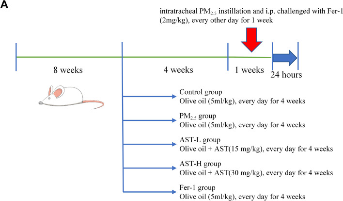
Methods: In vitro experiments, the H9C2 cells were subjected to pretreatment with varying concentrations of AST, and then cardiomyocyte injury model induced by PM2.5 was established. The cell viability and the ferroptosis-related proteins expression were measured in different groups. In vivo experiments, the rats were pretreated with different concentrations of AST for 21 days. Subsequently, a rat model of myocardial PM2.5 injury was established by intratracheal instillation every other day for 1 week. The effects of AST on myocardial tissue injury caused by PM2.5 indicating by histological, serum, and protein analyses were examined.
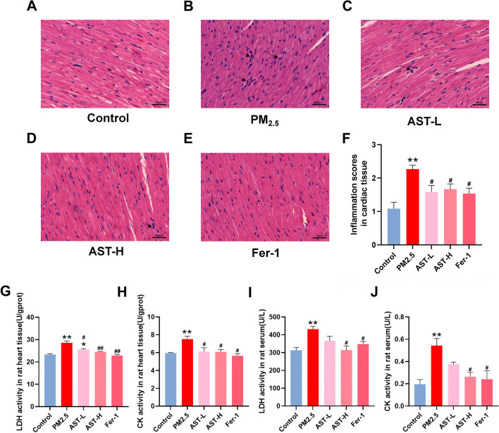
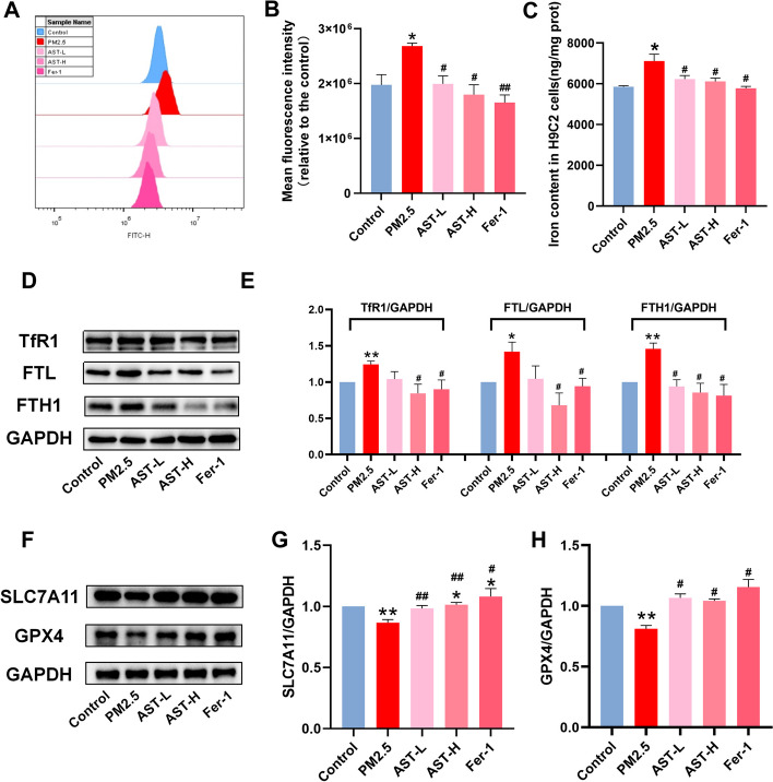
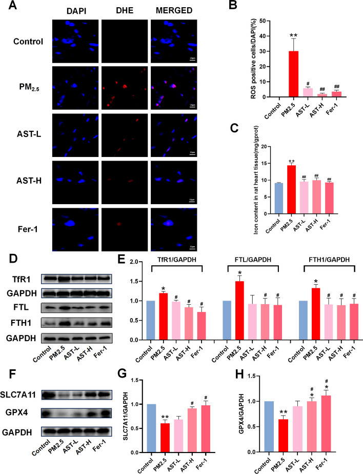
Results: AST significantly ameliorated PM2.5-induced myocardial tissue injury, inflammatory cell infiltration, the release of inflammatory factors, and cardiomyocyte H9C2 cell damage. Mechanistically, AST pretreatment increased the expression of SLC7A11, GPX4 and down-regulated the expression of TfR1, FTL and FTH1 in vitro and in vivo.
Conclusions: Our study suggest that ferroptosis plays a significant role in the pathogenesis of cardiomyocyte injury induced by PM2.5. AST may serve as a potential therapeutic agent for mitigating cardiomyocyte injury caused by PM2.5 through the inhibition of ferroptosis.
Keywords: Astaxanthin; Cardiomyocyte injury; Cardiovascular diseases; Ferroptosis; PM2.5.
© 2023. The Author(s).
Conflict of interest statement
The authors declare that they have no competing interests.
Figures


Similar articles
-
Astaxanthin mitigates doxorubicin-induced cardiotoxicity via inhibiting ferroptosis and autophagy: a study based on bioinformatic analysis and in vivo/vitro experiments.Front Pharmacol. 2025 Jan 21;16:1524448. doi: 10.3389/fphar.2025.1524448. eCollection 2025. Front Pharmacol. 2025. PMID: 39906141 Free PMC article.
-
Astaxanthin alleviates fine particulate matter (PM2.5)-induced lung injury in rats by suppressing ferroptosis and apoptosis.Food Funct. 2023 Dec 11;14(24):10841-10854. doi: 10.1039/d3fo03641c. Food Funct. 2023. PMID: 37982854
-
Astragaloside IV regulates the ferroptosis signaling pathway via the Nrf2/SLC7A11/GPX4 axis to inhibit PM2.5-mediated lung injury in mice.Int Immunopharmacol. 2022 Nov;112:109186. doi: 10.1016/j.intimp.2022.109186. Epub 2022 Sep 15. Int Immunopharmacol. 2022. PMID: 36115280 Review.
-
Sesamin attenuates PM2.5-induced cardiovascular injury by inhibiting ferroptosis in rats.Food Funct. 2021 Dec 13;12(24):12671-12682. doi: 10.1039/d1fo02913d. Food Funct. 2021. PMID: 34825691
-
Ferroptosis is involved in PM2.5-induced acute nasal epithelial injury via AMPK-mediated autophagy.Int Immunopharmacol. 2023 Feb;115:109658. doi: 10.1016/j.intimp.2022.109658. Epub 2023 Jan 4. Int Immunopharmacol. 2023. PMID: 36608444
Cited by
-
Short-term effects of extreme air pollutant concentrations on coronary heart disease hospitalization in Henan province: a time-stratified case-crossover study.Front Cardiovasc Med. 2025 Apr 24;12:1538788. doi: 10.3389/fcvm.2025.1538788. eCollection 2025. Front Cardiovasc Med. 2025. PMID: 40342980 Free PMC article.
-
Astaxanthin activated the SLC7A11/GPX4 pathway to inhibit ferroptosis and enhance autophagy, ameliorating dry eye disease.Front Pharmacol. 2024 Aug 19;15:1407659. doi: 10.3389/fphar.2024.1407659. eCollection 2024. Front Pharmacol. 2024. PMID: 39224780 Free PMC article.
-
Astaxanthin mitigates doxorubicin-induced cardiotoxicity via inhibiting ferroptosis and autophagy: a study based on bioinformatic analysis and in vivo/vitro experiments.Front Pharmacol. 2025 Jan 21;16:1524448. doi: 10.3389/fphar.2025.1524448. eCollection 2025. Front Pharmacol. 2025. PMID: 39906141 Free PMC article.
-
Ferroptosis induced by environmental pollutants and its health implications.Cell Death Discov. 2025 Jan 24;11(1):20. doi: 10.1038/s41420-025-02305-2. Cell Death Discov. 2025. PMID: 39856053 Free PMC article. Review.
-
PERK/Sestrin2 Signaling Pathway Mediated Autophagy Regulates Human Cardiomyocytes Apoptosis Induced by Traffic-Related PM2.5 and Diverse Constituents.Int J Mol Sci. 2025 Apr 17;26(8):3784. doi: 10.3390/ijms26083784. Int J Mol Sci. 2025. PMID: 40332408 Free PMC article.
References
-
- Tanaka M, Okuda T, Itoh K, Ishihara N, Oguro A, Fujii-Kuriyama Y, Nabetani Y, Yamamoto M, Vogel CFA, Ishihara Y. Polycyclic aromatic hydrocarbons in urban particle matter exacerbate movement disorder after ischemic stroke via potentiation of neuroinflammation. Part Fibre Toxicol. 2023;20:6. doi: 10.1186/s12989-023-00517-x. - DOI - PMC - PubMed
MeSH terms
Substances
Grants and funding
LinkOut - more resources
Full Text Sources
Miscellaneous
