

Animal models of acute exacerbation of pulmonary fibrosis
- PMID: 38007420
- PMCID: PMC10675932
- DOI: 10.1186/s12931-023-02595-z
Animal models of acute exacerbation of pulmonary fibrosis
Abstract
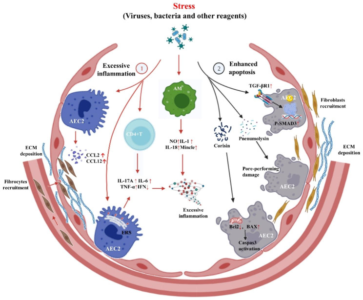
Idiopathic pulmonary fibrosis (IPF) is a chronic, progressive scarring interstitial lung disease with an unknown cause. Some patients may experience acute exacerbations (AE), which result in severe lung damage visible on imaging or through examination of tissue samples, often leading to high mortality rates. However, the etiology and pathogenesis of AE-IPF remain unclear. AE-IPF patients exhibit diffuse lung damage, apoptosis of type II alveolar epithelial cells, and an excessive inflammatory response. Establishing a reliable animal model of AE is critical for investigating the pathogenesis. Recent studies have reported a variety of animal models for AE-IPF, each with its own advantages and disadvantages. These models are usually established in mice with bleomycin-induced pulmonary fibrosis, using viruses, bacteria, small peptides, or specific drugs. In this review, we present an overview of different AE models, hoping to provide a useful resource for exploring the mechanisms and targeted therapies for AE-IPF.
Keywords: Acute exacerbation; Animal model; Pulmonary fibrosis.
© 2023. The Author(s).
Conflict of interest statement
None.
Figures

Similar articles
-
A New Model of Acute Exacerbation of Experimental Pulmonary Fibrosis in Mice.Cells. 2022 Oct 26;11(21):3379. doi: 10.3390/cells11213379. Cells. 2022. PMID: 36359778 Free PMC article.
-
CSF3 aggravates acute exacerbation of pulmonary fibrosis by disrupting alveolar epithelial barrier integrity.Int Immunopharmacol. 2024 Jun 30;135:112322. doi: 10.1016/j.intimp.2024.112322. Epub 2024 May 23. Int Immunopharmacol. 2024. PMID: 38788452
-
The overexpression of peroxiredoxin-4 affects the progression of idiopathic pulmonary fibrosis.BMC Pulm Med. 2019 Dec 30;19(1):265. doi: 10.1186/s12890-019-1032-2. BMC Pulm Med. 2019. PMID: 31888585 Free PMC article.
-
Association of the RAGE/RAGE-ligand axis with interstitial lung disease and its acute exacerbation.Respir Investig. 2022 Jul;60(4):531-542. doi: 10.1016/j.resinv.2022.04.004. Epub 2022 May 2. Respir Investig. 2022. PMID: 35504814 Review.
-
Acute exacerbation of idiopathic pulmonary fibrosis: a clinical review.Intern Emerg Med. 2015 Jun;10(4):401-11. doi: 10.1007/s11739-015-1204-x. Epub 2015 Feb 12. Intern Emerg Med. 2015. PMID: 25672832 Free PMC article. Review.
Cited by
-
Sivelestat sodium: a novel therapeutic agent in a mouse model of acute exacerbation pulmonary fibrosis through multiple mechanisms.J Thorac Dis. 2025 Jul 31;17(7):5024-5043. doi: 10.21037/jtd-2025-163. Epub 2025 Jul 29. J Thorac Dis. 2025. PMID: 40809237 Free PMC article.
-
A novel senolytic drug for pulmonary fibrosis: BTSA1 targets apoptosis of senescent myofibroblasts by activating BAX.Aging Cell. 2024 Sep;23(9):e14229. doi: 10.1111/acel.14229. Epub 2024 Jun 3. Aging Cell. 2024. PMID: 38831635 Free PMC article.
-
Autologous precision-cut lung slice co-culture models for studying macrophage-driven fibrosis.Front Physiol. 2025 Jan 31;16:1526787. doi: 10.3389/fphys.2025.1526787. eCollection 2025. Front Physiol. 2025. PMID: 39958688 Free PMC article.
-
Endothelial CD38-induced endothelial-to-mesenchymal transition is a pivotal driver in pulmonary fibrosis.Cell Mol Life Sci. 2024 Dec 27;82(1):30. doi: 10.1007/s00018-024-05548-x. Cell Mol Life Sci. 2024. PMID: 39725783 Free PMC article.
-
Comorbidities' Effect on IPF: Pathogenesis and Management.Biomedicines. 2025 Jun 1;13(6):1362. doi: 10.3390/biomedicines13061362. Biomedicines. 2025. PMID: 40564081 Free PMC article. Review.
References
-
- Raghu G, Remy-Jardin M, Richeldi L, Thomson CC, Inoue Y, Johkoh T, et al. Idiopathic Pulmonary Fibrosis (an update) and Progressive pulmonary fibrosis in adults: an Official ATS/ERS/JRS/ALAT Clinical Practice Guideline. Am J Respir Crit Care Med. 2022;205:e18–47. doi: 10.1164/rccm.202202-0399ST. - DOI - PMC - PubMed
Publication types
MeSH terms
Grants and funding
LinkOut - more resources
Full Text Sources
