. 2023 Nov 25;13(1):215.
doi: 10.1186/s13578-023-01159-4.

Modulation of cell cycle increases CRISPR-mediated homology-directed DNA repair

Affiliations

Modulation of cell cycle increases CRISPR-mediated homology-directed DNA repair

Guoling Li et al. Cell Biosci. .

Abstract

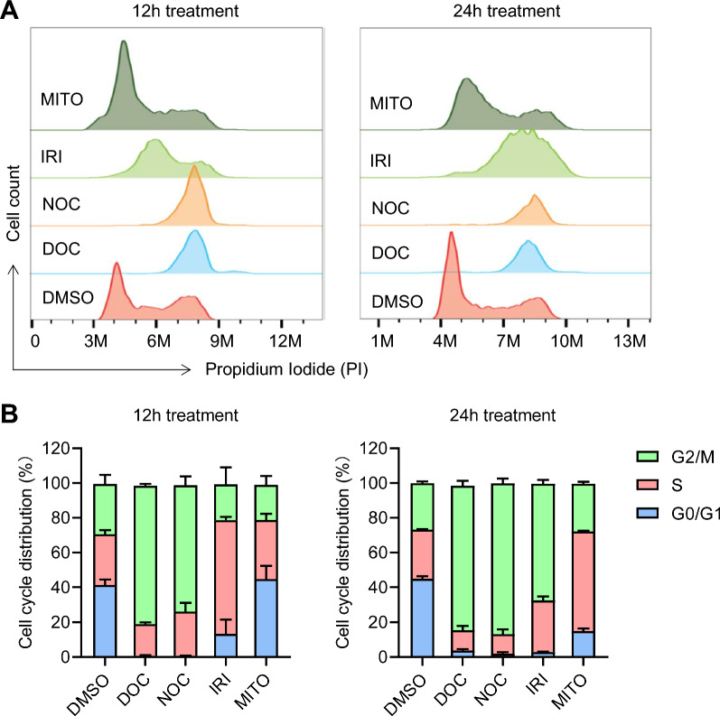
Background: Gene knock-in (KI) in animal cells via homology-directed repair (HDR) is an inefficient process, requiring a laborious work for screening from few modified cells. HDR tends to occur in the S and G2/M phases of cell cycle; therefore, strategies that enhance the proportion of cells in these specific phases could improve HDR efficiency.

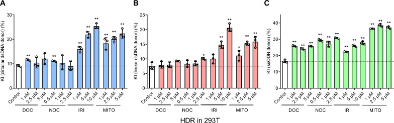
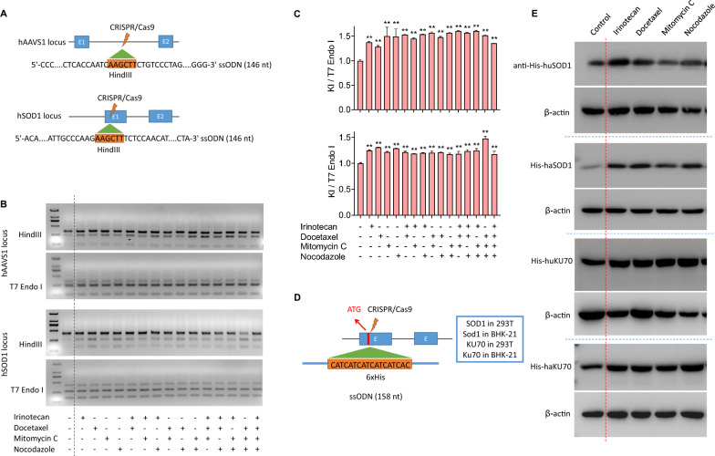
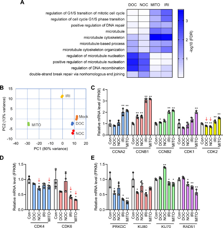
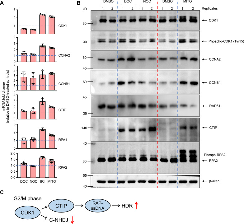
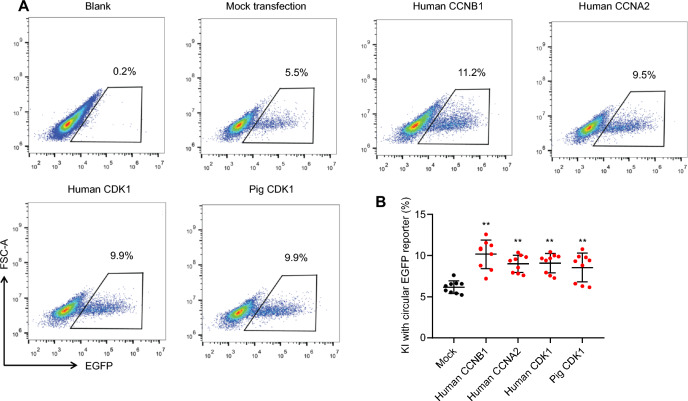
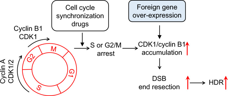
Results: We used various types of cell cycle inhibitors to synchronize the cell cycle in S and G2/M phases in order to investigate their effect on regulating CRISPR/Cas9-mediated HDR. Our results indicated that the four small molecules-docetaxel, irinotecan, nocodazole and mitomycin C-promoted CRISPR/Cas9-mediated KI with different homologous donor types in various animal cells. Moreover, the small molecule inhibitors enhanced KI in animal embryos. Molecular analysis identified common signal pathways activated during crosstalk between cell cycle and DNA repair. Synchronization of the cell cycle in the S and G2/M phases results in CDK1/CCNB1 protein accumulation, which can initiate the HDR process by activating HDR factors to facilitate effective end resection of CRISPR-cleaved double-strand breaks. We have demonstrated that augmenting protein levels of factors associated with the cell cycle via overexpression can facilitate KI in animal cells, consistent with the effect of small molecules.

Conclusion: Small molecules that induce cell cycle synchronization in S and G2/M phases promote CRISPR/Cas9-mediated HDR efficiency in animal cells and embryos. Our research reveals the common molecular mechanisms that bridge cell cycle progression and HDR activity, which will inform further work to use HDR as an effective tool for preparing genetically modified animals or for gene therapy.

Keywords: CCNB1; CDK1; CRISPR/Cas9; Cell cycle; Gene editing; Homology-directed repair.


Conflict of interest statement

The authors declare that they have no competing interests.

Figures

Fig. 1
Fig. 1
Cell cycle distribution analysis of 293T cells treated with small molecule inhibitors. A Analysis of 293T cells unsynchronized (DMSO) and synchronized by DOC (5 μM), NOC (2.5 μM), IRI (10 μM) and MITO (5 μM) for 12 (left panel) and 24 h (right panel) showing DOC and NOC arrested cells at G2/M stage and IRI and MITO increased the proportions at both S and G2/M phases of cell cycle. B Quantification of cell cycle distribution by measuring the area representing the specific cell cycle stage in FACS histogram. The mean values and error bars (SD) were calculated from three experiments
Fig. 2
Fig. 2
HDR-promoting effect of the four small molecules in 293T cells. The 293T cells were transfected with CRISPR/Cas9 and circular dsDNA (A), linear dsDNA (B) and ssODN donor (C)-mediated KI reporter system as shown in Additional file 3: Fig S1. After 12 h-transfection, the four small molecules were used to treat 293T cells for 48 h. The KI efficiency is shown by the percentage of EGFP-positive cells determined by flow cytometry. Data are mean ± SD. Each dot represents an independent experiment. **P < 0·01 and *P < 0·05 vs DMSO-treated control
Fig. 3
Fig. 3
Small molecule effects on ssODN-mediated KI in endogenous genes of cells. A The donor is a 146 nt ssODN that is homologous to the target sequence and contains a 6 nt insertion (HindIII restriction sequence) at the CRISPR cleavage site for a simple identification of positive KI alleles by HindIII digestion. B The KI frequency after 48 h-treatment with different small molecules was determined directly by HindIII digestion of PCR products covering the KI site. The ratio of cleaved products to total DNA substrate (cleaved PCR bands + uncleaved PCR band) is KI frequency, and a T7E1 digestion of the same PCR product was used an inner control to show all mutant alleles (NHEJ + HDR). C Quantification of KI frequency (up, AAVS1 locus; down, SOD1 locus) in 293T cells with different small molecule treatments by estimating band density shown in B by Image J software. The mean values and error bars (SD) were calculated from three experiments. **P < 0.01 compared to control group. D The strategy for gene tagging by CRISPR-induced ssODN-mediated KI. A 158 nt ssODN donor for tagging a 6 × His epitope in the N terminal of two genes (SOD1 and KU70) in 293T and BHK-21 cells. E The ssODN-mediated protein tagging effects were determined by western blot. Representative results showing increased 6 × His tagged proteins in small molecule-treated groups compared to DMSO-treated control
Fig. 4
Fig. 4
RNAseq analysis of 293T cells with cell cycle arrest with small molecules. A Gene Ontology (GO) analysis on RNAseq data showing significantly enriched GO terms in cell cycle, DNA repair and mitosis upon small molecule treatment. The differentially enriched GO terms implying the different action mechanism for cell cycle synchronization among the small molecules. B PCA showing IRI and MITO treatment groups clustered farther from the control than DOC and NOC groups, indicating the more profound change in transcriptome profile of cells with IRI and MITO treatment. C Cell cycle-associated genes, CCNA2, CCNB1/2, CDK1 and CDK2, are significantly upregulated in IRI and MITO treatment groups, whereas only CCNB1 had significant increase in mRNA level in DOC and NOC treatment groups. D CDK4 and CDK6 controlling G1 cell cycle phase showing insignificant difference or decrease in mRNA level of cells with small molecule treatment. E NHEJ factors (PRKDC, KU70 and KU80) and HDR factor (RAD51) showing decreased mRNA level in most groups with small molecule treatment. Gene expression levels were represented by normalized FPKM values. Data are mean ± SD from 3 independent samples. **P < 0·01 and *P < 0·05 vs DMSO-treated control
Fig. 5
Fig. 5
Analyzing the expression levels of genes in S-G2 cell cycle phases and HDR pathway by qPCR and WB. A qPCR assay showing the main factors controlling S and G2 progression (CDK1, CCNB1 and CCNA2) and HDR factors (CTIP, RPA1 and RPA2) exhibited greatly increased expression in 293T cells with IRI and MITO treatment compared to DMSO treatment control, whereas their up-regulation was less or insignificant in DOC and NOC treatment groups. Data are mean ± SD from 3 independent samples. B WB results showing similar gene expression changes as qPCR. CDK1 and RPA2 showing evident phosphorylation in all small molecule treated cells. C The data indicate cell cycle synchronization increases CDK1, CTIP and RPA2 activity to promote HDR. The small molecule inhibitors induce up-regulated CDK1 expression or activity, which promotes efficient DSB end resection by phosphorylating CITP and other nucleases and thus prevents DNA repair by NHEJ. Efficient break end resection generates sufficient ssDNA overhangs for RPA complex coating which is essential for HDR initiation
Fig. 6
Fig. 6
Overexpression of cell cycle factors increases KI frequency. A Overexpression of CCNA2, CCNB1 and CDK1 in 293T cells by transiently transfection of the eukaryotic expression vectors harboring cDNA of the genes. Enhanced KI frequency tested by further transfection of GAPDH-EGFP reporter shown in Additional file 3: Fig. S1 in overexpressing cells. B Overexpressing all tested genes in 293T cells showing promoting effect on KI, representing by increased proportion of EGFP-positive cells in CCNA2, CCNB1 and CDK1-transfected cells compared to mock transfection control. Data are mean ± SD of nine independent experiments. **P < 0·01 vs mock transfection control
Fig. 7
Fig. 7
Model for the role of the small molecule inhibitors in enhancing HDR efficiency. The explanation is shown in main text

Similar articles

Cited by

References

    1. Mao Z, Bozzella M, Seluanov A, Gorbunova V. Comparison of nonhomologous end joining and homologous recombination in human cells. DNA Repair. 2008;7:1765–1771. doi: 10.1016/j.dnarep.2008.06.018. - DOI - PMC - PubMed
    1. Sander JD, Joung JK. CRISPR-Cas systems for editing, regulating and targeting genomes. Nat Biotechnol. 2014;32:347–355. doi: 10.1038/nbt.2842. - DOI - PMC - PubMed
    1. West SC. Molecular views of recombination proteins and their control. Nat Rev Mol Cell Biol. 2003;4(6):435–445. doi: 10.1038/nrm1127. - DOI - PubMed
    1. Hendrickson EA. Cell-cycle regulation of mammalian DNA double-strand-break repair. Am J Hum Genet. 1997;61(4):795–800. doi: 10.1086/514895. - DOI - PMC - PubMed
    1. Saleh-Gohari N, Helleday T. Conservative homologous recombination preferentially repairs DNA double-strand breaks in the S phase of the cell cycle in human cells. Nucleic Acids Res. 2004;32(12):3683–3688. doi: 10.1093/nar/gkh703. - DOI - PMC - PubMed