. 2023 Nov 25;20(1):279.
doi: 10.1186/s12974-023-02952-1.

Protective role of IL-17-producing γδ T cells in a laser-induced choroidal neovascularization mouse model

Affiliations

Protective role of IL-17-producing γδ T cells in a laser-induced choroidal neovascularization mouse model

Yu-Hsien Chang et al. J Neuroinflammation. .

Abstract

Background: Vision loss in patients with wet/exudative age-related macular degeneration (AMD) is associated with choroidal neovascularization (CNV), and AMD is the leading cause of irreversible vision impairment in older adults. Interleukin-17A (IL-17A) is a component of the microenvironment associated with some autoimmune diseases. Previous studies have indicated that wet AMD patients have elevated serum IL-17A levels. However, the effect of IL-17A on AMD progression needs to be better understood. We aimed to investigate the role of IL-17A in a laser-induced CNV mouse model.

Methods: We established a laser-induced CNV mouse model in wild-type (WT) and IL-17A-deficient mice and then evaluated the disease severity of these mice by using fluorescence angiography. We performed enzyme-linked immunosorbent assay (ELISA) and fluorescence-activated cell sorting (FACS) to analyze the levels of IL-17A and to investigate the immune cell populations in the eyes of WT and IL-17A-deficient mice. We used ARPE-19 cells to clarify the effect of IL-17A under oxidative stress.

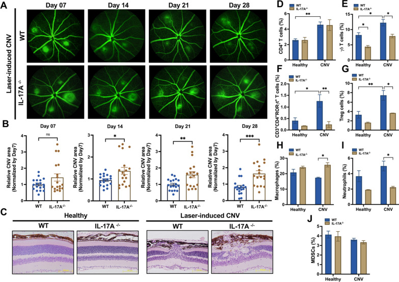
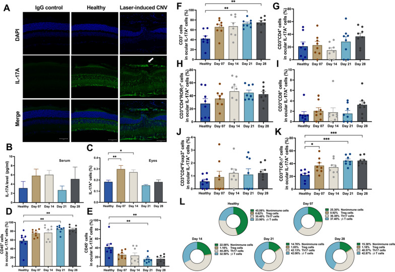
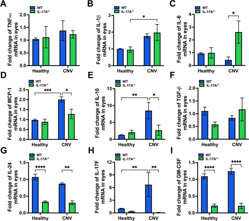
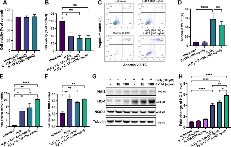
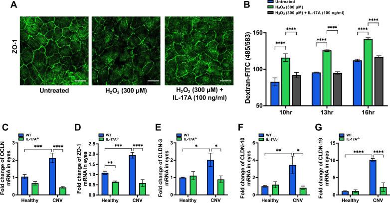
Results: In the laser-induced CNV model, the CNV lesions were larger in IL-17A-deficient mice than in WT mice. The numbers of γδ T cells, CD3+CD4+RORγt+ T cells, Treg cells, and neutrophils were decreased and the number of macrophages was increased in the eyes of IL-17A-deficient mice compared with WT mice. In WT mice, IL-17A-producing γδ T-cell numbers increased in a time-dependent manner from day 7 to 28 after laser injury. IL-6 levels increased and IL-10, IL-24, IL-17F, and GM-CSF levels decreased in the eyes of IL-17A-deficient mice after laser injury. In vitro, IL-17A inhibited apoptosis and induced the expression of the antioxidant protein HO-1 in ARPE-19 cells under oxidative stress conditions. IL-17A facilitated the repair of oxidative stress-induced barrier dysfunction in ARPE-19 cells.

Conclusions: Our findings provide new insight into the protective effect of IL-17A in a laser-induced CNV model and reveal a novel regulatory role of IL-17A-producing γδ T cells in the ocular microenvironment in wet AMD.

Keywords: AMD; Choroidal neovascularization; IL-17A; Inflammation; Oxidative stress.


Conflict of interest statement

The authors declare no competing interests.

Figures

Fig. 1
Fig. 1
IL-17A deficiency exacerbated disease severity in a laser-induced CNV mouse model. A Representative fundus fluorescein angiography (FFA) images from WT and IL-17A−/− mice at 7, 14, 21, and 28 days after laser photocoagulation. CNV lesions were analyzed by fluorescence angiography. B Quantification of the CNV area after laser photocoagulation at 7, 14, 21, and 28 days. Statistical differences were determined by unpaired t test. Data are presented as the mean ± SEM (*P < 0.05, ** < 0.01, *** < 0.001). N = 18 mice per group. C H&E staining of the retinal structure in healthy mice and WT and IL-17A−/− mice with laser-induced CNV at 28 days after laser injury. Magnification: 200X. Scale bar = 100 μm. DJ Representative flow cytometry data for the CD4+ T cell, γδ T cell, CD3+CD4+RORγt+ T cell, Treg cell, macrophage, neutrophil, and MDSC populations in the eyes of WT and IL-17A−/− mice 28 days after laser injury. N = 5 mice per group. Statistical differences were determined by two-way ANOVA and Sidak’s multiple comparisons test. Data are presented as the mean ± SEM (*P < 0.05, **P < 0.01)
Fig. 2
Fig. 2
Cellular source of IL-17A in WT mice with the laser-induced CNV. A Immunofluorescence staining of ocular tissue with an anti-IL-17A antibody in vivo. Rabbit IgG isotype was used as the negative control. Magnification: 200X. Scale bar = 50 μm. B Serum IL-17A levels were measured by using ELISA at 7, 14, 21 and 28 days after laser photocoagulation. N = 5 mice per group. CE Flow cytometry analysis of IL-17A-expressing cells among ocular cells and further gating of immune cells (CD45+) and nonimmune cells (CD45). Statistical differences were determined by one-way ANOVA and Tukey’s multiple comparisons test. Data are presented as the mean ± SEM (*P < 0.05, **P < 0.01). N = 8 mice per group. FK Representative flow cytometry data for the CD3+IL-17A+ T cell, CD3+CD4+IL-17A+ T cell, CD3+CD8+IL-17A+ T cell, γδ T cell, Th17 cell, and Treg cell populations in the eyes of WT mice at 7, 14, 21, and 28 days after laser injury in comparison with healthy control mice. N = 8 mice per group. Statistical differences were determined by one-way ANOVA and Tukey’s multiple comparisons test. Data are presented as the mean ± SEM (*P < 0.05, **P < 0.01, ***P < 0.001). The data shown are representative of three independent experiments with similar results. L The dynamic changes in the proportion of IL-17A-expressing immune cells and the percentages of nonimmune cells, Treg cells, Th17 cells, and γδ T cells were analyzed at 7, 14, 21, and 28 days after laser injury. N = 8 mice per group
Fig. 3
Fig. 3
IL-17A deficiency influenced ocular cytokine expression in a laser-induced CNV mouse model. Eyes were harvested from healthy mice and WT and IL-17A−/− mice with laser-induced CNV at 28 days after laser injury (N = 8). Both eyes were pooled for each animal. AI The mRNA expression of IL-10, MCP-1, IL-1β, TNF-α, IL-6, TGF-β, IL-24, IL-17F, and GM-CSF in the eyes was analyzed by using RT‒qPCR with specific primers. Statistical differences were determined by two-way ANOVA and Sidak’s multiple comparisons test. Data are presented as the mean ± SEM (*P < 0.05, **P < 0.01, ***P < 0.001, ****P < 0.0001). The data shown are representative of three independent experiments with similar results
Fig. 4
Fig. 4
IL-17A inhibited H2O2-induced apoptosis through HO-1 in ARPE-19 cells. A, B ARPE-19 cells were treated with IL-17A (10-100 ng/ml), H2O2 (300 μM), and IL-17A (100 ng/ml) plus H2O2 (300 μM) for 24 h. Cell viability was analyzed using the MTT assay. Statistical differences were determined by one-way ANOVA and Tukey’s multiple comparisons test. Data are presented as the mean ± SEM (*P < 0.05, **P < 0.01). C ARPE-19 cells were treated with or without IL-17A (100 ng/ml) for 24 h and then treated with H2O2 (300 μM) for 24 h, and cell apoptosis was analyzed using FACS. D Quantification of Annexin V+/ PI+ ratios in ARPE-19 cells after H2O2 and IL-17A treatment. Statistical differences were determined by one-way ANOVA and Tukey’s multiple comparisons test. Data are presented as the mean ± SEM (**P < 0.01, ****P < 0.0001). The data shown are representative of three independent experiments with similar results. E, F ARPE-19 cells were cotreated with H2O2 and IL-17A for 8 h, and the mRNA levels of HO-1 and NQO-1 were analyzed by RT‒qPCR with specific primers. N = 3. Statistical differences were determined by one-way ANOVA and Tukey’s multiple comparisons test. Data are presented as the mean ± SEM (*P < 0.05, **P < 0.01, ****P < 0.0001). The data shown are representative of three independent experiments with similar results. G, H ARPE-19 cells were cotreated with H2O2 and IL-17A for 24 h. The cell lysates were collected and analyzed using immunoblotting with specific antibodies against Nrf2, HO-1, and NQO-1. Tubulin was used as an internal control. Statistical differences were determined by one-way ANOVA and Tukey’s multiple comparisons test. Data are presented as the mean ± SEM of three independent experiments. (*P < 0.05, **P < 0.01, ****P < 0.0001)
Fig. 5
Fig. 5
IL-17A facilitated repair of oxidative stress-induced barrier dysfunction. ARPE-19 cells were cultured in DMEM/F12 with 1% FBS for 7 days until they reached the appropriate cell confluence. A ARPE-19 cells were cotreated with 300 μM H2O2 and IL-17A (100 ng/ml) in the Transwell insert for 16 h, and immunofluorescence staining was performed with an anti-ZO-1-specific antibody in ARPE-19 monolayers in vitro. Scale bar = 75 μm. B To analyze the permeability function, ARPE-19 cells were cotreated with 300 μM H2O2 and IL-17A (100 ng/ml) in the Transwell insert for 16 h. FITC-dextran (100 μg/ml) was added after treatment, and the fluorescent content was measured at 485/538 nm excitation/emission wavelengths using a luminometer at 10-16 h. Statistical differences were determined by one-way ANOVA and Tukey’s multiple comparisons test. Data are presented as the mean ± SEM (****P < 0.0001). The data shown are representative of three independent experiments with similar results. CG The mRNA expression of ZO-1, OCLN, CLDN-3, CLDN-10, and CLDN-19 in eyes isolated from WT and IL-17A−/− mice (N = 6) at 28 days after laser injury was analyzed using RT‒qPCR with specific primers. Statistical differences were determined by two-way ANOVA and Sidak's multiple comparisons test. Data are presented as the mean ± SEM (*P < 0.05, **P < 0.01, ***P < 0.001, ****P < 0.0001). The data shown are representative of three independent experiments with similar results

Similar articles

Cited by

References

    1. Mehta S. Age-related macular degeneration. Prim Care. 2015;42:377–391. doi: 10.1016/j.pop.2015.05.009. - DOI - PubMed
    1. Friedman DS, O'Colmain BJ, Muñoz B, Tomany SC, McCarty C, de Jong PT, Nemesure B, Mitchell P, Kempen J. Prevalence of age-related macular degeneration in the United States. Arch Ophthalmol. 2004;122:564–572. doi: 10.1001/archopht.122.4.564. - DOI - PubMed
    1. Tielsch JM, Javitt JC, Coleman A, Katz J, Sommer A. The prevalence of blindness and visual impairment among nursing home residents in Baltimore. N Engl J Med. 1995;332:1205–1209. doi: 10.1056/NEJM199505043321806. - DOI - PubMed
    1. Ferris FL, 3rd, Fine SL, Hyman L. Age-related macular degeneration and blindness due to neovascular maculopathy. Arch Ophthalmol. 1984;102:1640–1642. doi: 10.1001/archopht.1984.01040031330019. - DOI - PubMed
    1. Caprara C, Grimm C. From oxygen to erythropoietin: relevance of hypoxia for retinal development, health and disease. Prog Retin Eye Res. 2012;31:89–119. doi: 10.1016/j.preteyeres.2011.11.003. - DOI - PubMed