. 2023 Nov 25;14(11):773.
doi: 10.1038/s41419-023-06318-6.

Caspase-8 activation by cigarette smoke induces pro-inflammatory cell death of human macrophages exposed to lipopolysaccharide

Affiliations

Caspase-8 activation by cigarette smoke induces pro-inflammatory cell death of human macrophages exposed to lipopolysaccharide

Marta Cristaldi et al. Cell Death Dis. .

Abstract

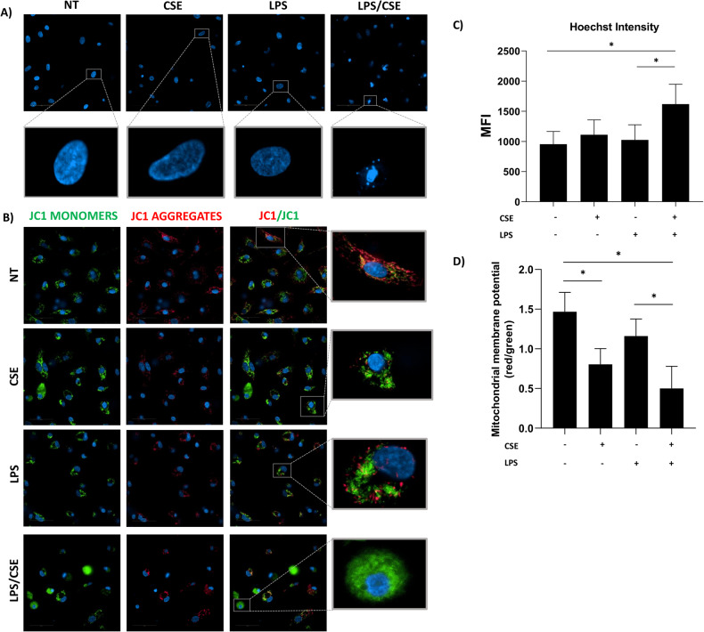
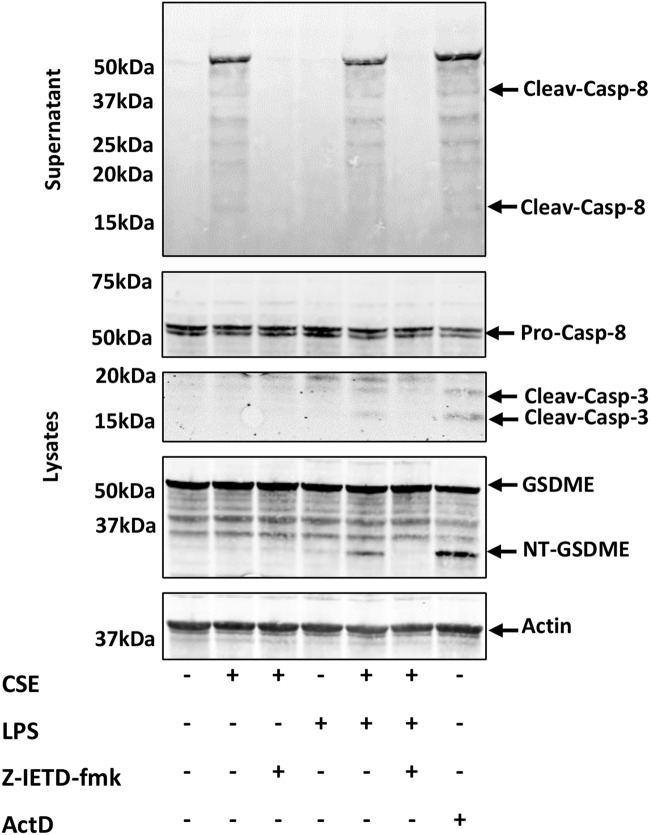
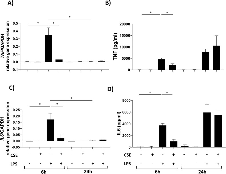
Cigarette smoking impairs the lung innate immune response making smokers more susceptible to infections and severe symptoms. Dysregulation of cell death is emerging as a key player in chronic inflammatory conditions. We have recently reported that short exposure of human monocyte-derived macrophages (hMDMs) to cigarette smoke extract (CSE) altered the TLR4-dependent response to lipopolysaccharide (LPS). CSE caused inhibition of the MyD88-dependent inflammatory response and activation of TRIF/caspase-8/caspase-1 pathway leading to Gasdermin D (GSDMD) cleavage and increased cell permeability. Herein, we tested the hypothesis that activation of caspase-8 by CSE increased pro-inflammatory cell death of LPS-stimulated macrophages. To this purpose, we measured apoptotic and pyroptotic markers as well as the expression/release of pro-inflammatory mediators in hMDMs exposed to LPS and CSE, alone or in combination, for 6 and 24 h. We show that LPS/CSE-treated hMDMs, but not cells treated with CSE or LPS alone, underwent lytic cell death (LDH release) and displayed apoptotic features (activation of caspase-8 and -3/7, nuclear condensation, and mitochondrial membrane depolarization). Moreover, the negative regulator of caspase-8, coded by CFLAR gene, was downregulated by CSE. Activation of caspase-3 led to Gasdermin E (GSDME) cleavage. Notably, lytic cell death caused the release of the damage-associated molecular patterns (DAMPs) heat shock protein-60 (HSP60) and S100A8/A9. This was accompanied by an impaired inflammatory response resulting in inhibited and delayed release of IL6 and TNF. Of note, increased cleaved caspase-3, higher levels of GSDME and altered expression of cell death-associated genes were found in alveolar macrophages of smoker subjects compared to non-smoking controls. Overall, our findings show that CSE sensitizes human macrophages to cell death by promoting pyroptotic and apoptotic pathways upon encountering LPS. We propose that while the delayed inflammatory response may result in ineffective defenses against infections, the observed cell death associated with DAMP release may contribute to establish chronic inflammation. CS exposure sensitizes human macrophages to pro-inflammatory cell death. Upon exposure to LPS, CS inhibits the TLR4/MyD88 inflammatory response, downregulating the pro-inflammatory genes TNF and IL6 and the anti-apoptotic gene CFLAR, known to counteract caspase-8 activity. CS enhances caspase-8 activation through TLR4/TRIF, with a partial involvement of RIPK1, resulting on the activation of caspase-1/GSDMD axis leading to increased cell permeability and DAMP release through gasdermin pores [19]. At later timepoints caspase-3 becomes strongly activated by caspase-8 triggering apoptotic events which are associated with mitochondrial membrane depolarization, gasdermin E cleavage and secondary necrosis with consequent massive DAMP release.


Conflict of interest statement

The authors declare no competing interests.

Figures

None
CS exposure sensitizes human macrophages to pro-inflammatory cell death. Upon exposure to LPS, CS inhibits the TLR4/MyD88 inflammatory response, downregulating the pro-inflammatory genes TNF and IL6 and the anti-apoptotic gene CFLAR, known to counteract caspase-8 activity. CS enhances caspase-8 activation through TLR4/TRIF, with a partial involvement of RIPK1, resulting on the activation of caspase-1/GSDMD axis leading to increased cell permeability and DAMP release through gasdermin pores [19]. At later timepoints caspase-3 becomes strongly activated by caspase-8 triggering apoptotic events which are associated with mitochondrial membrane depolarization, gasdermin E cleavage and secondary necrosis with consequent massive DAMP release.
Fig. 1
Fig. 1. Lytic cell death occurred in hMDMs stimulated with LPS and CSE and was associated with enhanced caspase-8 and -3/7 activation and CFLAR inhibition.
hMDMs were treated with 1 μg/ml of LPS and 20% CSE, alone or in combination. A LDH release and (B) extracellular activity of Caspase-8 and -3/7 (expressed as relative luminescence unit, RLU), were measured after 24 h stimulation. C CFLAR gene expression was measured after 6 and 24 h stimulation. Where indicated, cells were pre-treated for 1 h with 0.1 μM Z-IETD-fmk Caspase-8 inhibitor, 20 μM Z-VAD-fmk pan-caspase inhibitor, 50 μM Nec-1 RIPK1 inhibitor. The absorbance of LDH was measured at 490 nm. Data are presented as mean ± SEM (N = 3 independent donors).
Fig. 2
Fig. 2. hMDMs exposed to CSE and LPS display apoptotic hallmarks.
Representative images of hMDMs treated with 1 μg/ml of LPS and 20% CSE, alone or in combination, for 24 h and stained with (A) Hoechst 33342 for nuclear morphology evaluation and (B) JC-1 dye for MMP assessment. C Quantification of Hoechst 33342 intensity expressed as mean fluorescent intensity (MFI). D Quantification of the red (485/590 nm)/green (485/529 nm) fluorescence intensity ratio after JC1 staining. Scale bar: 50 μm. Data are presented as mean ± SEM (N = 3 independent donors).
Fig. 3
Fig. 3. LPS triggers the activation of the axis caspase-8/caspase-3/GSDME in hMDMs exposed to CSE.
Representative western blot images of Caspase-8, -3 and GSDME in hMDMs treated with 1 μg/ml of LPS and 20% CSE, alone or in combination for 24 h. Where indicated, hMDMs were pre-treated for 1 h with 0.1 μM Z-IETD-fmk Caspase-8 inhibitor (N = 3 independent donors). ActD 0.5 μg/ml was used as positive control. β-actin was used as loading control.
Fig. 4
Fig. 4. Cleaved Caspase-3 and GSDME gene expression in alveolar macrophages from smokers compared to non-smoking controls.
A Representative images of immunohistochemical staining of distal lung tissue sections of Smokers (N = 5) and Non-smoking controls (N = 6), using a specific antibody for cleaved Caspase-3. B Graph showing the percentage of cells positive for cleaved Caspase-3. Data are presented as median with interquartile range. The difference between the percentage of immunopositivity in Non-Smoking controls and Smokers was statistically evaluated with the Mann–Whitney test. C Box plot of log-normalized expression of GSDM family genes in Non-Smokers and Smokers samples. Data are presented as median with interquartile range. Differences of Smokers vs. Non-Smokers gene expression were evaluated in the whole microarray with the moderated t-test and FDR correction.
Fig. 5
Fig. 5. Exposure to CSE delayed the inflammatory response to LPS.
Gene expression (left) and cytokine release (right) of (A, B) TNF and (C, D) IL6 from hMDMs exposed to 1 μg/ml of LPS and 20% CSE, alone or in combination, for 6 and 24 h. Data are presented as mean ± SEM (N = 3 independent donors).
Fig. 6
Fig. 6. DAMPs are released from hMDMs exposed to LPS and CSE.
Release of HSP60, S100A8/A9, IL1 alpha and IL33 in hMDMs exposed to 1 μg/ml of LPS and 20% CSE, alone or in combination for 24 h, evaluated by multiplex analysis. Where indicated, cells were pre-treated for 1 h with 20 μM Z-VAD-fmk pan-caspase inhibitor and 50 μM Nec-1 RIPK1 inhibitor. Data are presented as mean ± SEM (N = 3 independent donors).

References

    1. Lugg ST, Scott A, Parekh D, Naidu B, Thickett DR. Cigarette smoke exposure and alveolar macrophages: mechanisms for lung disease. Thorax. 2022;77:94–101. doi: 10.1136/thoraxjnl-2020-216296. - DOI - PMC - PubMed
    1. Lugade AA, Bogner PN, Thatcher TH, Sime PJ, Phipps RP, Thanavala Y. Cigarette smoke exposure exacerbates lung inflammation and compromises immunity to bacterial infection. J Immunol. 2014;192:5226–35. doi: 10.4049/jimmunol.1302584. - DOI - PMC - PubMed
    1. Thomson NC, Chaudhuri R, Livingston E. Asthma and cigarette smoking. Eur Respir J. 2004;24:822–33. doi: 10.1183/09031936.04.00039004. - DOI - PubMed
    1. Christenson SA, Smith BM, Bafadhel M, Putcha N. Chronic obstructive pulmonary disease. Lancet. 2022;399:2227–42. doi: 10.1016/S0140-6736(22)00470-6. - DOI - PubMed
    1. Zuo L, He F, Sergakis GG, Koozehchian MS, Stimpfl JN, Rong Y, et al. Interrelated role of cigarette smoking, oxidative stress, and immune response in COPD and corresponding treatments. Am J Physiol Lung Cell Mol Physiol. 2014;307:L205–18. doi: 10.1152/ajplung.00330.2013. - DOI - PubMed

Publication types

MeSH terms