. 2023 Nov 25;13(1):20758.
doi: 10.1038/s41598-023-48086-5.

Phylogenetic structure of body shape in a diverse inland ichthyofauna

Affiliations

Phylogenetic structure of body shape in a diverse inland ichthyofauna

Kevin T Torgersen et al. Sci Rep. .

Abstract

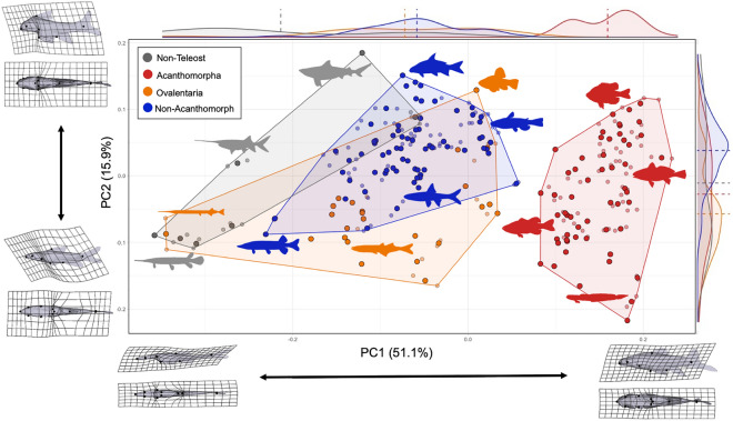
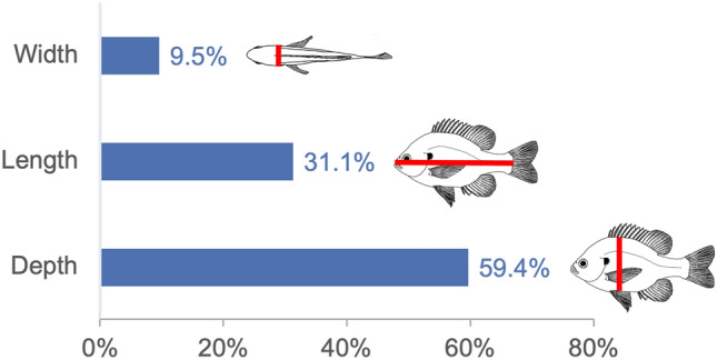
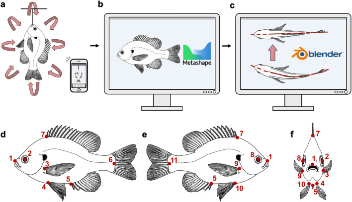
Body shape is a fundamental metric of animal diversity affecting critical behavioral and ecological dynamics and conservation status, yet previously available methods capture only a fraction of total body-shape variance. Here we use structure-from-motion (SFM) 3D photogrammetry to generate digital 3D models of adult fishes from the Lower Mississippi Basin, one of the most diverse temperate-zone freshwater faunas on Earth, and 3D geometric morphometrics to capture morphologically distinct shape variables, interpreting principal components as growth fields. The mean body shape in this fauna resembles plesiomorphic teleost fishes, and the major dimensions of body-shape disparity are similar to those of other fish faunas worldwide. Major patterns of body-shape disparity are structured by phylogeny, with nested clades occupying distinct portions of the morphospace, most of the morphospace occupied by multiple distinct clades, and one clade (Acanthomorpha) accounting for over half of the total body shape variance. In contrast to previous studies, variance in body depth (59.4%) structures overall body-shape disparity more than does length (31.1%), while width accounts for a non-trivial (9.5%) amount of the total body-shape disparity.


Conflict of interest statement

The authors declare no competing interests.

Figures

Figure 1
Figure 1
Phylogenetic diversity of the LMB fish fauna. (a) Time-calibrated phylogeny of extant ray-finned fishes (Actinopterygii), with family names and colored silhouettes illustrating 40 of 490 (8.1%) actinopterygian families represented in the LMB fish fauna. Colored polygons represent nested clades used in the analysis of morphospace occupancy: gray, non-teleosts; blue, non-acanthomorph teleosts, red, non-ovalentarian acanthomorphs; orange, ovalentarian acanthomorphs; purple, clades excluded from analysis due to lack of homologous landmarks. Colored silhouettes were generated from images of actual fish specimens from the LMB. (b) Map of Mississippi River basin with LMB study area highlighted in red box. (c) Species and family composition of the four assemblages within the LMB fish fauna. Taxonomic names follow accepted conventions for the field.
Figure 2
Figure 2
Common LMB fish species with exemplar body shapes. (a) The Pugnose Minnow Opsopoeodus emiliae (Leuciscidae), to 6.4 cm standard length (SL). (b) Longnose Gar Lepisosteus osseus (Lepisosteidae), to 122 cm SL. (c) Bluegill Lepomis macrochirus (Centrarchidae), to 30 cm SL. (d) Flathead Catfish Pylodictis olivaris (Ictaluridae), to 155 cm SL.
Figure 3
Figure 3
Morphospace of LMB fishes from PCA of 3D landmark data. Data for 232 specimens (smaller circles) in 166 species (larger circles) representing 37 family-level clades. PC1 on horizontal axis, PC2 on vertical axis. Relative frequency histograms along PC axes with mean values as dashed lines. Deformation grids illustrating extreme PC values for each axis in lateral and ventral views. Color scheme as in Fig. 1.
Figure 4
Figure 4
Morphospace analysis of LMB fishes from 3D landmark data in PCs 3–6. Data for 232 specimens (smaller circles) in 166 species (larger circles) representing 40 family-level clades present in the LMB fish fauna. PC1 on horizontal axis, PC3–6 on vertical axes. Relative frequency histograms along PC axes with mean values as dashed lines. Colors as in Fig. 3. (a) PC1 versus PC3. (b) PC1 versus PC4. (c) PC1 versus PC5. (d) PC1 versus PC6.
Figure 5
Figure 5
Summed variance of landmark deformations in each of the three spatial dimensions. Note the greater variance in dorsoventral (depth) than anteroposterior (length) or mediolateral (width) landmark positions. Note also the relative magnitude of variance among spatial dimensions differs from the absolute size of these dimensions; i.e. length > depth > width.
Figure 6
Figure 6
Digitalization of biological specimens for downstream multivariate analyses using 3D photogrammetry. (a) Specimen is prepared and suspended at an appropriate height to allow for 360° access and photography by the researcher in an evenly lit location. 150–600 overlapping photographs are taken from every angle using modern cell phone cameras; however, we note that more expensive cameras may provide 3D models with higher levels of surface detail. (b) Digital photographs of specimen are loaded into Agisoft Metashape for 3D reconstruction. 3D models were exported as .obj files with .jpg textures. (c) 3D models were then individually loaded into Blender to clean and carefully straighten while still maintaining their overall natural shape. The preservation and long-term storage of wet biological specimens often results in the specimen becoming permanently bent or twisted, confounding any GM analysis of shape, and thus requiring a method of unbending. Unbent and cleaned 3D models were then exported from Blender to be used in the GM analysis. (d) Landmark scheme of 11 homologous landmarks used in the GM analysis in left lateral view. (e) Landmark scheme in right lateral view. (f) Landmark scheme in anterior view to convey 3D nature of landmarks.

Similar articles

Cited by

  • An ecological trait matrix of Neotropical freshwater fishes.
    Albert JS, Abrahão V, Akin DR, Allen JG, Ândrade M, Arce M, Armbruster JW, Benine R, Bernt MJ, Bichuette ME, Birindelli J, Bragança P, Britto MR, Buckup PA, Burns MD, Calegari BB, Carvalho FG, Carvalho TP, Casatti L, Costa W, Crampton WGR, da Costa Ramos R, da Silva RG, Dagosta F, Egan J, Fernández L, Figueiredo-Filho JM, Fitzgerald D, Fontenelle JP, Haddad EB, Kolmann M, Hrbek T, Langeani F, Lehmann P, Leitão RP, Lima FCT, Loeb MV, Lucena CAS, Lucinda PHF, Lujan NK, Malabarba LR, Matamoros WA, Melo BF, Netto-Ferreira A, Pavanelli CS, Pelicice FM, Pereira EHL, Plasencia Iii X, Rocha M, Roxo F, Anna VS, Shibatta O, Slobodian V, Stange M, Tagliacollo VA, Tan M, Torgersen KT, Winemiller KO, Reis RE. Albert JS, et al. Sci Data. 2025 Jul 2;12(1):1127. doi: 10.1038/s41597-025-04674-w. Sci Data. 2025. PMID: 40603332 Free PMC article.

References

    1. Peters RH, Peters RH. The Ecological Implications Of Body Size. Cambridge Univ. Press; 1986.
    1. Raff RA. The Shape of Life: Genes, Development, and the Evolution of Animal Form. University of Chicago Press; 2012.
    1. Carmona CP, et al. Erosion of global functional diversity across the tree of life. Sci. Adv. 2021;7:eabf2675. doi: 10.1126/sciadv.abf2675. - DOI - PMC - PubMed
    1. Toussaint A, et al. Extinction of threatened vertebrates will lead to idiosyncratic changes in functional diversity across the world. Nat. Commun. 2021;12:5162. doi: 10.1038/s41467-021-25293-0. - DOI - PMC - PubMed
    1. Webb PW. Body form, locomotion and foraging in aquatic vertebrates. Am. Zool. 1984;24:107–120. doi: 10.1093/icb/24.1.107. - DOI