. 2023 Nov 25;6(1):1203.
doi: 10.1038/s42003-023-05564-w.

Biologically derived epicardial patch induces macrophage mediated pathophysiologic repair in chronically infarcted swine hearts

Affiliations

Biologically derived epicardial patch induces macrophage mediated pathophysiologic repair in chronically infarcted swine hearts

J J Lancaster et al. Commun Biol. .

Abstract

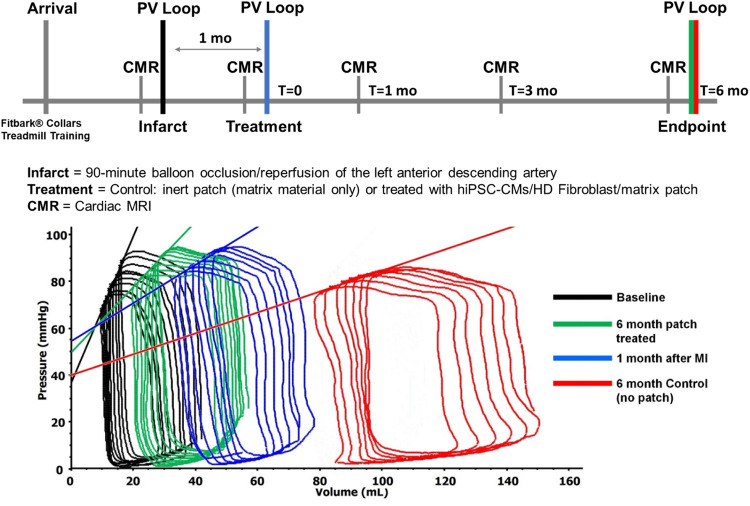
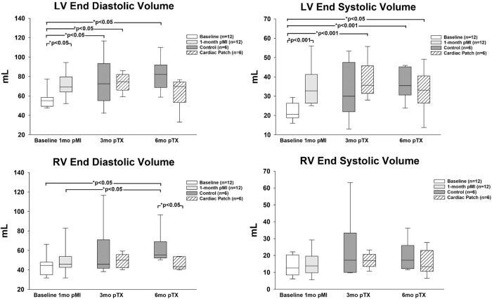
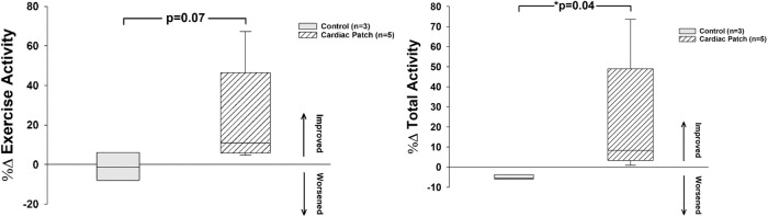
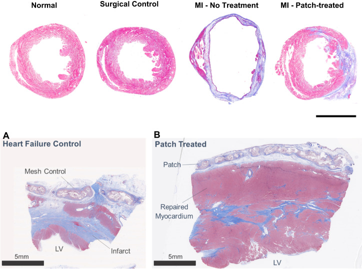
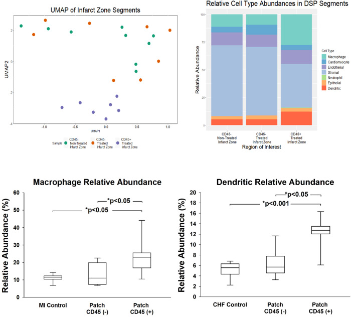
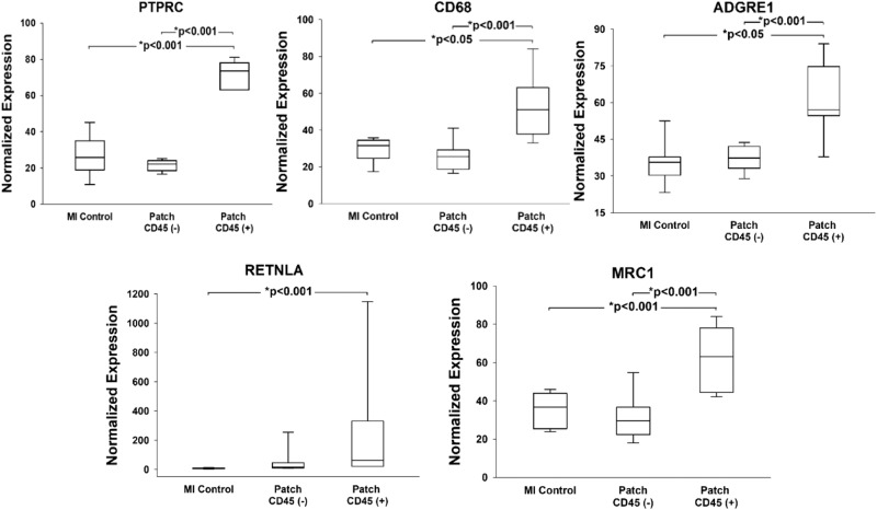
There are nearly 65 million people with chronic heart failure (CHF) globally, with no treatment directed at the pathologic cause of the disease, the loss of functioning cardiomyocytes. We have an allogeneic cardiac patch comprised of cardiomyocytes and human fibroblasts on a bioresorbable matrix. This patch increases blood flow to the damaged heart and improves left ventricular (LV) function in an immune competent rat model of ischemic CHF. After 6 months of treatment in an immune competent Yucatan mini swine ischemic CHF model, this patch restores LV contractility without constrictive physiology, partially reversing maladaptive LV and right ventricular remodeling, increases exercise tolerance, without inducing any cardiac arrhythmias or a change in myocardial oxygen consumption. Digital spatial profiling in mice with patch placement 3 weeks after a myocardial infarction shows that the patch induces a CD45pos immune cell response that results in an infiltration of dendritic cells and macrophages with high expression of macrophages polarization to the anti-inflammatory reparative M2 phenotype. Leveraging the host native immune system allows for the potential use of immunomodulatory therapies for treatment of chronic inflammatory diseases not limited to ischemic CHF.


Conflict of interest statement

Drs. Goldman, Koevary, Lancaster and Ms. Sherry Daugherty have disclosed a financial interest in Avery Therapeutics, Inc. to the University of Arizona. In addition, the University of Arizona has a financial interest in Avery Therapeutics, Inc. These interests have been reviewed and are being managed by the University of Arizona in accordance with its policies on outside interests. The work outlined in this report were the basis of forming a commercial entity, Avery Therapeutics. Drs. Goldman, Koevary, Lancaster and Ms. Sherry Daugherty have disclosed a financial interest in Avery Therapeutics, Inc. to the University of Arizona. In addition, the University of Arizona has a financial interest in Avery Therapeutics, Inc. These interests have been reviewed and are being managed by the University of Arizona in accordance with its policies on outside interests. All other authors declare no competing interests.

Figures

Fig. 1
Fig. 1. Chronologic swine study timeline and Pressure/Volume relationships.
The myocardial infarction was performed by cardiac catheterization occlusion/reperfusion, 4 weeks prior to thoracotomy to place patch with cells or inert-matrix placement. The swine underwent CMR imaging, hemodynamic measurements, conductance catheter pressure/volume assessments, activity monitoring, treadmill testing, ECG loop recorders 24/7 and histopathologic examination at necropsy. CMR; cardiac magnetic resonance. Conductance catheter composite LV P/V analyses showed a decrease in the slope of the LV end-systolic P/V or the Ees and increased LV volume at 1 month after MI. These changes worsened at 6months in the control group. The treated group showed an increase in Ees and decreased LV volume at 6 months, with no change in LV EDPVR. This is evidence of partial reversal of LV remodeling and an increase in LV contractility after patch treatment. P/V, pressure/volume; LV, left ventricle; Ees, end-systolic elastance; LV EDPVR, left ventricular end diastolic pressure/volume relationship.
Fig. 2
Fig. 2. Ventricular Volumes: Left ventricular (LV) and right ventricular (RV) volumes at 3 and 6 months showed similar trends with increases in both at 3 and 6 months in the control animals, while RV end-diastolic volume trends down at both 3 and 6 months after patch placement.
LV left ventricle, RV right ventricle. Statistics performed on the data set were based on the mean ± SEM for n = 12 baseline animals, n = 12 1-month post injury animals, n = 6 control animals, and n = 6 patch treated animals.
Fig. 3
Fig. 3. Exercise and Overall Activity: All swine in this study had activity monitored with FitBark® collars 24/7.
There was a trend for exercise activity level to increase (left) and an increase in total activity (right) in the patch treated swine. Statistics performed on the data were based on mean ± SEM for n = 3 control swine and n = 6 patch treated swine, with p = 0.07 for exercise activity level, and p = 0.04 for total activity.
Fig. 4
Fig. 4. Heart rate changes with exercise.
Heart rates increased with exercise (post run) in all the swine. Heart rate decreased at 3 months after patch placement at all time points. There were no differences in heart rate at any time point between the control and cardiac patch treated swine. T-test performed and where normality failed a Mann-Whitney Rank Sum test was performed. *P < 0.001; # P < 0.001 vs 1mo pMI; @ P < 0.001 vs Resting Heart Rate; ! P < 0.001 vs Post-Run Heart Rate. These analyses were performed for n = 6; baseline, n = 6; 1 month pMI, n = 2; 3 month pTx Control, n = 3; 3 month pTx Patch, n = 2; 6 month pTx Control, and n = 4; 6 month pTx Patch.
Fig. 5
Fig. 5. Trichrome stained cross-sections of porcine and murine hearts sections.
Masson’s trichrome histopathological sections of porcine control (A) and patch treated (B) swine at 6-month study endpoint. All swine received myocardial infarction by a 90-minute balloon occlusion followed by reperfusion. Heart failure was allowed to develop over 4-weeks prior to treatment. Swine received epicardial implant of either Control (bioresorbable mesh without cells) or patch. Control tissue had a higher scar burden and a thinner LV anterior wall. Patch treatment resulted in improved tissue composition (increased myocyte presence and decreased scar) and restored wall thickness. After fixation each murine heart was transversely cut approximately 3-5 millimeters above the apex. The apex was formalin fixed, paraffin-embedded and sectioned with subsequent Masson Trichrome staining to define infarcted myocardium (blue) and healthy myocardium (red). There is essentially no difference between normal and surgical control hearts, the MI heart has a dilated LV cavity with thinned scarred anterior wall, the MI- patch shows a smaller LV cavity and thicker anterior wall compared to the MI heart. Normal = no intervention; surgical control = thoracotomies without MI or patch treatment. Abbreviation: MI-myocardial infarction. Murine heart scale bar = 3 mm.
Fig. 6
Fig. 6. Immunohistochemistry for DSP and CD45 quantification.
Trichrome images next to the IHC stained tissue sections prepared for the DSP analysis: Desmin (red)-muscle/endothelial cells, CD 45-immune cells (green), αSMA-activated fibroblasts (yellow), SYTO-nuclear stain (blue). A The trichrome images show a dilated left ventricle with a thinned scarred anterior wall with decreased cardiomyocytes and minimal immune cell infiltrate. B The patch treated images show a smaller left ventricle with a thickened anterior wall and dramatically increased immune cell presence. The images confirm the structural changes and increased number of immune cells with patch treatment. C %CD45 expression per ROI is significantly elevated in Cardiac Patch treated group compared to control MI. Statistics performed on CD45 quantification were based on mean ± SEM for n = 4 AOIs and n = 6 AOIs, with p = 0.03.
Fig. 7
Fig. 7. Digital spatial profiling analyses with macrophage/dendritic cell abundance.
UMAP of MI-non-treated and MI-patch-treated infarct region transcriptome profiles with distinct clustered CD45pos regions [Left]. Cell deconvolution [Right] of the CD45neg and CD45pos segments of the infarcted region showing increases in macrophages and dendritic cells in CD45pos treated infarct zone. UMAP, uniform manifold approximation and projection; MI, Myocardial Infarction. Relative Abundance (left) and Dendritic Relative Abundance (right) increase in CD45pos treated infarct zones, compared to MI control and CD45neg regions. MI, Myocardial Infarction. Statistics performed on the data were based on mean ± SEM for n = 9 MI Control, n = 17 Patch CD45neg, and n = 16 Patch CD45pos, with p < 0.05 for all indicated groups, except for dendritic cells in patch treated CD45 negative and positive regions, where p < 0.05.
Fig. 8
Fig. 8. Immune cell characterization.
High macrophage abundance within CD45pos infarct region defined by increases in PTPCR, CD68, and ADGRE expression compared to lower expression in CD45neg non-treated and treated infarct zones. *P < 0.05, *P < 001. Abbreviations: PTPRC: protein tyrosine phosphatase receptor signaling molecules that regulate cell growth, differentiation, mitosis, and oncogenic transformation; CD68: Routinely used as a histochemical/cytochemical marker of inflammation associated with the involvement of monocytes/macrophages; ADGRE: Adhesion G protein coupled receptor, activated in dendritic cell development. The cardiac patch polarizes macrophages to anti-inflammatory states within the CD45pos regions as defined by increases in RETNLA and MRC1 expression, compared to CD45neg non-treated and treated infarct zones. Abbreviations: RETNLA: Alternatively activated macrophage marker for M2 phenotype; MRC1: Mannose Receptor C-Type 1 is a membrane receptor that mediates the endocytosis of glycoproteins by macrophages. Statistics performed on the data were based on mean ± SEM for n = 9 MI Control, n = 8 Patch CD45neg, and n = 7 Patch CD45pos, with p < 0.01 for all indicated groups, except for CD68 and ADGRE1 markers between MI and patch treated CD45pos regions, where p < 0.05.
Fig. 9
Fig. 9. Computer models are produced from CMR images and compared to intraoperative views of patch placement.
This figure includes a computer model of an infarcted swine heart that was generated from Cardiac Magnetic Resonance (CMR) imaging. The top left image shows the infarcted region in white. In intraoperative view of the same swine, top right, only the infarcted apex is visible. The cardiac patch is placed to cover as much of the scarred area as possible. The bottom images show the computer model placement [Left] and the actual placement on the heart [Right].

Similar articles

Cited by

  • Living Nanofiber-Enabled Cardiac Patches for Myocardial Injury.
    Bhullar SK, Thingnam R, Kirshenbaum E, Nematisouldaragh D, Crandall M, Willerth SM, Ramkrishna S, Rabinovich-Nikitin I, Kirshenbaum LA. Bhullar SK, et al. JACC Basic Transl Sci. 2025 Feb;10(2):227-240. doi: 10.1016/j.jacbts.2024.06.010. Epub 2024 Sep 4. JACC Basic Transl Sci. 2025. PMID: 40131159 Free PMC article. Review.

References

    1. Savarese G, Lund LH. Global public health burden of heart failure. Card. Fail Rev. 2017;3:7–11. doi: 10.15420/cfr.2016:25:2. - DOI - PMC - PubMed
    1. Roger VL. Epidemiology of heart failure: a contemporary perspective. Circ. Res. 2021;128:1421–1434. doi: 10.1161/CIRCRESAHA.121.318172. - DOI - PubMed
    1. van Berlo, J. H. et al. c-kit+ cells minimally contribute cardiomyocytes to the heart. Nature. 509, 337–341. (2014). - PMC - PubMed
    1. Bolli R, Tang XL, Guo Y, Li Q. After the storm: an objective appraisal of the efficacy of ckit+ cardiac progenitor cells in preclinical models of heart disease. Can. J. Physiol. Pharm. 2020;99:129–139. doi: 10.1139/cjpp-2020-0406. - DOI - PMC - PubMed
    1. Bolli R, Solankhi M, Tang XL, Kahlon A. Cell therapy in patients with heart failure: A comprehensive review and emerging concepts. Cardiovasc. Res. 2022;118:951–976. doi: 10.1093/cvr/cvab135. - DOI - PMC - PubMed

Publication types