

miRNA-143 expression is associated with inflammation and time of exposure to amniotic fluid in experimental gastroschisis
- PMID: 38008037
- PMCID: PMC10757286
- DOI: 10.1016/j.clinsp.2023.100311
miRNA-143 expression is associated with inflammation and time of exposure to amniotic fluid in experimental gastroschisis
Abstract
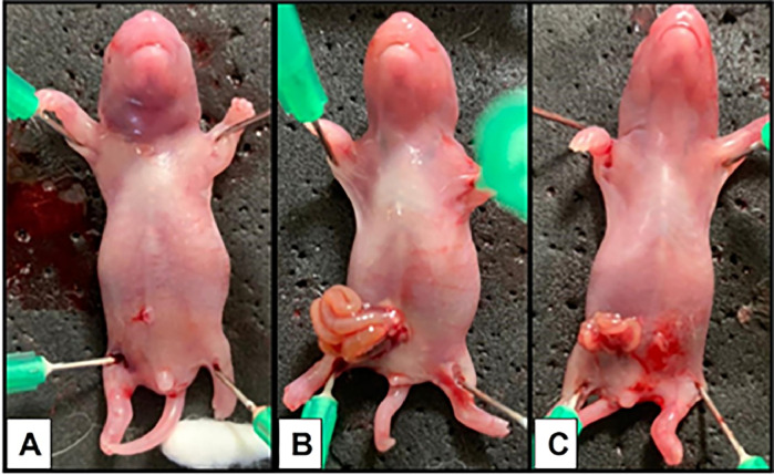
Objective: Gastroschisis (GS) is a congenital anomaly in the abdominal wall with the intestinal loops exiting laterally to the umbilicus. The contact of the loops with Amniotic Fluid (AF) causes an inflammatory process in the exposed part, leading to an extended hospital stay and an increased risk of morbidity due to alterations related to intestinal motility. The authors aimed to evaluate the time of exposure to the AF in the experimental GS and to search for potential biomarkers of intestinal inflammation by measuring microRNAs.
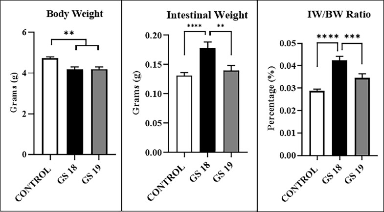
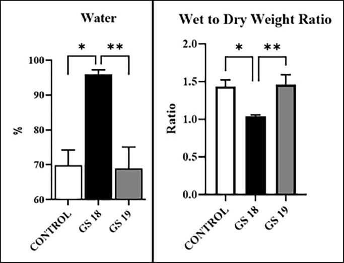
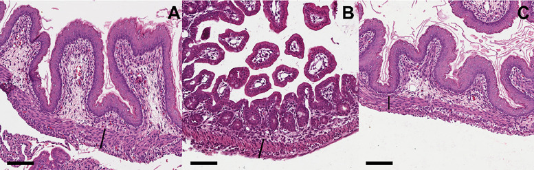
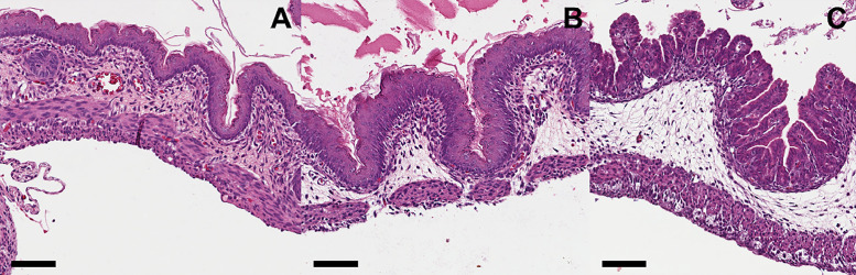
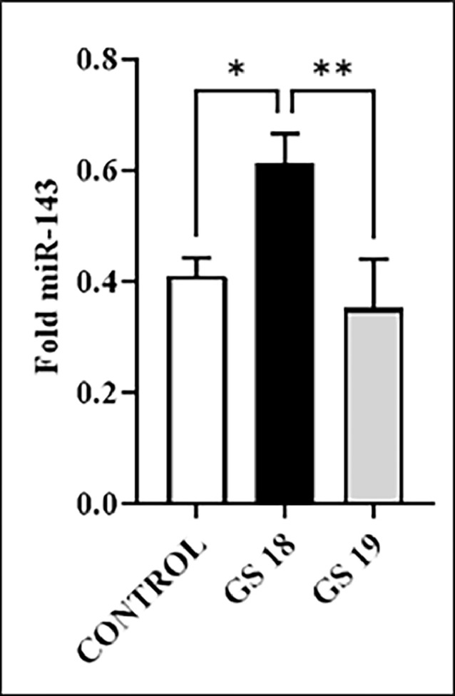
Methods: Rat fetuses were divided into three groups: a) CONTROL, b) GS reared on day 18 (GS = 18), and c) GS reared on day 19.5 (GS = 19) (term = 22 days). On day 21.5, the fetuses were removed for biometric parameters and biochemical analyses: 1) Biometrics: Body and Intestinal Weight (BW, IW), and intestinal-body weight ratio (IW/BW); 2) Descriptive histopathology and 3) miR-143 quantification by real-time Polymerase Chain Reaction (PCR).
Results: BW was higher in CONTROL than GS 18 and G19 (p < 0.05). IW, IW/BW, intestinal water, and mRNA-143 were higher in GS 18 and GS 19 than in CONTROL, and GS 18 was higher than GS 19 (p < 0.05). The average of the inflammation score from the intestinal wall with mucosal inflammation and intra-epithelial lymphocytes shows worst in GS 18 and GS 19 vs. CONTROL (p < 0.05).
Conclusions: The tissue expression of mRNA-143 and the morphological changes in the intestine of GS worsened according to the time of exposure to AF, which could be a possible marker of fetal intestinal damage.
Keywords: Amniotic fluid; Gastroschisis; Inflammation; Rat; mRNA-143.
Copyright © 2023 HCFMUSP. Published by Elsevier España, S.L.U. All rights reserved.
Conflict of interest statement
Declaration of Competing Interest The authors declare no conflicts of interest.
Figures
Similar articles
-
NTP Developmental and Reproductive Toxicity Technical Report on the Prenatal Development Studies of 2-((1-(4-Phenoxyphenoxy)propan-2-yl)oxy)pyridine (CASRN 95737-68-1) in Sprague Dawley (Hsd:Sprague Dawley® SD®) Rats and New Zealand White (Hra:NZW SPF) Rabbits: DART Report 07 [Internet].Research Triangle Park (NC): National Toxicology Program; 2022 Jan. Research Triangle Park (NC): National Toxicology Program; 2022 Jan. PMID: 35593777 Free Books & Documents. Review.
-
[Volume and health outcomes: evidence from systematic reviews and from evaluation of Italian hospital data].Epidemiol Prev. 2013 Mar-Jun;37(2-3 Suppl 2):1-100. Epidemiol Prev. 2013. PMID: 23851286 Italian.
-
Stem cell transplantation for induction of remission in medically refractory Crohn's disease.Cochrane Database Syst Rev. 2022 May 13;5(5):CD013070. doi: 10.1002/14651858.CD013070.pub2. Cochrane Database Syst Rev. 2022. PMID: 35556242 Free PMC article.
-
Sexual Harassment and Prevention Training.2024 Mar 29. In: StatPearls [Internet]. Treasure Island (FL): StatPearls Publishing; 2025 Jan–. 2024 Mar 29. In: StatPearls [Internet]. Treasure Island (FL): StatPearls Publishing; 2025 Jan–. PMID: 36508513 Free Books & Documents.
-
Elective preterm birth for fetal gastroschisis.Cochrane Database Syst Rev. 2013 Jun 5;2013(6):CD009394. doi: 10.1002/14651858.CD009394.pub2. Cochrane Database Syst Rev. 2013. PMID: 23737031 Free PMC article.
References
-
- Beaudoin S. Insights into the etiology and embryology of gastroschisis. Semin Pediatr Surg. 2018;27(5):283–288. - PubMed
-
- Morrison JJ, Klein N, Chitty LS, Kocjan G, Walshe D, Goulding M, et al. Intra-amniotic inflammation in human gastroschisis: possible aetiology of postnatal bowel dysfunction. Br J Obstet Gynaecol. 1998;105(11):1200–1204. - PubMed
-
- Sydorak RM, Nijagal A, Sbragia L, Hirose S, Tsao K, Phibbs RH, et al. Gastroschisis: small hole, big cost. J Pediatr Surg. 2002;37(12):1669–1672. - PubMed
-
- Wilson RD, Johnson MP. Congenital abdominal wall defects: an update. Fetal Diagn Ther. 2004;19(5):385–398. - PubMed
-
- Rittler M, Castilla EE, Chambers C, Lopez-Camelo JS. Risk for gastroschisis in primigravidity, length of sexual cohabitation, and change in paternity. Birth Defects Res A Clin Mol Teratol. 2007;79(6):483–487. - PubMed
Publication types
MeSH terms
Substances
LinkOut - more resources
Full Text Sources
