. 2024 Jan 2:339:199275.
doi: 10.1016/j.virusres.2023.199275. Epub 2023 Nov 30.

Drug repurposing screens identify Tubercidin as a potent antiviral agent against porcine nidovirus infections

Affiliations

Drug repurposing screens identify Tubercidin as a potent antiviral agent against porcine nidovirus infections

Tianliang Wang et al. Virus Res. .

Abstract

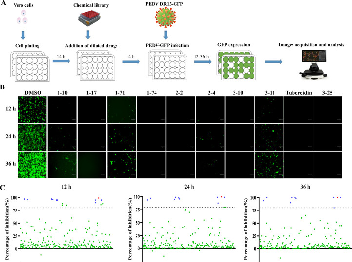
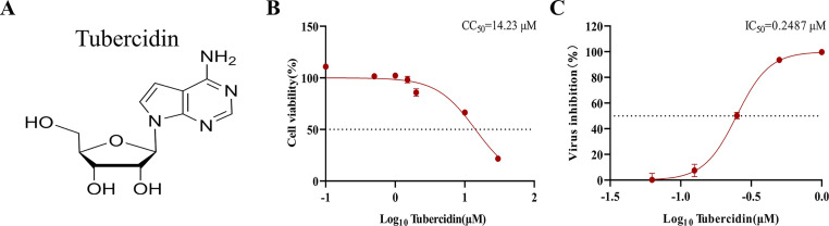
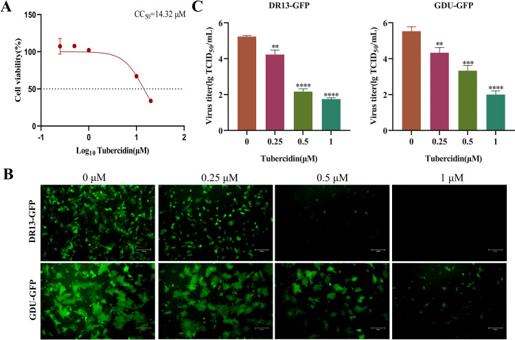
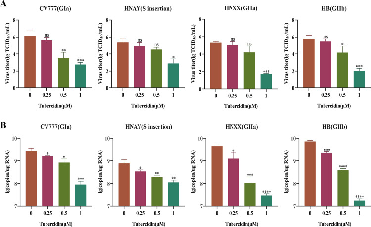
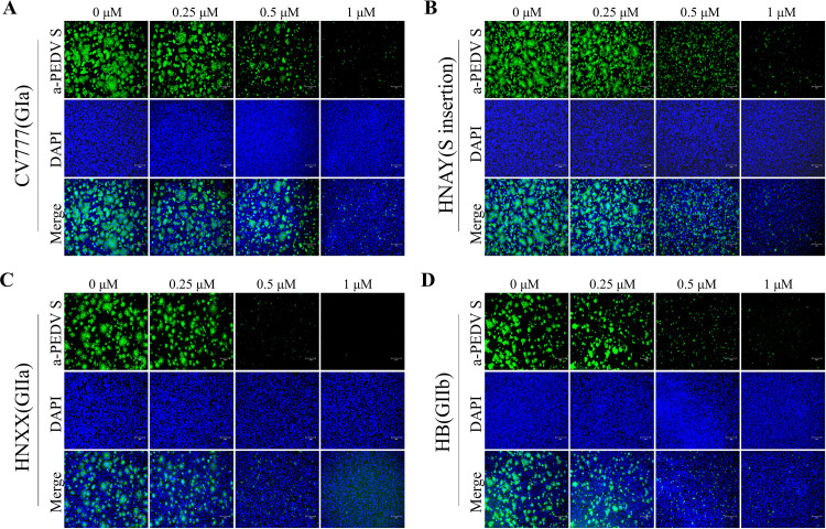
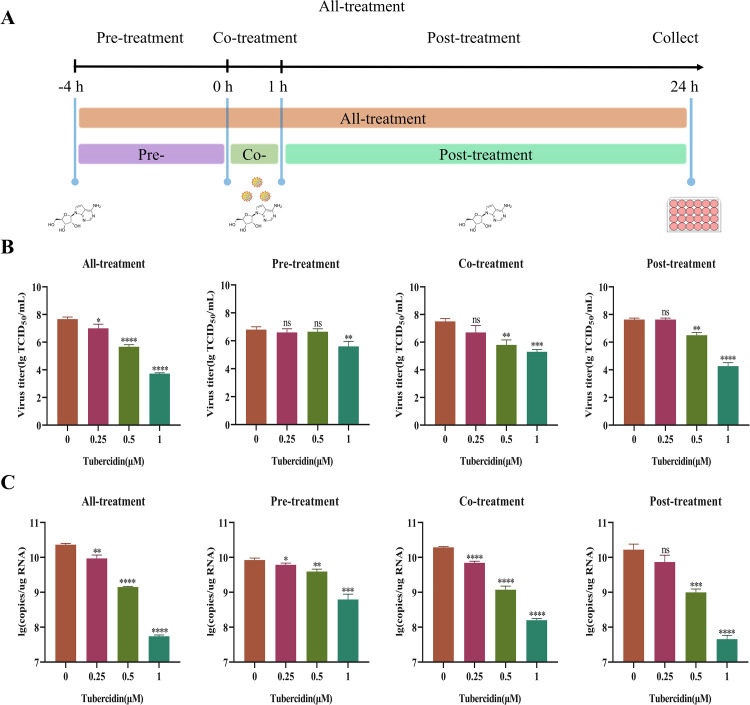
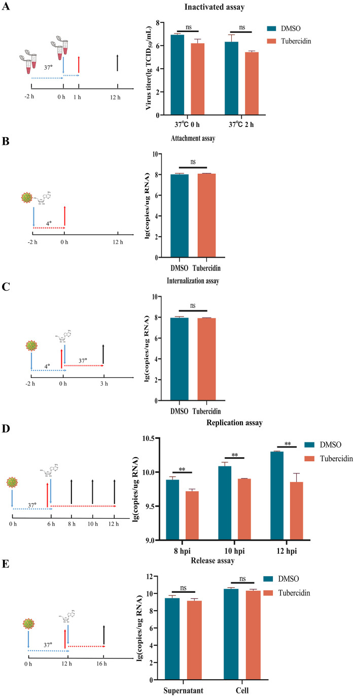
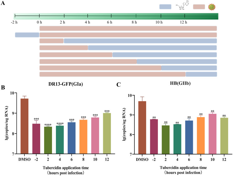
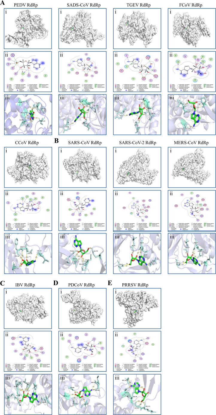
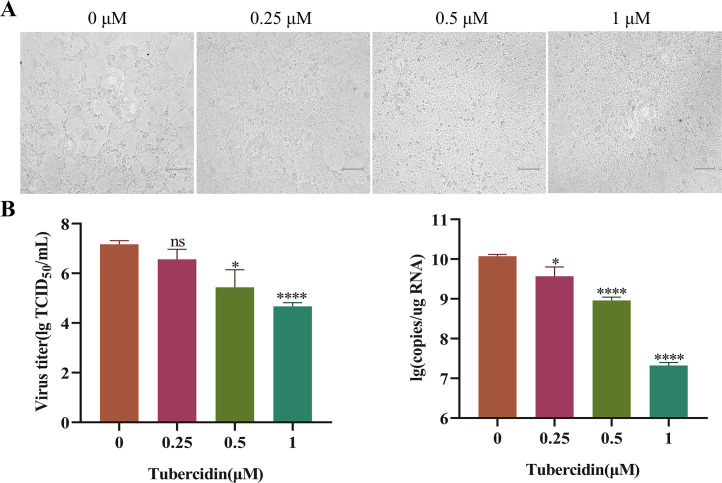
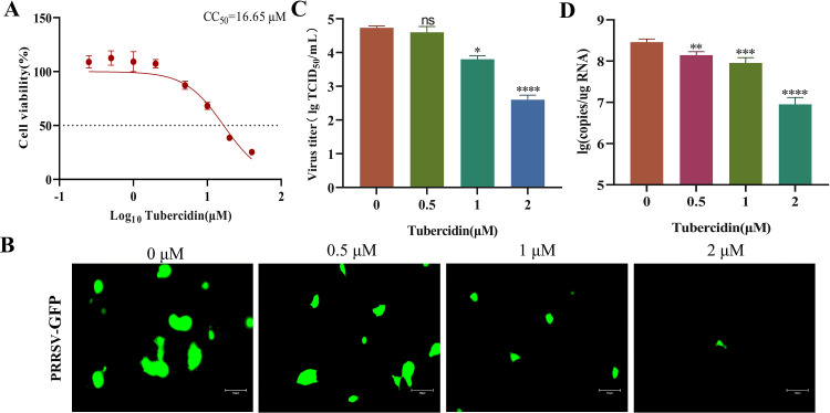
The emergence of new coronaviruses poses a significant threat to animal husbandry and human health. Porcine epidemic diarrhea virus (PEDV) is considered a re-emerging porcine enteric coronavirus, which causes fatal watery diarrhea in piglets. Currently, there are no effective drugs to combat PEDV. Drug repurposing screens have emerged as an attractive strategy to accelerate antiviral drug discovery and development. Here, we screened 206 natural products for antiviral activity using live PEDV infection in Vero cells and identified ten candidate antiviral agents. Among them, Tubercidin, a nucleoside analog derived from Streptomyces tubercidicus, showed promising antiviral activity against PEDV infection. Furthermore, we demonstrated that Tubercidin exhibited significant antiviral activity against both classical and variant PEDV. Time of addition assay showed that Tubercidin displayed a significant inhibitory effect on viral post-entry events but not during other periods. Molecular docking analysis indicated that Tubercidin had better docking efficiency and formed hydrophobic interactions with the active pocket of RNA-dependent RNA polymerase (RdRp) of PEDV and other nidoviruses. Additionally, Tubercidin can effectively suppress other porcine nidoviruses, such as SADS-CoV and PRRSV, demonstrating its broad-spectrum antiviral properties. In summary, our findings provide valuable evidence for the antiviral activity of Tubercidin and offer insights into the development of new strategies for the prevention and treatment of coronavirus infections.

Keywords: Antiviral; Coronavirus; Porcine epidemic diarrhea virus; RNA-dependent RNA polymerase; Tubercidin.


Conflict of interest statement

Declaration of Competing Interest The authors declare that they have no known competing financial interests or personal relationships that could have appeared to influence the work reported in this paper.

Figures

Fig 1
Fig. 1
Schematic of PEDV inhibitors screening from a natural product library. Vero cells were pre-treated with natural compounds or 0.1 % DMSO for 4 h, and then infected with DR13-GFP in the presence of compounds. The CPE was observed and recorded at 12, 24 and 36 hpi, scale bars = 150 μm. (A) Diagram of the initial antiviral screening model. (B) Ten drugs, including Oleanolic acid (1–10), Moxidectin (1–17), Octyl gallate (1–71), 1,2,3,4,6-Pentagalloylglucose (1–74), Theaflavin 3,3′- digallate (2–2), Curcumin (2–4), Baicalin (3–10), Xanthohumol (3–11), Tubercidin (3–19), Rubitecan (3–25), displayed inhibitory effects on PEDV DR13-GFP infection. (C) PEDV replication inhibition rates after treatment. The blue dot represents the natural compounds with effective antiviral activity, while the red dot represents Tubercidin.
Fig 2
Fig. 2
Evaluation of cytotoxicity on Vero cells and antiviral efficacy of Tubercidin against PEDV. (A) Tubercidin structural formula. (B) Cytotoxicity of Tubercidin on Vero cells. Evaluation of cell viability using CCK-8 assay. Vero cells were treated or not treated with different concentrations of Tubercidin for 16 h. (C) IC50 determination of Tubercidin. Vero cells were pre-treated or not treated with Tubercidin, infected with DR13-GFP at 0.1 MOI in the presence or absence of Tubercidin. After 16 h, flow cytometry was used to assess the proportion of GFP-positive cells in the cell population. The IC50 and CC50 values were calculated by a best-fit Log(dose)-response curve-fitting using GraphPad Prism 8.0.2.
Fig 3
Fig. 3
Evaluation of cytotoxicity on LLC-PK1 cells and antiviral efficacy of Tubercidin against PEDV. (A) Cytotoxicity of Tubercidin in LLC-PK1 cells. The cytotoxicity was determined by CCK-8 assay. The LLC-PK1 cells were treated or not treated with different concentrations of Tubercidin for 16 h. (B and C) Microscopic images of LLC-PK1 cells infected with DR13-GFP and GDU-GFP (0.1 MOI) treated with different concentrations of Tubercidin (0.25–1 μM). At the same time, viral fluids were collected and virus titer was determined by TCID50 assay. The experiment was performed three times independently, images were representative of results obtained from three independent experiments, scale bars = 150 μm. Differences were considered significant at P < 0.05 (*), P < 0.01 (**), P < 0.001 (***) and P < 0.0001 (****).
Fig 4
Fig. 4
Effect of Tubercidin on different genotypes of PEDV strains. Vero cells were infected with CV777, HNAY, HNXX, or HB strains at 0.1 MOI with Tubercidin (0.25–1 μM) treatment. At 16 hpi, viral fluids were collected. (A) The PEDV titers were measured by TCID50 assay. (B) The genomic RNA level of PEDV was measured by RT-qPCR assay. All experiments were performed with three independent replicates. Differences were considered significant at P < 0.05 (*), P < 0.01 (**), P < 0.001 (***) and P < 0.0001 (****).
Fig 5
Fig. 5
Effect of Tubercidin on S protein Synthesis of PEDV strains. (A-D) Vero cells were infected with CV777, HNAY, HNXX or HB strains (0.1 MOI) with Tubercidin (0.25–1 μM) treatment. At 12 hpi, the S protein expression of four PEDV strains on Vero cells was determined by IFA. Images were representative of results obtained from three independent experiments, scale bars = 150 μm.
Fig 6
Fig. 6
Antiviral activity of Tubercidin on DR13-GFP infections at different kinds of drug-addition approaches. (A) Schematics of Tubercidin addition experiments. Different concentrations of Tubercidin (0.25–1 μM) were added to Vero cells prior to infection with DR13-GFP (−4 h), as well as at 0 or 1 h post-infection. At 16 hpi, the samples were collected. (B) The PEDV titers were measured by TCID50 assay. (C) The genomic RNA level of PEDV was measured by RT-qPCR assay. All experiments were performed with three independent replicates. Differences were considered significant at P < 0.05 (*), P < 0.01 (**), P < 0.001 (***) and P < 0.0001 (****).
Fig 7
Fig. 7
Antiviral activity of Tubercidin at different stages of PEDV infection. Vero cells were infected with PEDV and treated with Tubercidin at indicated time points, which represented the stage of viral inactivation (A), attachment (B), internalization (C), replication (D) or release (E), respectively. All experiments were performed with three independent replicates. After 16 h, the samples were collected for determining the RNA expression level of PEDV by RT-qPCR.
Fig 8
Fig. 8
Effect of Tubercidin on virus replication at various intervals pre- and post-PEDV infection. (A) Add Tuberculin to Vero cells pre- and post-infection with PEDV at the specific time points. Light brown bars represent PEDV infection, light blue bars represent Tubercidin treatment. (B and C) Vero cells were infected with DR13-GFP and HB variant and treated with Tubercidin (1 μM) or 0.1 % DMSO at 2 h before infection, as well as at 2, 4, 6, 8, 10 and 12 h post-infection. After 16 h, the samples were collected for determining viral RNA copies by RT-qPCR assay. All experiments were performed with three independent replicates. Differences were considered significant at P < 0.05 (*), P < 0.01 (**), P < 0.001 (***) and P < 0.0001 (****).
Fig 9
Fig. 9
Molecular docking of Tubercidin with RdRp of nidoviruses. Tubercidin was docked with RdRp proteins from viruses including (A) α-coronaviruses (PEDV, SADS-CoV, TGEV, FCoV and CCoV), (B) β-coronaviruses (SARS-CoV, SARS-CoV-2, and MERS-CoV), (C) γ-coronavirus (IBV), (D) δ-coronavirus (PDCoV) and (E) Arteritis virus (PRRSV) using Autodock. (i) Cartoon representation, overlay of the crystal structures of Tubercidin and viruses RdRp protein were illustrated. (ii) 2D interactions of Tubercidin and viruses RdRp protein. (iii) Three-dimensional structures of the binding pockets were showed by PyMOL software. The proteins and compound are represented as cartoons and sticks, respectively. The RdRp proteins' three-dimensional structures are colored light white. The Tubercidin's two-dimensional structure is colored green, red, and blue. The Pymol software displays the sticks structure (Pale cyan) of amino acids residues in the virus RdRp protein. These amino acids residues are connected through H-bonds (yellow dotted lines) to Tubercidin, which is stabilized in the active pocket. The binding energy of the Tubercidin–viruses RdRp protein, calculated using Autodock, is listed (Table 1).
Fig 10
Fig. 10
Antiviral effect of Tubercidin on SADS-CoV. (A) Vero cells were pre-treated with Tubercidin (0.25–1 μM) for 4 h, and then infected them with SADS-CoV in the presence or absence of Tubercidin. At 16 hpi, CPE results were observed and viral fluids were collected. (B) The SADS-CoV titers were measured by TCID50 assay. (C) The genomic RNA levels of SADS-CoV were measured by RT-qPCR assay. The experiment was performed three times independently, images were representative of results obtained from three independent experiments, scale bars = 150 μm. Differences were considered significant at P < 0.05 (*), P < 0.01 (**), P < 0.001 (***) and P < 0.0001 (****).
Fig 11
Fig. 11
Antiviral effect of Tubercidin on PRRSV. (A) Cytotoxicity of Tubercidin on Marc145 cells. Marc145 cells were treated or not treated with different concentrations of Tubercidin for 16 h. The cytotoxicity was determined by CCK-8 assay. (B) Microscopic images of Marc145 cells infected with PRRSV-GFP (0.1 MOI) treated with different concentrations of Tubercidin (0.5–2 μM). (B) The PRRSV titers were measured by TCID50 assay. (C) The genomic RNA levels of PRRSV were measured by RT-qPCR assay. Each experiment was performed three times independently, images were representative of results obtained from three independent experiments, scale bars = 150 μm. Differences were considered significant at P < 0.05 (*), P < 0.01 (**), P < 0.001 (***) and P < 0.0001 (****).

Similar articles

Cited by

References

    1. Amirian E.S., Levy J.K. Current knowledge about the antivirals remdesivir (GS-5734) and GS-441524 as therapeutic options for coronaviruses. One Health. 2020;9 - PMC - PubMed
    1. Bergant V., Yamada S., Grass V., Tsukamoto Y., Lavacca T., Krey K., Muhlhofer M.T., Wittmann S., Ensser A., Herrmann A., Vom Hemdt A., Tomita Y., Matsuyama S., Hirokawa T., Huang Y., Piras A., Jakwerth C.A., Oelsner M., Thieme S., Graf A., Krebs S., Blum H., Kummerer B.M., Stukalov A., Schmidt-Weber C.B., Igarashi M., Gramberg T., Pichlmair A., Kato H. Attenuation of SARS-CoV-2 replication and associated inflammation by concomitant targeting of viral and host cap 2′-O-ribose methyltransferases. EMBO J. 2022;41(17) - PMC - PubMed
    1. Cao L., Li Y., Yang S., Li G., Zhou Q., Sun J., Xu T., Yang Y., Liao R., Shi Y., Yang Y., Zhu T., Huang S., Ji Y., Cong F., Luo Y., Zhu Y., Luan H., Zhang H., Chen J., Liu X., Luo R., Liu L., Wang P., Yu Y., Xing F., Ke B., Zheng H., Deng X., Zhang W., Lin C., Shi M., Li C.M., Zhang Y., Zhang L., Dai J., Lu H., Zhao J., Zhang X., Guo D. The adenosine analog prodrug ATV006 is orally bioavailable and has preclinical efficacy against parental SARS-CoV-2 and variants. Sci. Transl. Med. 2022;14(661):eabm7621. - PMC - PubMed
    1. Chen C., Breslin M.B., Guidry J.J., Lan M.S. 5′-Iodotubercidin represses insulinoma-associated-1 expression, decreases cAMP levels, and suppresses human neuroblastoma cell growth. J. Biol. Chem. 2019;294(14):5456–5465. - PMC - PubMed
    1. Chen D., Oezguen N., Urvil P., Ferguson C., Dann S.M., Savidge T.C. Regulation of protein-ligand binding affinity by hydrogen bond pairing. Sci. Adv. 2016;2(3) - PMC - PubMed