. 2024 Feb;39(1):113-122.
doi: 10.1016/j.virs.2023.11.008. Epub 2023 Nov 24.

Hsp90 β is critical for the infection of severe fever with thrombocytopenia syndrome virus

Affiliations

Hsp90 β is critical for the infection of severe fever with thrombocytopenia syndrome virus

Bo Wang et al. Virol Sin. 2024 Feb.

Abstract

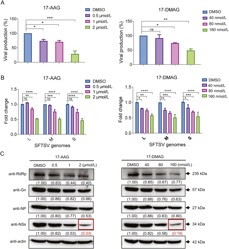
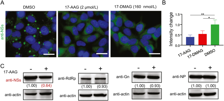
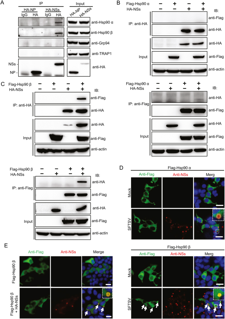
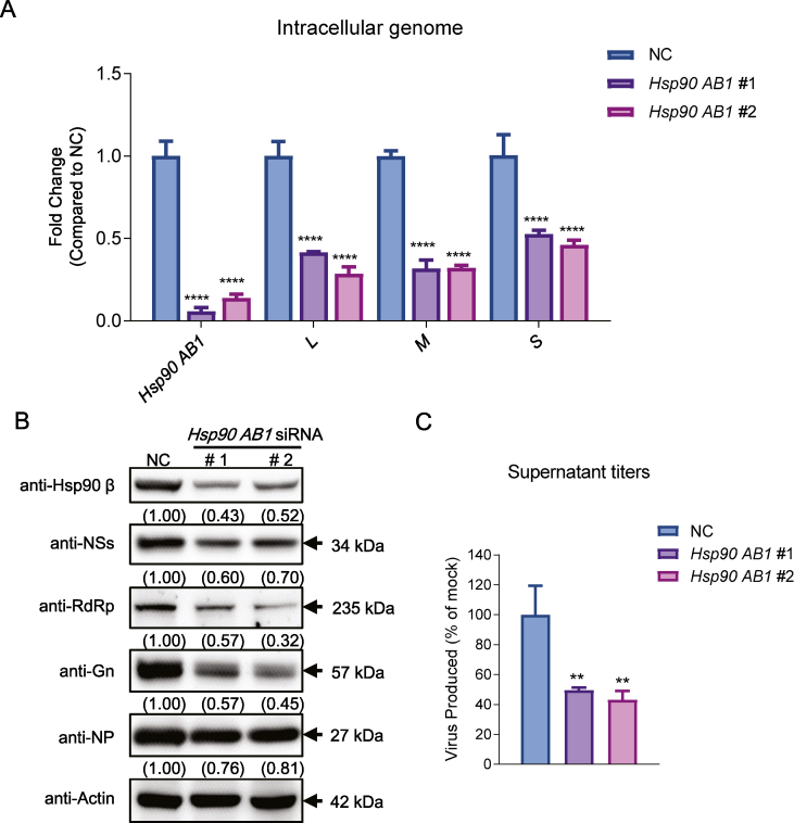
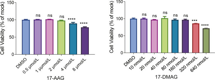
Severe fever with thrombocytopenia syndrome (SFTS) caused by the SFTS virus (SFTSV) is an emerging disease in East Asia with a fatality rate of up to 30%. However, the viral-host interaction of SFTSV remains largely unknown. The heat-shock protein 90 (Hsp90) family consists of highly conserved chaperones that fold and remodel proteins and has a broad impact on the infection of many viruses. Here, we showed that Hsp90 is an important host factor involved in SFTSV infection. Hsp90 inhibitors significantly reduced SFTSV replication, viral protein expression, and the formation of inclusion bodies consisting of nonstructural proteins (NSs). Among viral proteins, NSs appeared to be the most reduced when Hsp90 inhibitors were used, and further analysis showed that their translation was affected. Co-immunoprecipitation of NSs with four isomers of Hsp90 showed that Hsp90 β specifically interacted with them. Knockdown of Hsp90 β expression also inhibited replication of SFTSV. These results suggest that Hsp90 β plays a critical role during SFTSV infection and could be a potential target for the development of drugs against SFTS.

Keywords: Heat-shock protein 90; Host-virus interaction; Hsp90 β; Nonstructural protein; Severe fever with thrombocytopenia syndrome virus (SFTSV).


Conflict of interest statement

Conflict of interest The authors declare that they have no conflict of interest. Prof. Fei Deng is an editorial board member for Virologica Sinica and was not involved in the editorial review or the decision to publish this article.

Figures

Fig. 1
Fig. 1
Heat-shock protein 90 (Hsp90) inhibitors influence severe fever with thrombocytopenia syndrome virus (SFTSV) replication. HEK293 cells were infected with SFTSV at a multiplicity of infection (MOI) of 1 and treated with the indicated drugs or dimethyl sulfoxide (DMSO). At 24 ​h post-infection, the viral production, genome copy number and viral protein expression level were determined. The viral titers in supernatant were determined using an endpoint dilution assay and normalized to that of DMSO group (A). Cells were lysed to either quantify the SFTSV genome using RT-qPCR (B) or detect SFTSV protein expression using Western blotting (C). For analysis of the viral production and genome replication, three in-dependent experiments were performed. For detection of viral protein expression, two in-dependent experiments were performed, and representative immunoblot results are shown in the figure. Data are shown as the mean ​± ​SD in (A) and (B). Statistical significance was determined using one-way (A) or two-way (B) ANOVA followed by Dunnett's multiple comparisons test. ns, not significant; ∗, P ​< ​0.05; ∗∗, P ​< ​0.01; ∗∗∗, P ​< ​0.001, and ∗∗∗∗, P ​< ​0.0001.
Fig. 2
Fig. 2
Nonstructural proteins (NSs) are affected by Hsp90 inhibitors. A 17-AAG and 17-DMAG inhibit the formation of inclusion bodies induced by NSs. SFTSV-infected HEK293 ​cells were treated with 17-AAG or 17-DMAG for 24 ​h and then stained using anti-NSs antibodies for immunofluorescence analysis. Infected cells treated with DMSO were used as controls and nuclei stained with DAPI. Scale bars, 20 ​μm. B The fluorescence intensity analysis of the NSs protein in Fig. 2A. The mean fluorescence intensity of the NSs protein was calculated through ImageJ software, and the intensity change was obtained as the fluorescence intensity in 17-AAG/17-DMAG treated group normalized to DMSO group. Two independent experiments were performed. Data are shown as the mean ​± ​SD (n ​> ​20 ​cells). Statistical significance was determined using one-way ANOVA followed by Dunnett's multiple comparisons test. ∗P ​< ​0.05 and ∗∗P ​< ​0.01. C Hsp90 inhibitors specifically suppressed the overexpression of NSs. HEK293T cells were transfected with plasmids expressing NSs, RNA-dependent RNA polymerase (RdRp), Gn, or nucleoproteins (NPs), respectively. At 6 ​h post-transfection, the cells were treated with 2 ​μmol/L 17-AAG for another 24 ​h. Thereafter, the cell lysates were subjected to Western blotting analysis with anti-NSs, anti-NP, anti-Gn, and anti-RdRp antibodies. The relative levels of viral and host proteins were assessed using ImageJ software according to the immunoblot and showed under the corresponding image. Two independent experiments were performed, and representative results are shown in the figure.
Fig. 3
Fig. 3
Suppression of protein degradation pathways could not recover the reduced expression of NSs caused by the Hsp90 inhibitor, 17-AAG. A Workflow of the experiment. HEK293 cells were infected with SFTSV with an MOI of 5 for 24 ​h, thereafter the supernatants were replaced with fresh medium containing 17-AAG (2 ​μmol/L), MG-132 (500 ​nmol/L), 3-methyladenine (3-MA; 5 ​μmol/L), and bafilomycin A1 (Baf-A1; 100 ​nmol/L), individually or in combination, for 6 ​h. Fresh medium containing DMSO was used as a control. Then, viral proteins were detected using Western blotting with anti-NSs, anti-RdRp, anti-Gn, anti-NP, and anti-β-actin antibodies. B Results of the Western blotting analyses. The relative levels of viral and host proteins were assessed using ImageJ software according to the immunoblot and showed under the corresponding image. Two independent experiments were performed, and representative results are shown in the figure.
Fig. 4
Fig. 4
Hsp90 inhibitor, 17-AAG, likely affects the protein synthesis of NSs. A 17-AAG inhibited the protein synthesis of NSs in infected cells. HEK293T cells were infected with SFTSV at an MOI of 2 for 24 ​h. Then, the supernatants were replaced with fresh medium containing 17-AAG (2 ​μmol/L), and/or cycloheximide (CHX; 50 ​μmol/L). Fresh medium containing DMSO was used as a control. The cells were harvested at 12 ​h post-treatment for Western blotting analysis. Two independent experiments were performed, and representative results are shown in figure. B 17-AAG effected the protein synthesis of NSs in transfected cells. HEK293T cells were transfected with plasmids expressing NSs or nucleoproteins (NPs), respectively. At 6 ​h post-transfection, the cells were treated with 2 ​μmol/L 17-AAG, and/or cycloheximide (CHX; 50 ​μmol/L) for another 24 ​h. Thereafter, the cell lysates were subjected to Western blotting analysis. The relative levels of viral and host proteins were assessed using ImageJ software according to the immunoblot and showed under the corresponding image.
Fig. 5
Fig. 5
Hsp90 β interacts with NSs of SFTSV. A Co-immunoprecipitation (Co-IP) results of NSs with endogenous Hsp90 isomers. HEK293T cells were transfected with plasmids expressing HA-NP or HA-NSs. After 48 ​h, cells were lysed for Co-IP analysis with hemagglutinin (HA) antibodies or mouse IgG (control). The immunoprecipitated or input proteins were analyzed using Western blotting with anti-HA, anti-Hsp90 α1, anti-Hsp90 β, anti-GRP94, or anti-TRAP1 antibodies. The protein bands are indicated. (B, C) NSs interact with exogenous Hsp90 β. HEK293T cells were transfected or co-transfected with plasmids expressing HA-NSs, Flag-HSP90α, and Flag-HSP90β as indicated. After 48 ​h, cells were lysed for Co-IP analysis with anti-HA or anti-Flag antibodies. The input and immunoprecipitated samples were immunoblotted with indicated antibodies. (D, E) Hsp90 β colocalized with NSs. D HEK293 ​cells transfected with plasmids expressing Flag-Hsp90 α (upper panel) or Flag-Hsp90 β (lower panel) were super-infected with SFTSV at 24 ​h post-transfection. E HEK293 ​cells transfected with plasmids expressing Flag-Hsp90 β with or without HA-NSs. At 24 ​h post-infection/transfection, cells were immunostained with anti-Flag (mouse-derived) and anti-NSs (rabbit-derived) antibodies, followed by staining with AF488-conjugated anti-mouse IgG and AF594-conjugated anti-rabbit IgG, respectively. Cell nuclei were stained with DAPI. The small image in the upper right corner is the enlarged image of box region in (D) and (E). Scale bars, 20 ​μm (D) or 50 ​μm (E).
Fig. 6
Fig. 6
Knockdown of Hsp90 β influences SFTSV replication. HEK293 cells were transfected with HSP90AB1-derived siRNA. After 48 ​h, cells were infected with SFTSV at an MOI of 1 for an additional 48 ​h. The levels of intracellular viral genome were quantified using quantitative real-time PCR (A), expression of viral proteins detected through immunoblotting (B), and titers in supernatant determined using an endpoint dilution assay and normalized to NC (C). NC, negative control. For analysis of the viral production and genome replication, three in-dependent experiments were performed. For detection of viral protein expression, two in-dependent experiments were performed, and representative immunoblot results are shown in the figure. Data are shown as the mean ​± ​SD. Statistical significance was determined using two-way (A) or one-way ANOVA (C) followed by Dunnett's multiple comparisons test. ∗∗, P ​< ​0.01; and ∗∗∗∗, P ​< ​0.0001.
figs1
figs1
figs2
figs2

Similar articles

References

    1. Abudurexiti A., Adkins S., Alioto D., Alkhovsky S.V., Avšič-Županc T., Ballinger M.J., Bente D.A., Beer M., Bergeron É., Blair C.D., Briese T., Buchmeier M.J., Burt F.J., Calisher C.H., Cháng C., Charrel R.N., Choi I.R., Clegg J.C.S., De La Torre J.C., De Lamballerie X., Dèng F., Di Serio F., Digiaro M., Drebot M.A., Duàn X., Ebihara H., Elbeaino T., Ergünay K., Fulhorst C.F., Garrison A.R., Gāo G.F., Gonzalez J.J., Groschup M.H., Günther S., Haenni A.L., Hall R.A., Hepojoki J., Hewson R., Hú Z., Hughes H.R., Jonson M.G., Junglen S., Klempa B., Klingström J., Kòu C., Laenen L., Lambert A.J., Langevin S.A., Liu D., Lukashevich I.S., Luò T., Lǚ C., Maes P., De Souza W.M., Marklewitz M., Martelli G.P., Matsuno K., Mielke-Ehret N., Minutolo M., Mirazimi A., Moming A., Mühlbach H.P., Naidu R., Navarro B., Nunes M.R.T., Palacios G., Papa A., Pauvolid-Corrêa A., Pawęska J.T., Qiáo J., Radoshitzky S.R., Resende R.O., Romanowski V., Sall A.A., Salvato M.S., Sasaya T., Shěn S., Shí X., Shirako Y., Simmonds P., Sironi M., Song J.W., Spengler J.R., Stenglein M.D., Sū Z., Sūn S., Táng S., Turina M., Wáng B., Wáng C., Wáng H., Wáng J., Wèi T., Whitfield A.E., Zerbini F.M., Zhāng J., Zhāng L., Zhāng Y., Zhang Y.Z., Zhāng Y., Zhou X., Zhū L., Kuhn J.H. Taxonomy of the order Bunyavirales: update 2019. Arch. Virol. 2019;164:1949–1965. - PMC - PubMed
    1. Albornoz A., Hoffmann A.B., Lozach P.Y., Tischler N.D. Early bunyavirus-host cell interactions. Viruses. 2016;8:143. - PMC - PubMed
    1. Basta S., Stoessel R., Basler M., Van Den Broek M., Groettrup M. Cross-presentation of the long-lived lymphocytic choriomeningitis virus nucleoprotein does not require neosynthesis and is enhanced via heat shock proteins. J. Immunol. 2005;175:796–805. - PubMed
    1. Burch A.D., Weller S.K. Herpes simplex virus type 1 DNA polymerase requires the mammalian chaperone hsp90 for proper localization to the nucleus. J. Virol. 2005;79:10740–10749. - PMC - PubMed
    1. Chen B., Piel W.H., Gui L., Bruford E., Monteiro A. The HSP90 family of genes in the human genome: insights into their divergence and evolution. Genomics. 2005;86:627–637. - PubMed

MeSH terms

Supplementary concepts

LinkOut - more resources