

Image quality of DWI at breast MRI depends on the amount of fibroglandular tissue: implications for unenhanced screening
- PMID: 38008743
- PMCID: PMC11213722
- DOI: 10.1007/s00330-023-10321-y
Image quality of DWI at breast MRI depends on the amount of fibroglandular tissue: implications for unenhanced screening
Abstract
Objectives: To compare image quality of diffusion-weighted imaging (DWI) and contrast-enhanced breast MRI (DCE-T1) stratified by the amount of fibroglandular tissue (FGT) as a measure of breast density.
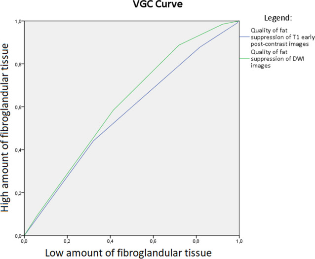
Methods: Retrospective, multi-reader, bicentric visual grading analysis study on breast density (A-D) and overall image and fat suppression quality of DWI and DCE-T1, scored on a standard 5-point Likert scale. Cross tabulations and visual grading characteristic (VGC) curves were calculated for fatty breasts (A/B) versus dense breasts (C/D).
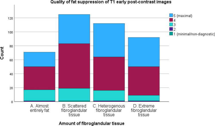
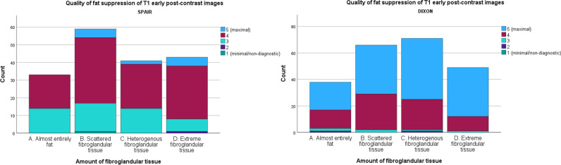
Results: Image quality of DWI was higher in the case of increased breast density, with good scores (score 3-5) in 85.9% (D) and 88.4% (C), compared to 61.6% (B) and 53.5% (A). Overall image quality of DWI was in favor of dense breasts (C/D), with an area under the VGC curve of 0.659 (p < 0.001). Quality of DWI and DCE-T1 fat suppression increased with higher breast density, with good scores (score 3-5) for 86.9% and 45.7% of density D, and 90.2% and 42.9% of density C cases, compared to 76.0% and 33.6% for density B and 54.7% and 29.6% for density A (DWI and DCE-T1 respectively).
Conclusions: Dense breasts show excellent fat suppression and substantially higher image quality in DWI images compared with non-dense breasts. These results support the setup of studies exploring DWI-based MR imaging without IV contrast for additional screening of women with dense breasts.
Clinical relevance statement: Our findings demonstrate that image quality of DWI is robust in women with an increased amount of fibroglandular tissue, technically supporting the feasibility of exploring applications such as screening of women with mammographically dense breasts.
Key points: • Image and fat suppression quality of diffusion-weighted imaging are dependent on the amount of fibroglandular tissue (FGT) which is closely connected to breast density. • Fat suppression quality in diffusion-weighted imaging of the breast is best in women with a high amount of fibroglandular tissue. • High image quality of diffusion-weighted imaging in women with a high amount of FGT in MRI supports that the technical feasibility of DWI can be explored in the additional screening of women with mammographically dense breasts.
Keywords: Area under the curve; Breast density; Diffusion magnetic resonance imaging; Early detection of cancer; Neoplasms.
© 2023. The Author(s).
Conflict of interest statement
The authors of this manuscript declare no relationships with any companies whose products or services may be related to the subject matter of the article.
Figures

Comment in
-
Dense breasts at breast cancer screening: can DWI-based breast MRI without contrast help us in the pursuit of personalized screening?Eur Radiol. 2024 Jul;34(7):4727-4729. doi: 10.1007/s00330-023-10323-w. Epub 2023 Nov 24. Eur Radiol. 2024. PMID: 38940855 No abstract available.
References
-
- Kang JW, Shin HJ, Shin KC, et al. Unenhanced magnetic resonance screening using fused diffusion-weighted imaging and maximum-intensity projection in patients with a personal history of breast cancer: role of fused DWI for postoperative screening. Breast Cancer Res Treat. 2017;165:119–128. doi: 10.1007/s10549-017-4322-5. - DOI - PubMed
MeSH terms
Substances
LinkOut - more resources
Full Text Sources
Medical
