. 2023 Nov 27;27(1):122.
doi: 10.1186/s40824-023-00448-w.

Mild heat treatment in vitro potentiates human adipose stem cells: delayed aging and improved quality for long term culture

Affiliations

Mild heat treatment in vitro potentiates human adipose stem cells: delayed aging and improved quality for long term culture

Chiseon Ryu et al. Biomater Res. .

Abstract

Background: Mesenchymal stem cells (MSCs) have gained significant attention for diverse biomedical applications, including cell-based therapy. Hence, in vitro expansion of MSCs is critical; however, in vitro MSC culture, especially long-term culture, inevitably leads to significant loss of stemness, growth, and differentiation potential.

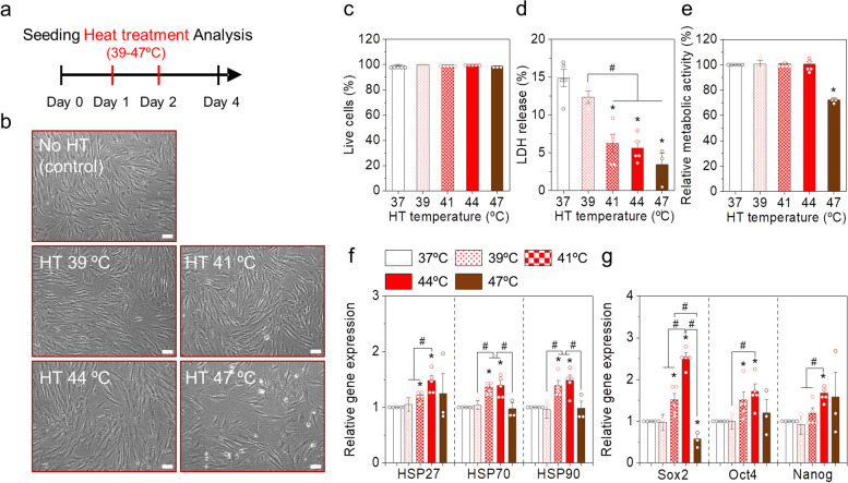
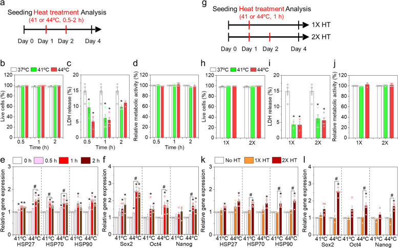
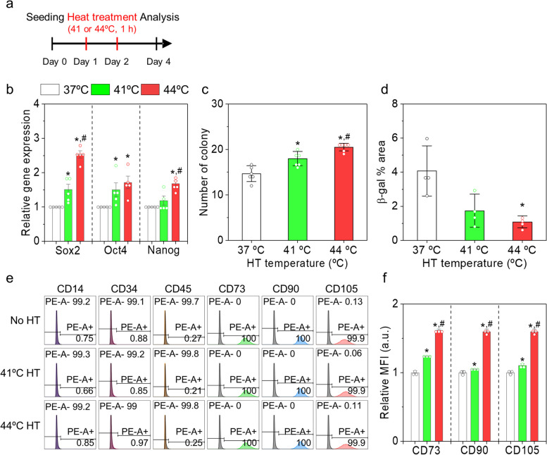
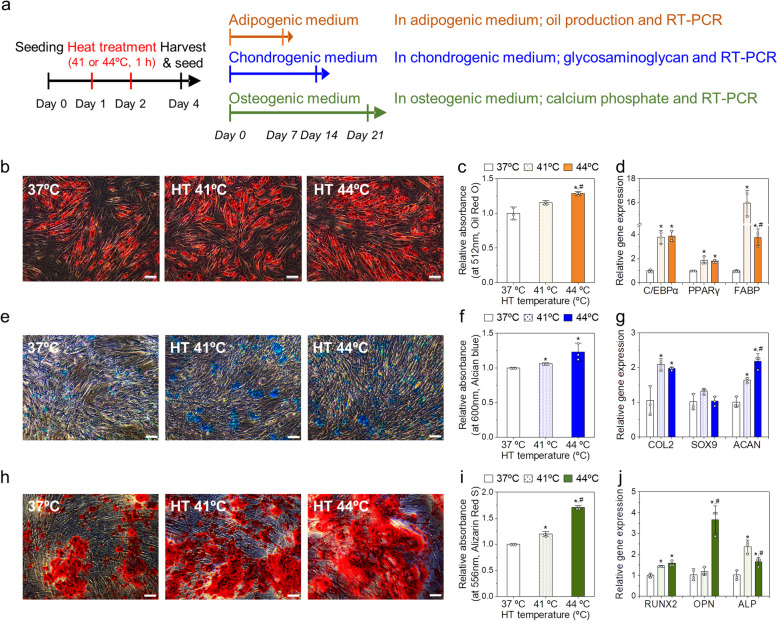
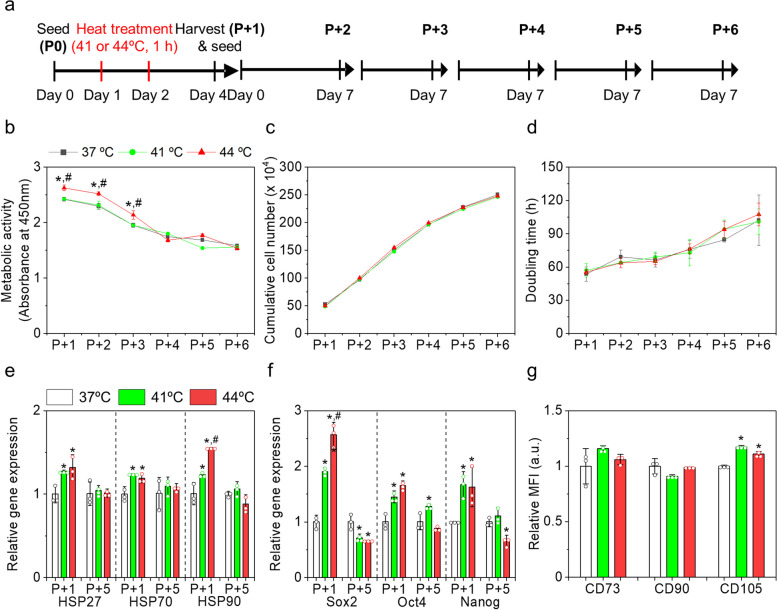
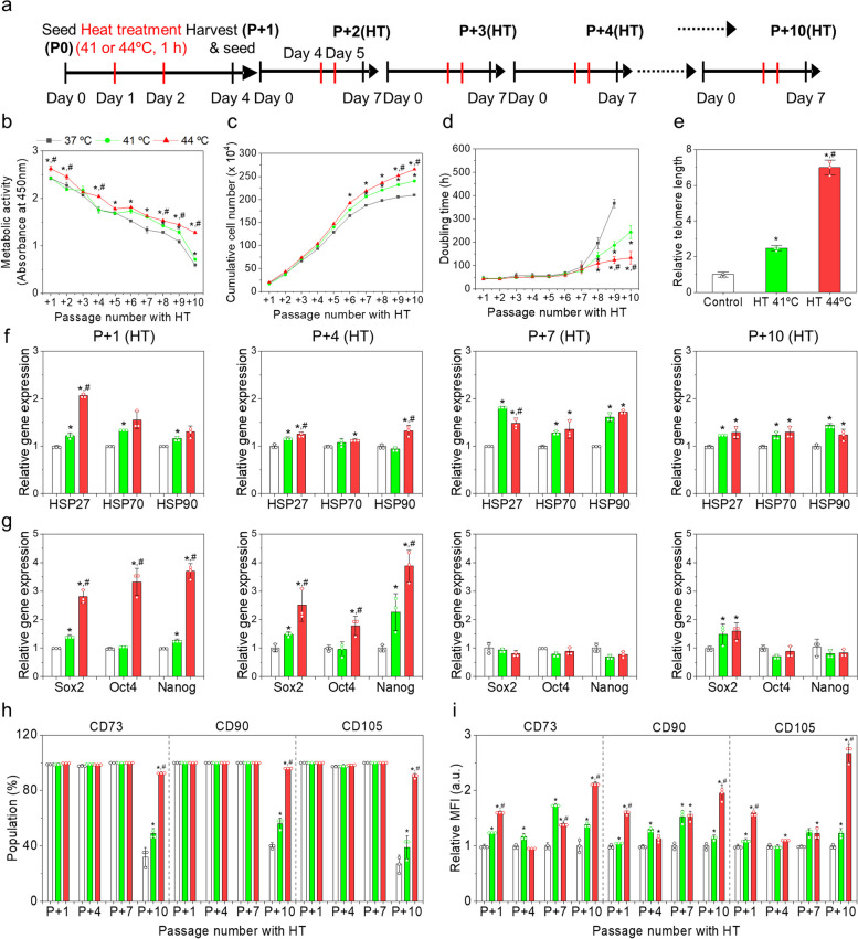
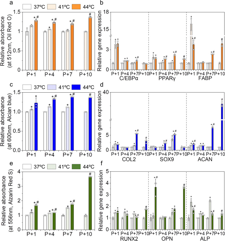
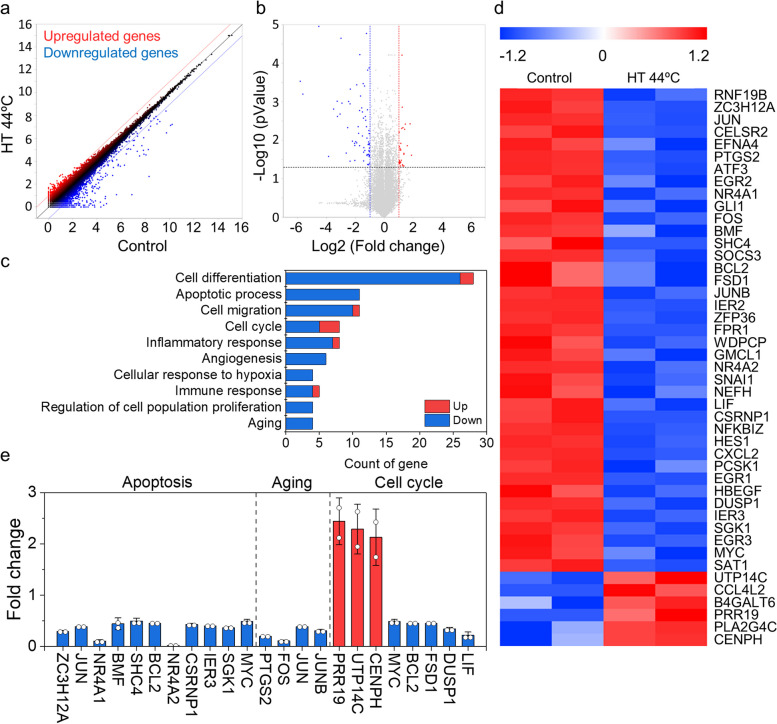
Method: Effects of mild heat treatment (HT) conditions (temperature, duration, and repetition) on the characteristics of adipose tissue-derived MSCs in vitro were systematically investigated. Characteristics of the MSCs subjected to the predetermined HT conditions (41 or 44ºC, 1 h, and 2X HT) were first analyzed in a single passage using various assays. In addition, the feasibility of HT for long-term MSC culture was studied. The RNA sequencing analyses were performed to elucidate the mechanism of HT effects on MSCs.

Results: A comprehensive exploration of various HT conditions revealed that specific mild HT at 41ºC or 44ºC for 1 h upregulated the expression of heat shock proteins and stemness markers and enhanced differentiation potentials. Furthermore, periodic mild HT extended the maintenance of growth rate and stemness of MSCs up to an additional 10 passages, which substantially retarded their spontaneous aging during subsequent in vitro culture. RNA sequencing analyses unveiled that HT downregulated genes associated with aging and apoptosis.

Conclusion: Our study successfully demonstrated that mild HT of MSCs has positive effects on their application in various biomedical fields, enhancing their capabilities and slowing down the aging process.

Keywords: Aging; Heat treatment; In vitro culture; Mesenchymal stem cell; Stemness.


Conflict of interest statement

The authors declare that they have no known competing financial interests that could have appeared to influence the work reported in this paper.

Figures

Fig. 1
Fig. 1
Mild heat treatment (HT) of human adipose tissue-derived mesenchymal stem cells (AD-MSCs) in a single passage at different temperatures. a Schematic representation of the experimental design. b Optical micrographs of the MSCs in each group on day 4. Scale bar is 200 μm. c Live cell percentage, d lactate dehydrogenase (LDH) levels, and e relative metabolic activities of the MSCs in each group. Gene expression levels of f heat shock proteins (HSPs) and g stemness markers. Gene expression levels were normalized with the control (37ºC). Error bars represent the average ± the standard error of the mean from three to five experiments. *p < 0.05 compared to the control group, and #p < 0.05 compared to two groups
Fig. 2
Fig. 2
Mild HT of AD-MSCs for varying durations (af) and repetition (gl) in a single passage. a An experimental scheme to study the effects of HT duration. b Live cell percentage, c LDH levels, and d relative metabolic activities of MSCs in each group. Gene expression levels of e HSPs and f stemness markers. g An experimental scheme to study the effects of HT repetition. h Live cell percentage, i LDH levels, and j relative metabolic activities of MSCs in each group. Gene expression levels of k HSPs and l stemness markers. Gene expression levels were normalized with the control (37ºC). Error bars represent the average ± the standard error of the mean from three to five experiments. *p < 0.05 compared to the control group, and #p < 0.05 compared to two groups
Fig. 3
Fig. 3
Effects of mild HT on stemness of human AD-MSCs. a Schematic representation of the experimental design. b Relative gene expression of SOX2, OCT4, and NANOG, c the number of colonies, and d β-galactosidase (β-gal) stained area in each group. e Flow cytometry of the MSCs stained with negative markers (CD14, CD34, and CD45) and positive markers (CD73, CD90, and CD105). f Mean fluorescence intensity (MFI) of the positive marker-stained MSCs in each group. *p < 0.05 compared to the control group, and #p < 0.05 compared to the HT 41ºC group
Fig. 4
Fig. 4
Effects of mild HT on the differentiation capacity of human AD-MSCs. a Schematic representation for the experimental design. b Oil red-staining images, c relative absorbance at 512 nm for Oil Red, and d relative expression levels of adipogenic genes (C/EBPα, PPARγ, and FABP) of the MSCs thermally stimulated and cultured in adipogeneic medium for 7 days. e Alcian blue-staining images, f relative absorbance at 600 nm for Alcian blue, and g relative expression levels of chondrogenic genes (COL2, SOX9, and ACAN) of the MSCs thermally stimulated and cultured in chondrogenic medium for 14 days. h Alizarin red-staining images, i relative absorbance at 556 nm for Alizarin red, and j relative expression levels of osteogenic genes (RUNX2, OPN, and ALP) of the MSCs thermally stimulated and cultured in osteogenic medium for 21 days. *p < 0.05 compared to the control group, and #p < 0.05 compared to the HT 41ºC group
Fig. 5
Fig. 5
Lasting effects of mild HT on human AD-MSCs during subculturing without additional HT. a Schematic representation of the experimental design. b metabolic activity, c cumulative cell number, and d doubling time of the MSCs in each group during the subculture. The relative gene expressions of e HSPs (HSP27, HSP70, and HSP90) and f stemness markers (SOX2, OCT4, and NANOG) in each group. g Relative MFI of the positive marker-stained MSCs in each group at P + 6. MFI was obtained using flow cytometry. *p < 0.05 compared to the control group, and #p < 0.05 compared to the HT 41ºC group
Fig. 6
Fig. 6
Effects of successive mild HT on human AD-MSCs during subculturing with HT. a Schematic representation of the experimental study design. b Metabolic activity, c cumulative cell number, and d doubling time of the MSCs in each group during the subculture. e Telomere lengths of the MSCs at P + 10(HT) in each group. Relative gene expressions of f HSPs (HSP27, HSP70, and HSP90) and g stemness markers (SOX2, OCT4, and NANOG) at P + 1(HT), P + 4(HT), P + 7(HT), and P + 10(HT) in each group. Gene expression levels were normalized with those of the control at each passage. h Populations and i MFI of the positive marker-stained MSCs in each group. *p < 0.05 compared to the control group, and #p < 0.05 compared to the HT 41ºC group
Fig. 7
Fig. 7
Effects of mild HT on the differentiation capacity of human AD-MSCs during subculturing with HT. a Relative absorbance of oil red and b relative expression levels of adipogenic genes (C/EBPα, PPARγ, and FABP) of the MSCs in each group during the subculture at P + 1(HT), P + 4(HT), P + 7(HT), and P + 10(HT). c Relative absorbance of Alcian blue and d relative expression levels of chondrogenic genes (COL2, SOX9, and ACAN) of the MSCs in each group during the subculture. e Relative absorbance of Alizarin red and f relative expression levels of osteogenic genes (RUNX2, OPN, and ALP) of the MSCs in each group during the subculture. The absorbance and gene expression levels were normalized with those of the control at each passage. *p < 0.05 compared to the control group, and #p < 0.05 compared to the HT 41ºC group
Fig. 8
Fig. 8
Total RNA sequencing analysis. a Scatter plot. The red and blue dots indicate up-regulated and down-regulated genes, respectively, of the 44ºC heat treated-MSCs compared to the control. b Volcano plot. c Ten major gene ontologies closely related with the genes, which showed significantly altered expressions induced by HT. d Heat map of the two repeated analysis for each gene which are involved in the listed gene ontologies. e Specific gene expression profiles of each gene ontology; apoptosis, aging, and cell cycle

Similar articles

Cited by

References

    1. Han Y, Li X, Zhang Y, Han Y, Chang F, Ding J. Mesenchymal stem cells for regenerative medicine. Cells. 2019;8:886. doi: 10.3390/cells8080886. - DOI - PMC - PubMed
    1. Liang X, Ding Y, Zhang Y, Tse H-F, Lian Q. Paracrine mechanisms of mesenchymal stem cell-based therapy: current status and perspectives. Cell Transplant. 2014;23:1045–1059. doi: 10.3727/096368913X667709. - DOI - PubMed
    1. Yu B, Zhang X, Li X. Exosomes derived from mesenchymal stem cells. IJMS. 2014;15:4142–4157. doi: 10.3390/ijms15034142. - DOI - PMC - PubMed
    1. Al-Azab M, Safi M, Idiiatullina E, Al-Shaebi F, Zaky MY. Aging of mesenchymal stem cell: machinery, markers, and strategies of fighting. Cell Mol Biol Lett. 2022;27:69. doi: 10.1186/s11658-022-00366-0. - DOI - PMC - PubMed
    1. Beane OS, Fonseca VC, Cooper LL, Koren G, Darling EM. Impact of Aging on the Regenerative Properties of Bone Marrow-, Muscle-, and Adipose-Derived Mesenchymal Stem/Stromal Cells. Shi X-M, editor. PLoS ONE. 2014;9:e115963. - PMC - PubMed