. 2023 Dec;20(12):1513-1526.
doi: 10.1038/s41423-023-01107-9. Epub 2023 Nov 27.

Integrated NLRP3, AIM2, NLRC4, Pyrin inflammasome activation and assembly drive PANoptosis

Affiliations

Integrated NLRP3, AIM2, NLRC4, Pyrin inflammasome activation and assembly drive PANoptosis

SuHyeon Oh et al. Cell Mol Immunol. 2023 Dec.

Abstract

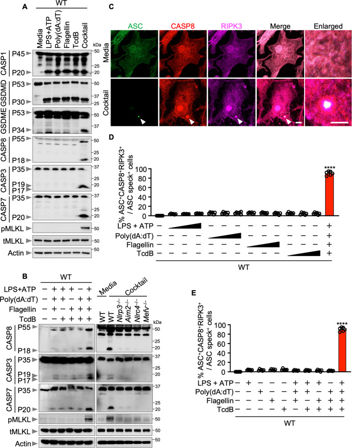
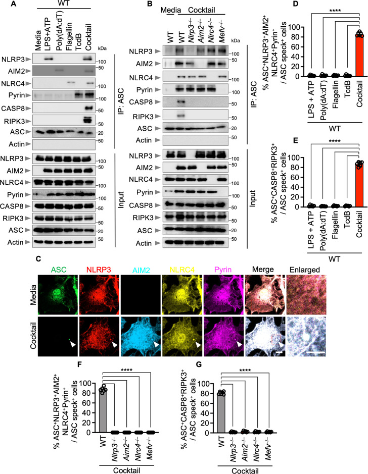
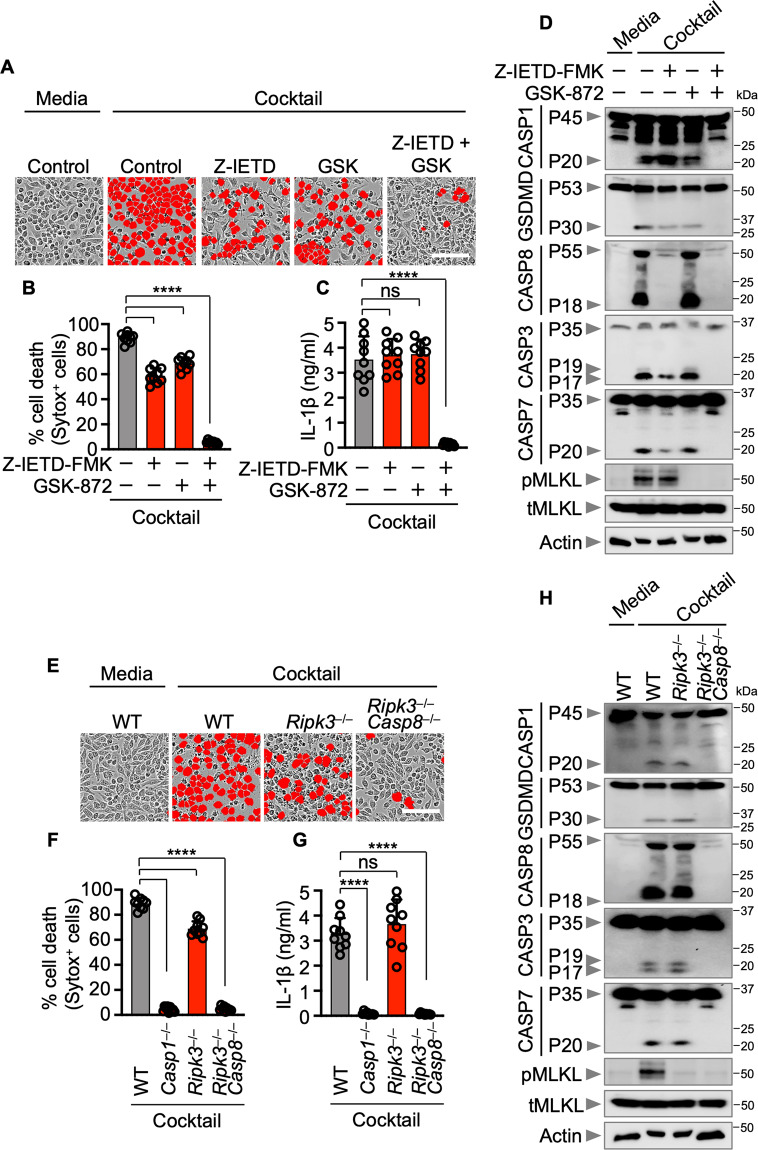
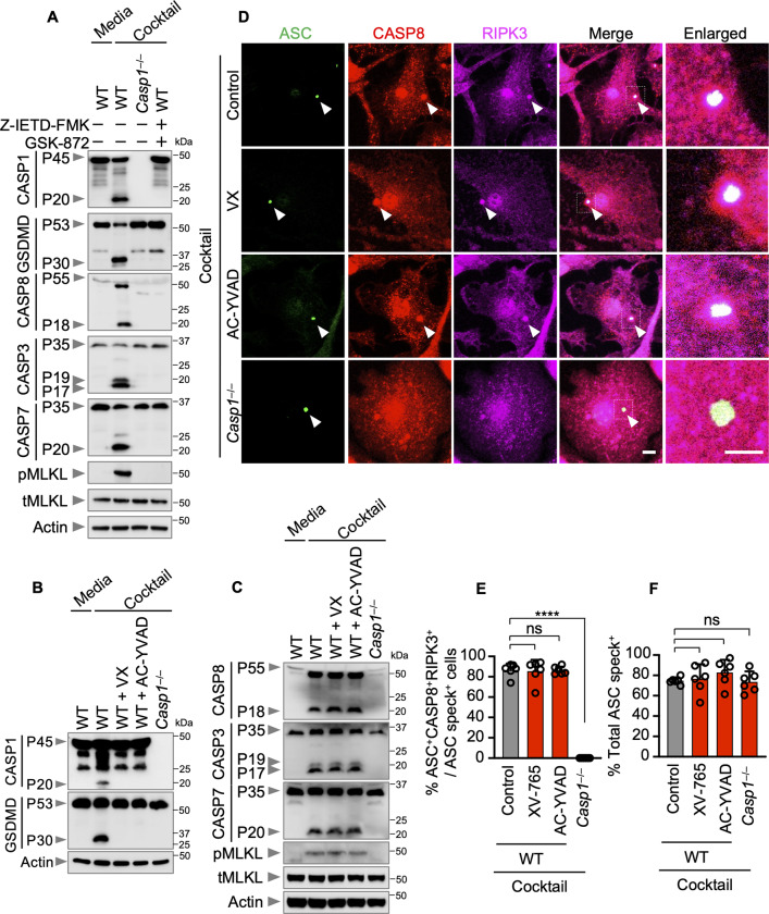
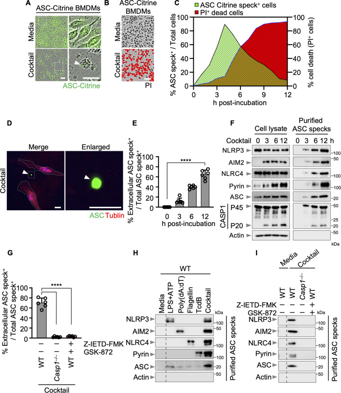
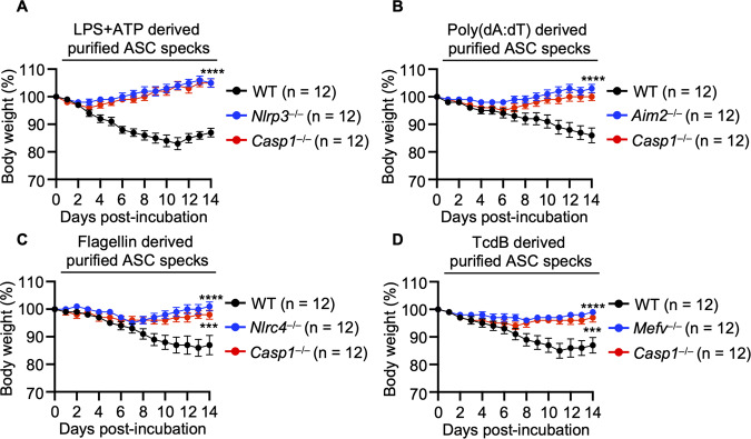
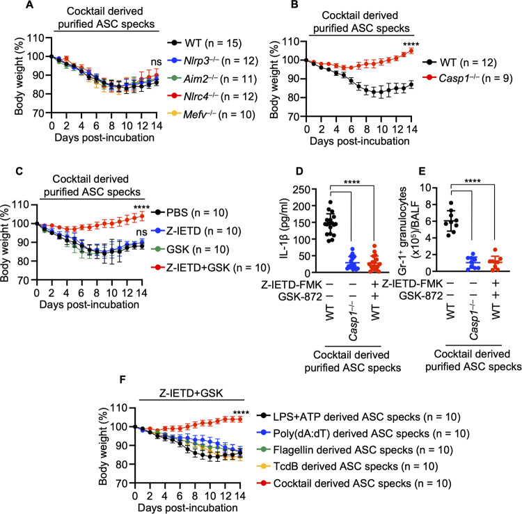
Inflammasomes are important sentinels of innate immune defense; they sense pathogens and induce the cell death of infected cells, playing key roles in inflammation, development, and cancer. Several inflammasome sensors detect and respond to specific pathogen- and damage-associated molecular patterns (PAMPs and DAMPs, respectively) by forming a multiprotein complex with the adapters ASC and caspase-1. During disease, cells are exposed to several PAMPs and DAMPs, leading to the concerted activation of multiple inflammasomes. However, the molecular mechanisms that integrate multiple inflammasome sensors to facilitate optimal host defense remain unknown. Here, we discovered that simultaneous inflammasome activation by multiple ligands triggered multiple types of programmed inflammatory cell death, and these effects could not be mimicked by treatment with a pure ligand of any single inflammasome. Furthermore, NLRP3, AIM2, NLRC4, and Pyrin were determined to be members of a large multiprotein complex, along with ASC, caspase-1, caspase-8, and RIPK3, and this complex drove PANoptosis. Furthermore, this multiprotein complex was released into the extracellular space and retained as multiple inflammasomes. Multiple extracellular inflammasome particles could induce inflammation after their engulfment by neighboring macrophages. Collectively, our findings define a previously unknown regulatory connection and molecular interaction between inflammasome sensors, which drives the assembly of a multiprotein complex that includes multiple inflammasome sensors and cell death regulators. The discovery of critical interactions among NLRP3, AIM2, NLRC4, and Pyrin represents a new paradigm in understanding the functions of these molecules in innate immunity and inflammasome biology as well as identifying new therapeutic targets for NLRP3-, AIM2-, NLRC4- and Pyrin-mediated diseases.

Keywords: Excellular ASC; Inflammatory Cell Death; Multiple Inflammasome; PANoptosis; PANoptosome.


Conflict of interest statement

The authors declare no competing interests.

Figures

Fig. 1
Fig. 1
Multiple inflammasome ligands promote multiple programmed cell death pathways. A Immunoblotting analysis of pro- (P45) and activated (P20) caspase-1 (CASP1), pro- (P53) and activated (P30) gasdermin D (GSDMD), pro- (P53) and activated (P34) gasdermin E (GSDME), pro- (P55) and cleaved (P18) caspase-8 (CASP8), pro- (P35) and cleaved (P17/P19) caspase-3 (CASP3), pro- (P35) and cleaved (P20) caspase-7 (CASP7), phosphorylated MLKL (pMLKL), total MLKL (tMLKL) expression wild type (WT) bone marrow-derived macrophages (BMDMs) treated with a single ligand (LPS + ATP, poly(dA:dT), flagellin, or TcdB) or a combination of LPS + ATP, poly(dA:dT), flagellin, and TcdB (Cocktail). B Immunoblotting analysis of CASP8, CASP3, CASP7, pMLKL, and tMLKL expression in WT BMDMs treated with the indicated treatments or in WT, Nlrp3−/−, Aim2−/−, Nlrc4−/−, or Mefv−/− BMDMs treated with the Cocktail. Data are representative of at least three independent experiments (A, B). C Immunofluorescence images of WT BMDMs 6 h after Cocktail treatment. Scale bars, 5 μm. Arrowheads indicate ASC specks. Images are representative of three independent experiments. D Quantification of the percentage of cells with ASC+CASP8+RIPK3+ specks among the ASC speck+ cells after treatment with varying concentrations of the individual ligands: 500 ng/ml LPS + 1 μM, 5 μM, or 25 μM ATP; 0.2 ng, 1 ng, or 5 ng poly(dA:dT); 2 ng, 10 ng, or 50 ng flagellin; 0.1 μg/ml, 0.5 μg/ml, or 2.5 μg/ml TcdB; or Cocktail (500 ng/ml LPS + 5 μM ATP + 1 ng poly(dA:dT) + 10 ng flagellin + 0.5 μg/ml TcdB). Data are mean ± s.e.m. ****P < 0.0001 (one-way ANOVA with Dunnett’s multiple comparisons test; n = 6 from three biologically independent samples). E Quantification of the percentage of cells with ASC+CASP8+RIPK3+ specks among the ASC speck+ WT BMDMs after the indicated treatment (5 μM ATP, 1 ng poly(dA:dT), 10 ng flagellin, and/or 0.5 μg/ml TcdB). Data are mean ± s.e.m. ****P < 0.0001 (one-way ANOVA with Dunnett’s multiple comparisons test; n = 6 from three biologically independent samples)
Fig. 2
Fig. 2
Multiple inflammasome ligands induce the assembly of integrated NLRP3, AIM2, NLRC4, and Pyrin complexes. A Immunoprecipitation (IP) of wild-type (WT) bone marrow-derived macrophages (BMDMs) with anti-ASC antibodies after treatment with LPS + ATP, poly(dA:dT), flagellin, or TcdB alone or treatment with a combination of LPS + ATP, poly(dA:dT), flagellin, and TcdB (Cocktail). Data are representative of three independent experiments. B IP of WT, Nlrp3−/−, Aim2−/−, Nlrc4−/−, or Mefv−/− BMDMs with anti-ASC antibodies after Cocktail treatment. Data are representative of three independent experiments. C Immunofluorescence images of WT BMDMs 6 h after Cocktail treatment. Scale bars, 5 μm. Arrowheads indicate ASC specks. Images are representative of three independent experiments. Quantification of the percentage of cells with ASC+NLRP3+AIM2+NLRC4+Pyrin+ specks among the ASC speck+ WT BMDMs (D) or cells with ASC+CASP8+RIPK3+ specks among the ASC speck+ WT BMDMs (E) after the indicated treatment. Quantification of the percentage of cells with ASC+NLRP3+AIM2+NLRC4+Pyrin+ specks among the ASC speck+ cells (F) or ASC+CASP8+RIPK3+ specks among the ASC speck+ cells (G) after treatment of the indicated BMDMs with the Cocktail. Data are mean ± s.e.m. ns, not significant, ****P < 0.0001 (one-way ANOVA with Dunnett’s multiple comparisons test; n = 6 from three biologically independent samples) (D, E, F, G)
Fig. 3
Fig. 3
Multiple inflammasome ligands promote multiple programmed cell death pathways in a RIPK3/caspase-8-dependent manner. A Cell death of wild-type (WT) bone marrow-derived macrophages (BMDMs) treated with Z-IETD-FMK, GSK-872, or a combination of Z-IETD-FMK and GSK-872 after treatment with the combination of combination LPS + ATP, poly(dA:dT), flagellin, and TcdB (Cocktail). B Quantification of cell death in (A). C Assessment of IL-1β levels. D Immunoblotting analysis of pro- (P45) and activated (P20) caspase-1 (CASP1), pro- (P53) and activated (P30) gasdermin D (GSDMD), pro- (P55) and cleaved (P18) caspase-8 (CASP8), pro- (P35) and cleaved (P17/P19) caspase-3 (CASP3), pro- (P35) and cleaved (P20) caspase-7 (CASP7), phosphorylated MLKL (pMLKL), total MLKL (tMLKL) expression in WT BMDMs treated with Z-IETD-FMK, GSK-872, or a combination of Z-IETD-FMK and GSK-872 after Cocktail treatment. E Cell death in WT, Casp1−/−, Ripk3−/−, and Ripk3−/−Casp8−/− immortalized bone marrow-derived macrophages (iBMDMs) after Cocktail treatment. F Quantification of cell death in (E). G Assessment of IL-1β levels. H Immunoblotting analysis of CASP1, GSDMD, CASP8, CASP3, CASP7, pMLKL, and tMLKL expression in WT, Casp1−/−, Ripk3−/−, and Ripk3−/−Casp8−/− iBMDMs after Cocktail treatment. Images are representative of at least three independent experiments; red indicates dead cells; scale bar, 50 μm (A, E). Data are mean ± s.e.m. ****P < 0.0001 (one-way ANOVA with Dunnett’s multiple comparisons test; n = 9 from three biologically independent samples) (B, C, F, G). Data are representative of at least three independent experiments (D, H)
Fig. 4
Fig. 4
Catalytic activity of caspase-1 is not needed for recruitment of caspase-8 and RIPK3 to ASC speck. A Immunoblotting analysis of pro- (P45) and activated (P20) caspase-1 (CASP1), pro- (P53) and activated (P30) gasdermin D (GSDMD), pro- (P55) and cleaved (P18) caspase-8 (CASP8), pro- (P35) and cleaved (P17/P19) caspase-3 (CASP3), pro- (P35) and cleaved (P20) caspase-7 (CASP7), phosphorylated MLKL (pMLKL), total MLKL (tMLKL) expression in wild type (WT), Casp1−/−, and WT bone marrow-derived macrophages (BMDMs) treated with the combination of Z-IETD-FMK and GSK-872 after Cocktail treatment. B Immunoblotting analysis of CASP1 and GSDMD expression in Cocktail-treated WT, Casp1−/−, and WT BMDMs treated with either VX-765 or Ac-YAVD-cmk. C Immunoblotting analysis of pro- (P55) and cleaved (P18) caspase-8 (CASP8), pro- (P35) and cleaved (P17/P19) caspase-3 (CASP3), pro- (P35) and cleaved (P20) caspase-7 (CASP7), phosphorylated MLKL (pMLKL), and total MLKL (tMLKL) expression in WT, Casp1−/−, and WT BMDMs treated with either VX-765 or Ac-YAVD-cmk after Cocktail treatment. D Immunofluorescence images of the indicated BMDMs 6 h after Cocktail treatment. Scale bars, 5 μm. Arrowheads indicate ASC specks. Images are representative of three independent experiments. E Quantification of the percentage of cells with ASC specks among total cells in the indicated groups of BMDM after Cocktail treatment. F Quantification of the percentage of cells with ASC+CASP8+RIPK3+ specks among the ASC speck+ cells in the indicated groups of BMDMs after Cocktail treatment. Data are representative of at least three independent experiments (A, B, C). Data are mean ± s.e.m. ns, not significant, ****P < 0.0001 (one-way ANOVA with Dunnett’s multiple comparisons test; n = 6 from 3 biologically independent samples) (D, E, F)
Fig. 5
Fig. 5
Activation of the multiple inflammasome sensor-mediated multiprotein complex formation induces the release of NLRP3, AIM2, NLRC4, Pyrin, and ASC. Live-cell imaging of ASC-citrine bone marrow-derived macrophages (BMDMs) after treatment with the combination of LPS + ATP, poly(dA:dT), flagellin, and TcdB (Cocktail). Arrowheads indicate ASC specks (A). Red indicates dead cells (B). Scale bar, 25 μm. Quantification of ASC specks and cell death in ASC-citrine BMDMs over time after Cocktail treatment (C). Data are representative of at least three independent experiments. D Immunofluorescence images of WT BMDMs after Cocktail treatment. Scale bars, 5 μm. Arrowheads indicate extracellular ASC specks. Images are representative of three independent experiments. E Quantification of the percentage of extracellular ASC specks among total ASC specks in WT BMDMs over time after Cocktail treatment. F Immunoblotting analysis of NLRP3, AIM2, NLRC4, Pyrin, ASC, pro-caspase-1 (CASP1; P45), and cleaved CASP1 (P20) expression in cell lysates of WT BMDMs or 1 × 107 particles of purified ASC-Citrine specks obtained using a Percoll gradient after Cocktail treatment. G Quantification of the percentage of extracellular ASC specks among total ASC specks in WT, Casp1−/−, or WT BMDMs treated with Z-IETD-FMK and GSK-872 after Cocktail treatment. H Immunoblotting analysis of NLRP3, AIM2, NLRC4, Pyrin, and ASC expression in 1 × 107 particles of purified ASC-Citrine specks after the indicated treatment. I Immunoblotting analysis of NLRP3, AIM2, NLRC4, Pyrin, and ASC expression in 1 × 107 particles of purified ASC-Citrine specks after Cocktail treatment. Data are mean ± s.e.m. ****P < 0.0001 (one-way ANOVA with Dunnett’s multiple comparisons test; n = 6 from three biologically independent samples) (E, G). Data are representative of three independent experiments (F, H, I)
Fig. 6
Fig. 6
Single inflammasome sensors and caspase-1 are needed for changes in body weight in response to extracellular ASC specks induced by a single inflammasome ligand. A Body weights of 6- to 8-week-old wild-type (WT), Nlrp3−/−, or Casp1−/− mice after the intranasal administration of 5 × 106 particles of purified ASC-Citrine specks after LPS + ATP treatment. B Body weights of 6- to 8-week-old WT, Aim2−/−, or Casp1−/− mice after the intranasal administration of 5 × 106 particles of purified ASC-Citrine specks after poly(dA:dT) treatment. C Body weights of 6- to 8-week-old WT, Nlrc4−/−, or Casp1−/− mice after the intranasal administration of 5 × 106 particles of purified ASC-Citrine specks after flagellin treatment. D Body weights of 6- to 8-week-old WT, Mefv−/−, or Casp1−/− mice after the intranasal administration of 5 × 106 particles of purified ASC-Citrine specks after TcdB treatment. Data are shown as the mean ± SEM (A, B, C, D). Data are pooled from three independent experiments (A, B, C, D)
Fig. 7
Fig. 7
Extracellular multiple inflammasome particle-driven inflammation depends on caspase-1 or the caspase-8/RIPK3 axis. A Body weights of 6- to 8-week-old wild-type (WT), Nlrp3−/−, Aim2−/−, Nlrc4−/−, or Mefv−/− mice after the intranasal administration of 5 × 10⁶ particles of purified ASC-Citrine specks after treatment with the combination of LPS + ATP, poly(dA:dT), flagellin, and TcdB (Cocktail). B Body weights of 6- to 8-week-old WT or Casp1−/− mice after the intranasal administration of 5 × 106 particles of purified ASC-Citrine specks after Cocktail treatment. C Body weights of 6- to 8-week-old WT mice intranasally administered PBS, Z-IETD-FMK, GSK-872, or a combination of Z-IETD-FMK and GSK-872 24 h before the intranasal administration of 5 × 106 particles of purified ASC-Citrine specks after Cocktail treatment. D The amounts of IL-1β in bronchoalveolar lavage fluid (BALF) from the indicated mice were measured using ELISA 5 days after the intranasal administration of 5 × 106 particles of purified ASC-Citrine specks after Cocktail treatment. Data are mean ± s.e.m. ****P < 0.0001 (one-way ANOVA with Dunnett’s multiple comparisons test; n = 18 mice per group). E Number of Gr-1+ granulocytes in the BALF from the indicated mice 5 days after the intranasal administration of 5 × 106 particles of purified ASC-Citrine specks after Cocktail treatment. Data are mean ± s.e.m. ****P < 0.0001 (one-way ANOVA with Dunnett’s multiple comparisons test; n = 9 mice per group). F Body weights of 6- to 8-week-old WT mice intranasally administered a combination of Z-IETD-FMK and GSK-872 24 h before the intranasal administration of 5 × 106 particles of purified ASC-Citrine specks after the indicated treatment. Data are shown as the mean ± SEM (A, B, C, F). Data are pooled from three independent experiments (A, B, C, D, E, F)

Similar articles

Cited by

References

    1. Man SM, Karki R, Kanneganti TD. Molecular mechanisms and functions of pyroptosis, inflammatory caspases and inflammasomes in infectious diseases. Immunol Rev. 2017;277:61–75. doi: 10.1111/imr.12534. - DOI - PMC - PubMed
    1. Boyden ED, Dietrich WF. Nalp1b controls mouse macrophage susceptibility to anthrax lethal toxin. Nat Genet. 2006;38:240–4. doi: 10.1038/ng1724. - DOI - PubMed
    1. Sandstrom A, Mitchell PS, Goers L, Mu EW, Lesser CF, Vance RE. Functional degradation: a mechanism of NLRP1 inflammasome activation by diverse pathogen enzymes. Science. 2019;364:eaau1330. - PMC - PubMed
    1. Chui AJ, Okondo MC, Rao SD, Gai K, Griswold AR, Johnson DC, et al. N-terminal degradation activates the NLRP1B inflammasome. Science. 2019;364:82–5. doi: 10.1126/science.aau1208. - DOI - PMC - PubMed
    1. Robinson KS, Teo DET, Tan KS, Toh GA, Ong HH, Lim CK, et al. Enteroviral 3C protease activates the human NLRP1 inflammasome in airway epithelia. Science. 2020;370:eaay2002. - PubMed

Publication types

MeSH terms

LinkOut - more resources