. 2023 Nov 26;80(12):374.
doi: 10.1007/s00018-023-05019-9.

E3-ubiquitin ligase, FBXW7 regulates mitotic progression by targeting BubR1 for ubiquitin-mediated degradation

Affiliations

E3-ubiquitin ligase, FBXW7 regulates mitotic progression by targeting BubR1 for ubiquitin-mediated degradation

Vishnu M Nair et al. Cell Mol Life Sci. .

Abstract

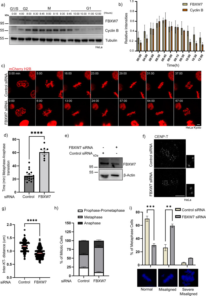
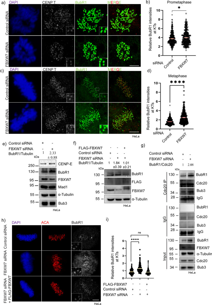
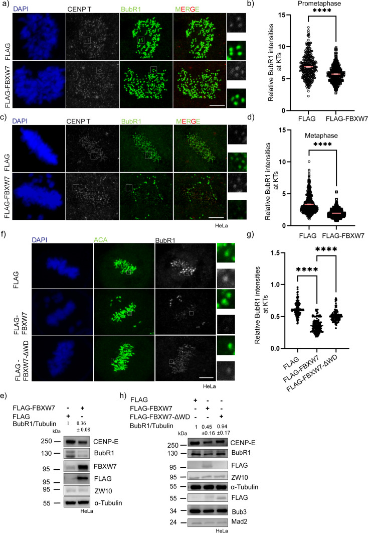
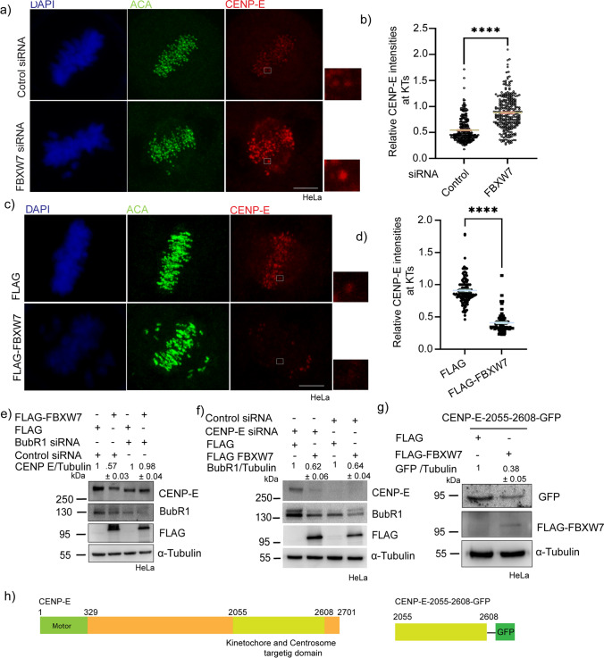
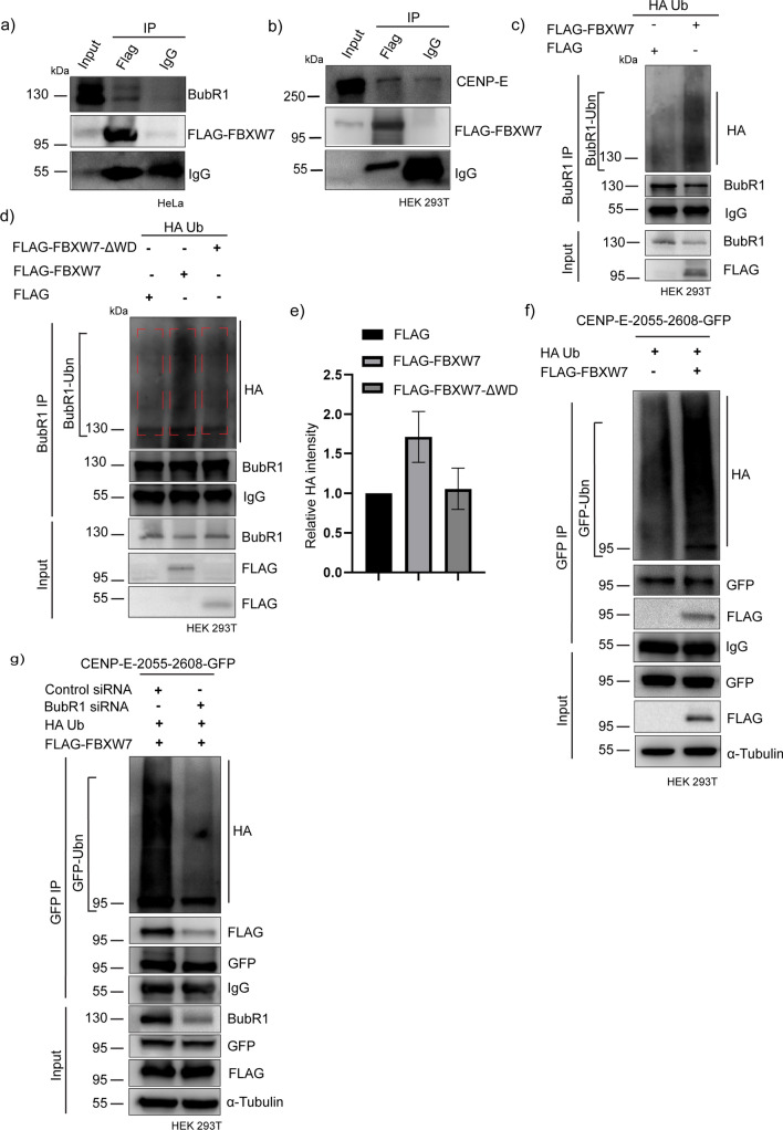
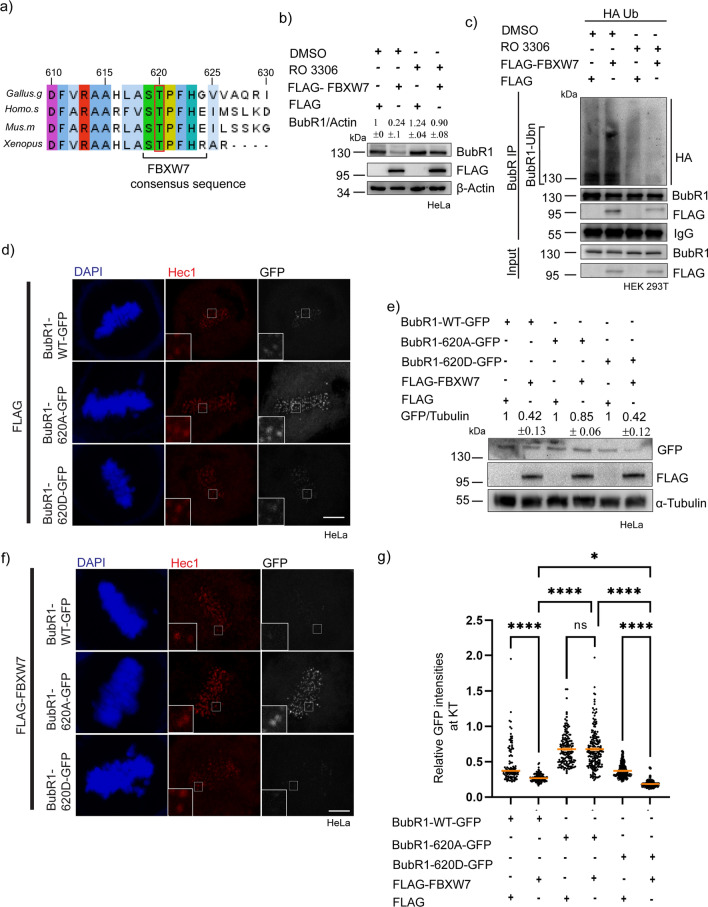
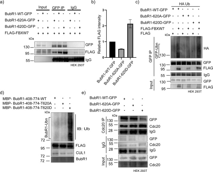
Faithful chromosome segregation requires correct attachment of kinetochores with the spindle microtubules. Erroneously-attached kinetochores recruit proteins to activate Spindle assembly checkpoint (SAC), which senses the errors and signals cells to delay anaphase progression for error correction. Temporal control of the levels of SAC activating-proteins is critical for checkpoint activation and silencing, but its mechanism is not fully understood. Here, we show that E3 ubiquitin ligase, SCF-FBXW7 targets BubR1 for ubiquitin-mediated degradation and thereby controls SAC in human cells. Depletion of FBXW7 results in prolonged metaphase arrest with increased stabilization of BubR1 at kinetochores. Similar kinetochore stabilization is also observed for BubR1-interacting protein, CENP-E. FBXW7 induced ubiquitination of both BubR1 and the BubR1-interacting kinetochore-targeting domain of CENP-E, but CENP-E domain degradation is dependent on BubR1. Interestingly, Cdk1 inhibition disrupts FBXW7-mediated BubR1 targeting and further, phospho-resistant mutation of Cdk1-targeted phosphorylation site, Thr 620 impairs BubR1-FBXW7 interaction and FBXW7-mediated BubR1 ubiquitination, supporting its role as a phosphodegron for FBXW7. The results demonstrate SCF-FBXW7 as a key regulator of spindle assembly checkpoint that controls stability of BubR1 and its associated CENP-E at kinetochores. They also support that upstream Cdk1 specific BubR1 phosphorylation signals the ligase to activate the process.

Keywords: BubR1; CENP-E; Checkpoint; FBXW7; Kinetochore; Mitosis; SCF complex.


Conflict of interest statement

The authors declare no conflict of interest.

Figures

Fig. 1
Fig. 1
FBXW7 depletion leads to a delay in metaphase to anaphase transition a Expression analysis of FBXW7 during the cell cycle. HeLa cells were synchronized with double thymidine treatment and then released to enter the S phase. Cells were collected at constant intervals and then probed for endogenous FBXW7 levels. b Histogram of FBXW7/cyclin B levels normalized to tubulin showed peaks during mitosis. Data = mean ± SEM from three independent experiments. c Selected frames of mCherry-tagged HeLa H2B cells after 48 h post-transfection of FBXW7 siRNA (lower panel) or control siRNA (upper panel). Cells were monitored upon entry to mitosis and mitotic timing was checked by live cell imaging. Images were acquired with a time interval of 1 min for 120 min. scale bar: 5 µm d Plot of mitotic timing FBXW7-depleted cells showed a delay of 45—65 min in metaphase to anaphase transition, whereas control cells enter anaphase within 15–25 min.****p < 0.0001 by student’s t-test. Each dot represent a single cell. e Lysate of HeLa cells showing FBXW7 levels in FBXW7 siRNA vs control siRNA. f Representative image of HeLa cells treated with FBXW7 siRNA (left panel) or control siRNA (right panel) probed with inner-kinetochore protein CENP -T as a marker. scale bar 5 µm. g Individual distances are marked on the dot plot Data = mean ± SEM ****p < 0.0001 by t-test h Percentage of mitotic cells in asynchronous HeLa cells were plotted in FBXW7 siRNA v/s Control siRNA condition. FBXW7 siRNA-treated cells showed increased metaphase cells as compared to Control siRNA. ~ 300–350 cells were analyzed. Data = mean ± SEM. i Graph showing the percentage of HeLa cells having congression defects in FBXW7 siRNA v/s Control siRNA condition. Data = mean ± SEM. ***p = 0.0001 by 2-way ANOVA
Fig. 2
Fig. 2
BubR1 levels were increased substantially in FBXW7-depleted cells. a, c Representative images of HeLa cells after 18 h of FBXW7 siRNA or control siRNA treatment followed by synchronization at prometaphase and metaphase by treating with thymidine followed by its release at appropriate times prior to stain for BubR1 and inner kinetochore marker, CENP T. b, d Dot Plots representing intensities of kinetochore-localized BubR1 in prometaphase and metaphase cells in FBXW7 siRNA vs. control HeLa cells. BubR1 intensities were normalized with respect to that of CENP T in all cases. ~ 300–400 kinetochores were analyzed from three independent experiments. Data = mean ± SEM. ****p < 0.0001, *p = 0.0178 by unpaired t-test e Lysates of mitotic synchronized HeLa cells after 18 h of FBXW7 siRNA or control siRNA were assessed for the levels of BubR1. Tubulin was probed as a control. BubR1 showed 2.33 ± 0.88 times increased overall levels in FBXW7-depleted cells as compared to control cells. f Lysates of mitotic synchronized HeLa cells after 18 h of FBXW7 siRNA or control siRNA or FBXW7 siRNA treatment rescued with FLAG-FBXW7 expression were assessed for the overall levels of BubR1. Tubulin was probed as a control. g Lysates of mitotic synchronized HeLa cells transfected with control siRNA or FBXW7 siRNA were subjected to Cdc20 immunoprecipitation and probed for BubR1 and Bub3 proteins. h Images of HeLa cells transfected with FBXW7 siRNA or control siRNA or FBXW7 siRNA rescued with FLAG-FBXW7 followed by synchronization at metaphase using double thymidine treatment followed by its release and fixed at the appropriate time. Cells were probed for BubR1 and inner kinetochore marker ACA. i Dot plot of intensities of kinetochore-localized BubR1 normalized with corresponding ACA intensity at the metaphase plate. ~ 300–400 kinetochores were analyzed. Data = mean ± SEM. ****p < 0.0001, and ns = 0.4774 from one-way ANOVA
Fig. 3
Fig. 3
FBXW7 over-expression reduces overall levels of BubR1 a, c Representative images of HeLa cells after 12 h of FLAG-tagged FBXW7α or control FLAG plasmid expression followed by synchronization at prometaphase and metaphase by treating with thymidine followed by its release and fixed at appropriate times prior to stain for BubR1 and inner kinetochore marker, CENP-T. b, d Plots of intensities of kinetochore-localized BubR1 at prometaphase and metaphase cells in FLAG-FBXW7-expressed vs. control cells. BubR1 intensities were normalized with respect to that of CENP-T in all cases. ~ 300–400 kinetochores were analyzed from three independent experiments. Data = mean ± SEM.****p < 0.0001 from unpaired t-test. e Lysates of mitotic synchronized HeLa cells expressed with control FLAG plasmid or FLAG-tagged FBXW7, were assessed for the CENP-E and BubR1 levels. Tubulin was probed as a control. BubR1 and CENP-E levels were reduced substantially in FBXW7-overexpressed cells. f Confocal images of HeLa cells after 12 h of FLAG-tagged FBXW7 or control FLAG or FLAG-FBXW7-ΔWD expression followed by synchronization at metaphase by treating with thymidine followed by its release and fixed at appropriate times prior to stain for BubR1 and inner kinetochore marker, ACA g Plot of intensities of kinetochore localized BubR1 at metaphase in control vs FLAG-FBXW7 expressed vs FLAG-FBXW7-ΔWD-expressed cells. BubR1 intensities were normalized with respect to that of ACA in all cases. ~ 300 kinetochores were analyzed from three independent experiments. Data = mean ± SEM. ****p < 0.0001 from one-way ANOVA h Lysates of mitotic synchronized cells expressed with Control FLAG vs FLAG-FBXW7 vs FLAG-FBXW7-ΔWD were assessed for overall BubR1 and CENP-E levels. Tubulin was probed as a control
Fig. 4
Fig. 4
FBXW7-mediated CENP-E degradation is BubR1 dependent. a Representative images of HeLa cells after 18 h of FBXW7 siRNA or control siRNA treatment followed by synchronization by treating with thymidine and released at the appropriate time prior to stain for CENP-E and inner kinetochore marker, ACA. b Dot Plots representing intensities of kinetochore localized BubR1 at metaphase cells in FBXW7 siRNA vs. control cells. CENP-E intensities were normalized with respect to that of ACA in all cases. ~ 300–400 kinetochores were analyzed from three independent experiments. Data = mean ± SEM. ****p < 0.0001 unpaired t-test. c Representative images of HeLa cells after 12 h of FLAG-tagged FBXW7α or control FLAG plasmid expression followed by synchronization at prometaphase and metaphase by treating with thymidine followed by its release and fixed at appropriate times prior to stain for BubR1 and inner kinetochore marker, CENP-T. d Plots of intensities of kinetochore localized CENP-E at metaphase cells in FLAG-FBXW7 expressed vs. control cells. CENP-E intensities were normalized with respect to that of ACA in all cases. ~ 300–400 kinetochores were analyzed. Data = mean ± SEM.****p < 0.0001 from unpaired t-test. e Overall levels of CENP-E were unaffected in cells depleted of BubR1 followed by FBXW7 overexpression vs control siRNA followed by FBXW7 overexpression. f Overall BubR1 levels were significantly reduced in CENP-E-depleted cells overexpressed with FLAG-FBXW7 with respect to control cells. g Overall levels of CENP-E-2055–2608-GFP showed a significant reduction in the FLAG-FBXW7-expressed cells with respect to control cells h Schematic representation of the amino acid regions of CENP-E-WT and CENP-E-2055–2608-GFP
Fig. 5
Fig. 5
BubR1 is a novel substrate of FBXW7. a HeLa cells overexpressed with FLAG-FBXW7 were synchronized at mitosis followed by the addition of MG132 treatment and then cell lysate was subjected to FLAG pull down by using FLAG antibody and then probed for BubR1 b HEK293T cells expressed with FLAG-FBXW7 were synchronized at mitosis followed by the addition of MG132, were subjected to FLAG pull down by using FLAG antibody and then probed for CENP-E. c HEK293T cells were expressed with FLAG-FBXW7 and HA-tagged Ubiquitin (HA-Ub) for 18 h followed by mitotic synchronization using thymidine and addition of MG132 and BubR1 was immunoprecipitated from the cell lysate. The BubR1 immunoprecipitate from the lysates of FLAG-FBXW7-expressed cells were probed for HA-Ub and compared to the BubR1 immunoprecipitate from control cell lysate. d HEK293T cells were expressed with FLAG-FBXW7 or FLAG-FBXW7-ΔWD together with HA-Followed by mitotic synchronization using thymidine and MG132. BubR1 was co immunoprecipitated and its ubiquitination was compared by probing with an HA antibody. e Plot of BubR1 ubiquitination levels in different condition as of d. f GFP immunoprecipitate of mitotic synchronized HEK293T cells expressed with FLAG-FBXW7 and HA-Ub along with CENP-E-2055–2608-GFP was probed for HA-Ub and the levels of ubiquitinated proteins were compared with respect to control CENP-E-2055–2608-GFP cells expressed with FLAG control plasmid. g HEK293T cells were co-expressed with CENP-E-2055–2608-GFP together with FLAG-FBXW7 and HA-Ub in BubR1 siRNA or Control siRNA transfected condition for 18 h followed by mitotic synchronization using thymidine followed by the addition of MG132. Then CENP-E-2055–2608-GFP was immunoprecipitated by GFP antibody and probed for HA
Fig. 6
Fig. 6
Cdk1-mediated phosphorylation at Thr 620in BubR1 acts as phosphodegron for FBXW7-mediated degradation. a Conservation of Thr620 (T620) of BubR1 among different species. b Overall levels of BubR1 were unaffected in Cdk1 inhibitor-treated cells in control vs FLAG-FBXW7 overexpressed cells. c Levels of ubiquitinated BubR1were reduced in Cdk1 inhibitor-treated HEK293T cells expressed with FLAG-FBXW7 and HA-Ub as compared to control conditions. d, f Representative images of Mitotic synchronized HeLa cells expressed with BubR1-WT-GFP or BubR1-T620A-GFP or BubR1 T620D-GFP together with control FLAG-plasmid or FLAG-FBXW7 g Dot plot of intensities of GFP-tagged BubR1 WT and mutants at kinetochores normalized to Hec1 as marker in metaphase cells expressed with BubR1-WT-GFP or BubR1-T620A-GFP or BubR1-T620D-GFP together with FLAG-FBXW7 vs. FLAG empty vector. ~ 200–300 kinetochores were analyzed. Data = mean ± SEM. ****p < 0.0001, *p = 0.211, ns > 0.9999 by one-way ANOVA e The overall cellular levels of BubR1-T620D-GFP showed increased degradation, whereas the BubR1-T620A-GFP showed reduced degradation in FLAG-FBXW7 expressed cells
Fig. 7
Fig. 7
BubR1 phosphorylation at Thr 620 promotes its ubiquitination by FBXW7 a HEK293T cells were co-expressed withBubR1-WT-GFP or BubR1-T620A-GFP or BubR1-T620D-GFP together with FLAG-FBXW7 for 18 h followed by mitotic synchronization using thymidine followed by the addition of MG132. Then GFP-tagged BubR1 was immunoprecipitated with GFP antibody and levels of FLAG-FBXW7 were compared by immunoblotting with FLAG antibody. b The histogram plot shows the relative intensity of FLAG-FBXW7 normalized to corresponding BubR1-WT-GFP or BubR1-T620A-GFP or BubR1-T620D-GFP level. c HEK293T cells were co-expressed with BubR1-WT-GFP or BubR1-T620A-GFP or BubR1-T620D-GFP together with FLAG-FBXW7 and HA-Ub and the lysates of cells after mitotic synchronization by thymidine release followed by MG132 were probed for HA-Ub. d Recombinant MBP-tagged BubR1-408–774-WT or MBP-tagged BubR1-408–774-T620A or MBP-tagged BubR1-408–774-T620D bound with amylose resin was incubated separately with a mixture of SCFFBXW7 components, Myc-Skp-1, Myc-Cul-1, HA-Rbx-1, and FLAG-FBXW7, followed by E1, E2, Ub, and ATP. SCFFBXW7component were isolated from HEK293T cells as described in Methods. Samples were immunoblotted for BubR1 and ubiquitin by antibodies. e Lysates of mitotic synchronized HEK293T cells transfected with BubR1-WT-GFP or BubR1-T620A-GFP or BubR1-T620D-GFP for 18 h followed by mitotic synchronization by thymidine release followed by MG132 addition were subjected to Cdc20 immunoprecipitation and probed for GFP-tagged BubR1 protein levels

Similar articles

Cited by

References

    1. Holland AJ, Cleveland DW. Boveri revisited: chromosomal instability, aneuploidy and tumorigenesis. Nat Rev Mol Cell Biol. 2009;10:478–487. doi: 10.1038/nrm2718. - DOI - PMC - PubMed
    1. Ricke RM, Van Ree JH, Van Deursen JM. Whole chromosome instability and cancer: a complex relationship. Trends Genet. 2008;24:457–466. doi: 10.1016/j.tig.2008.07.002. - DOI - PMC - PubMed
    1. Schvartzman JM, Sotillo R, Benezra R. Mitotic chromosomal instability and cancer: mouse modelling of the human disease. Nat Rev Cancer. 2010;10:102–115. doi: 10.1038/nrc2781. - DOI - PMC - PubMed
    1. Daniel P, Cahill CL, Jian Yu, Riggins GJ, Willson JKV, Markowitz SD, Kinzler KW, Vogelstein B. Mutations of mitotic checkpoint genes in human cancers. Nature. 1998;392:300–303. doi: 10.1038/32688. - DOI - PubMed
    1. Masuda A, Takahashi T. Chromosome instability in human lung cancers: possible underlying mechanisms and potential consequences in the pathogenesis. Oncogene. 2002;21:6884–6897. doi: 10.1038/sj.onc.1205566. - DOI - PubMed

MeSH terms

LinkOut - more resources