. 2023 Dec;90(6):e13797.
doi: 10.1111/aji.13797.

Validation of vaginal microbiome proxies for in vitro experiments that biomimic Lactobacillus-dominant vaginal cultures

Affiliations

Validation of vaginal microbiome proxies for in vitro experiments that biomimic Lactobacillus-dominant vaginal cultures

Abir Zahra et al. Am J Reprod Immunol. 2023 Dec.

Abstract

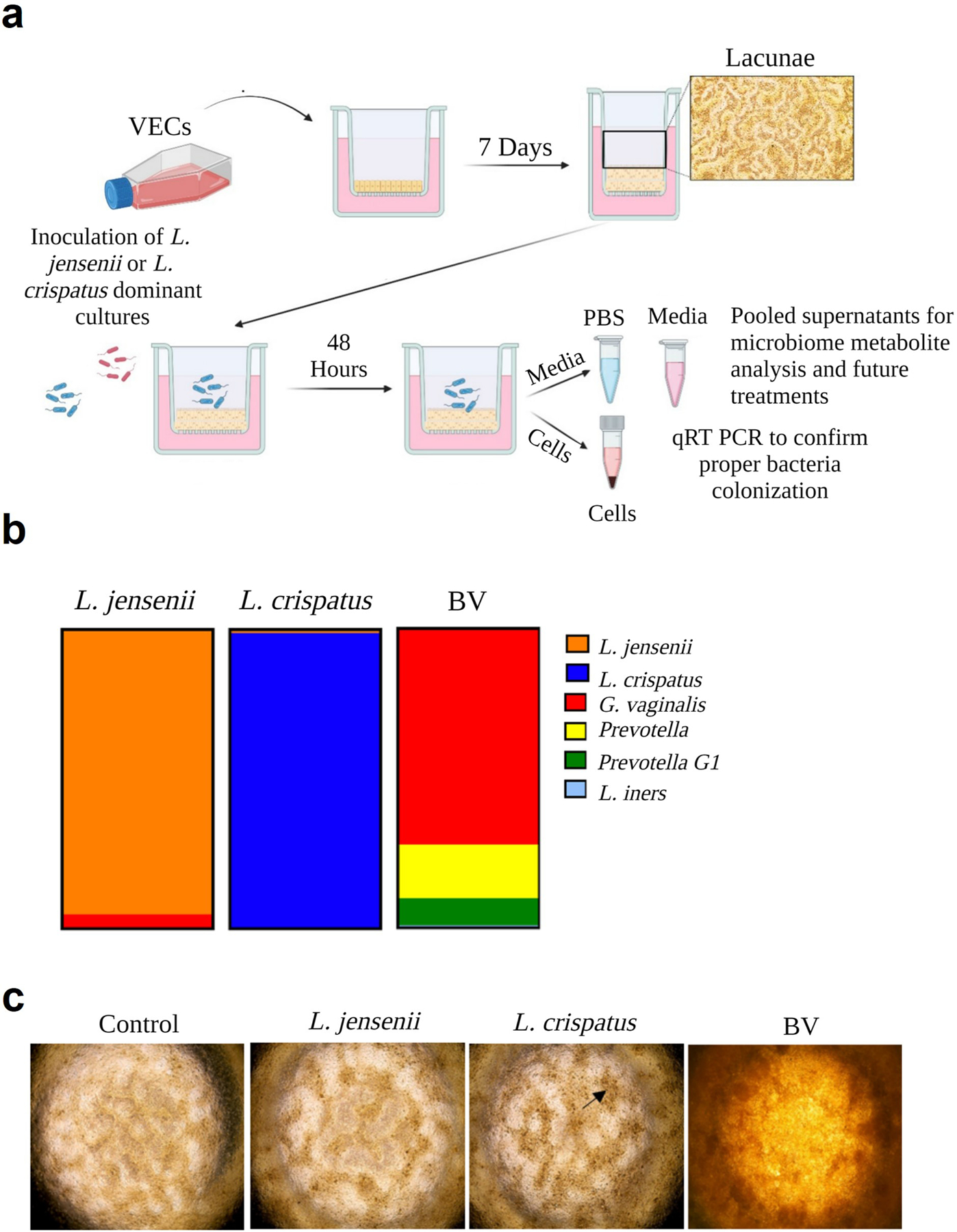
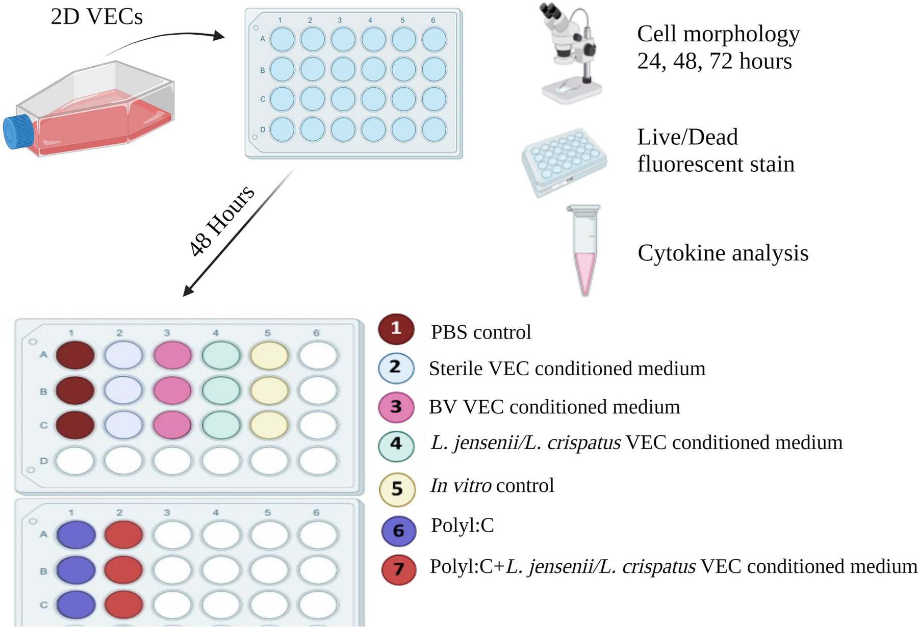
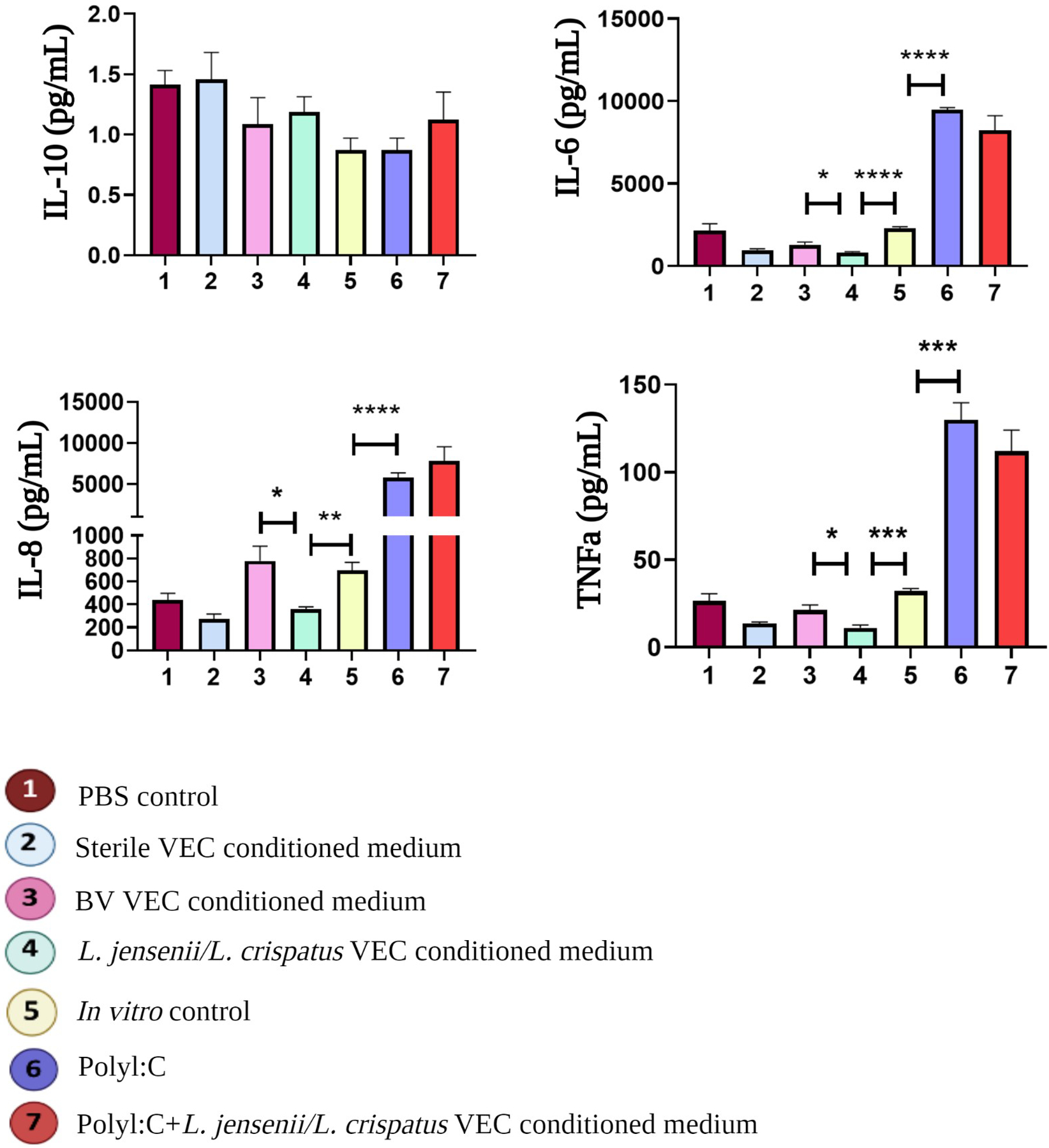
The vaginal microbiome includes diverse microbiota dominated by Lactobacillus [L.] spp. that protect against infections, modulate inflammation, and regulate vaginal homeostasis. Because it is challenging to incorporate vaginal microbiota into in vitro models, including organ-on-a-chip systems, we assessed microbial metabolites as reliable proxies in addition to traditional vaginal epithelial cultures (VECs). Human immortalized VECs cultured on transwells with an air-liquid interface generated stratified cell layers colonized by transplanted healthy microbiomes (L. jensenii- or L. crispatus-dominant) or a community representing bacterial vaginosis (BV). After 48-h, a qPCR array confirmed the expected donor community profiles. Pooled apical and basal supernatants were subjected to metabolomic analysis (untargeted mass spectrometry) followed by ingenuity pathways analysis (IPA). To determine the bacterial metabolites' ability to recreate the vaginal microenvironment in vitro, pooled bacteria-free metabolites were added to traditional VEC cultures. Cell morphology, viability, and cytokine production were assessed. IPA analysis of metabolites from colonized samples contained fatty acids, nucleic acids, and sugar acids that were associated with signaling networks that contribute to secondary metabolism, anti-fungal, and anti-inflammatory functions indicative of a healthy vaginal microbiome compared to sterile VEC transwell metabolites. Pooled metabolites did not affect cell morphology or induce cell death (∼5.5%) of VEC cultures (n = 3) after 72-h. However, metabolites created an anti-inflammatory milieu by increasing IL-10 production (p = .06, T-test) and significantly suppressing pro-inflammatory IL-6 (p = .0001), IL-8 (p = .009), and TNFα (p = .0007) compared to naïve VEC cultures. BV VEC conditioned-medium did not affect cell morphology nor viability; however, it induced a pro-inflammatory environment by elevating levels of IL-6 (p = .023), IL-8 (p = .031), and TNFα (p = .021) when compared to L.-dominate microbiome-conditioned medium. VEC transwells provide a suitable ex vivo system to support the production of bacterial metabolites consistent with the vaginal milieu allowing subsequent in vitro studies with enhanced accuracy and utility.

Keywords: anti-inflammatory; metabolites; vaginal epithelial cells.


Conflict of interest statement

Conflict of Interest: The authors declare no conflict of interest.

Figures

Figure 1.
Figure 1.. Generation of vaginal microbiome metabolites from healthy and dysbiosis states
a) Human immortalized vaginal epithelial cells (VEC) were cultured in KSFM medium on transwell inserts for seven days to generate stratified epithelial cultures. Medium was changed every 48-hours for 7 days to allow for the formation of Lacunae. The three vaginal microbiome samples from previously collected specimens were added to the VEC transwells to colonize for 48-hours. (right). b) Previously collected vaginal microbiome swab samples—L. jensenii, L. crispatus, and bacterial vaginosis (BV) dominant samples were selected for seeding into the VEC ex vivo transwells. These samples were cultured, cryopreserved, and subsequently revived for the purpose of this project. c) Phase contrast microscopy showed colonization on top of lacunae (black arrow) (2x images), and proper colonization of the microbiome was confirmed through qPCR analysis.
Figure 2.
Figure 2.. Characterization of cell and microbiome metabolites
The apical+basal supernatants dominated by L. jensenii + L. crispatus microbiomes were collected, filtered, and analyzed for metabolites through untargeted mass spectrometry analysis. The resulting data were further analyzed using IPA to identify the specific metabolites produced in these samples. a) Compounds were associated with canonical pathways such as tRNA turnover, creatine and putrescine biosynthesis, and DNA methylation (Z-score 0: Not significant [white bars as indicated by IPA]). b) signaling networks (AKT/ERK, nitric oxide, creatine), that contribute to secondary metabolism, anti-fungal, and anti-inflammatory functions of a healthy vaginal microbiome. Green ovals highlight the dominate upstream activators, signalers, and downstream related compounds in each network.
Figure 3.
Figure 3.. Experimental workflow assessing VEC+Microbiome induced function
To determine the effect of microbiome metabolites on 2D VEC cultures, VECs were colonized in a 2D monolayer for 72-hours with various treatments consisting of positive controls (Poly I:C [0.1mg/ml] or BV), experimental controls (VEC medium or PBS), and the experimental treatments at a 1:1 ratio of VEC medium. After 72-hours cells were assessed for morphological changes and cell viability. Medium was collected a processed for pro- and anti-inflammatory cytokine analysis.
Figure 4.
Figure 4.. Functional assessment of VEC+Microbiome metabolites effects on cell morphology
Brightfield microscopy documented the cell morphology of VECs after they were cultured with various treatments for 72-hours. Microbiome-conditioned medium did not change VEC cell morphology after 72-hours in traditional culture. PolyI:C induced a punctate cell morphology by 24-hours (black box) that increased over 72-hours, microbiome treatment recovered this phenotype (red box). 10x images.
Figure 5.
Figure 5.. Functional assessment of VEC+Microbiome metabolites effects on cell viability
VECs cultured with microbiome-conditioned medium after 72-hours showed comparably low levels of cell death (~5.5%) compared to controls (~3%). PolyI:C conditioned medium induced higher levels of cell death (~21%) compared to L. jensenii/L. crispatus conditioned (~5.5%) and control medium (~3%) after 72-hours. PolyI:C +microbiome conditioned medium showed lower levels of cell death (12%) when compared to PolyI:C conditioned medium only (21%). Viability marker – live-Calcein AM – Green. Cell death marker – dead-ethidium homodimer-1- Red. 10x images collected.
Figure 6.
Figure 6.. Cytokine Production of 2D VECs after different treatments
A cytokine multiplex assay was used to quantify pro-inflammatory (Interleukin [IL]-6, IL-8, Tumor Necrosis Factor-alpha [TNFα], and anti-inflammatory (IL-10) cytokines. Analysis showed that L. jensenii/L. crispatus metabolites changed the inflammatory milieu by increasing anti-inflammatory IL-10 production and significantly suppressing pro-inflammatory IL-6 (p<0.0001), IL-8 (p=0.004), and TNFα (p=0.0004) when compared to standard culture conditions (lane 4 vs. 5). PolyI:C significantly induced a pro-inflammatory environment noted by high levels of IL-6 (p<0.0001), IL-8 (p=0.0005), and TNFα (p=0.0003) when compared to standard culture conditions (lane 5 vs. 6).

References

    1. Ravel J, Gajer P, Abdo Z, et al. Vaginal microbiome of reproductive-age women. Proc Natl Acad Sci U S A. Mar 15 2011;108 Suppl 1(Suppl 1):4680–7. doi:10.1073/pnas.1002611107 - DOI - PMC - PubMed
    1. Chen X, Lu Y, Chen T, Li R. The Female Vaginal Microbiome in Health and Bacterial Vaginosis. Front Cell Infect Microbiol. 2021;11:631972. doi:10.3389/fcimb.2021.631972 - DOI - PMC - PubMed
    1. Lewis FMT, Bernstein KT, Aral SO. Vaginal Microbiome and Its Relationship to Behavior, Sexual Health, and Sexually Transmitted Diseases. Obstet Gynecol. Apr 2017;129(4):643–654. doi:10.1097/aog.0000000000001932 - DOI - PMC - PubMed
    1. Lehtoranta L, Ala-Jaakkola R, Laitila A, Maukonen J. Healthy Vaginal Microbiota and Influence of Probiotics Across the Female Life Span. Front Microbiol. 2022;13:819958. doi:10.3389/fmicb.2022.819958 - DOI - PMC - PubMed
    1. Chee WJY, Chew SY, Than LTL. Vaginal microbiota and the potential of Lactobacillus derivatives in maintaining vaginal health. Microb Cell Fact. Nov 7 2020;19(1):203. doi:10.1186/s12934-020-01464-4 - DOI - PMC - PubMed

Publication types

LinkOut - more resources