

Serial fractional flow reserve, coronary flow reserve and index of microcirculatory resistance after percutaneous coronary intervention in patients treated for stable angina pectoris assessed with PET
- PMID: 38009377
- PMCID: PMC10833199
- DOI: 10.1097/MCA.0000000000001308
Serial fractional flow reserve, coronary flow reserve and index of microcirculatory resistance after percutaneous coronary intervention in patients treated for stable angina pectoris assessed with PET
Abstract
Background: Cardiac 15 O-water PET is a noninvasive method to evaluate epicardial and microvascular dysfunction and further quantitate absolute myocardial blood flow (MBF).
Aim: The aim of this study was to assess the impact of revascularization on MBF and myocardial flow reserve (MFR) assessed with 15 O-water PET and invasive flow and pressure measurements.
Methods: In 21 patients with single-vessel disease referred for percutaneous coronary intervention (PCI), serial PET perfusion imaging and fractional flow reserve (FFR), coronary flow reserve (CFR) and index of microcirculatory resistance (IMR) were performed during PCI and after 3 months.
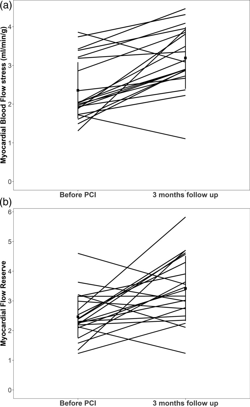
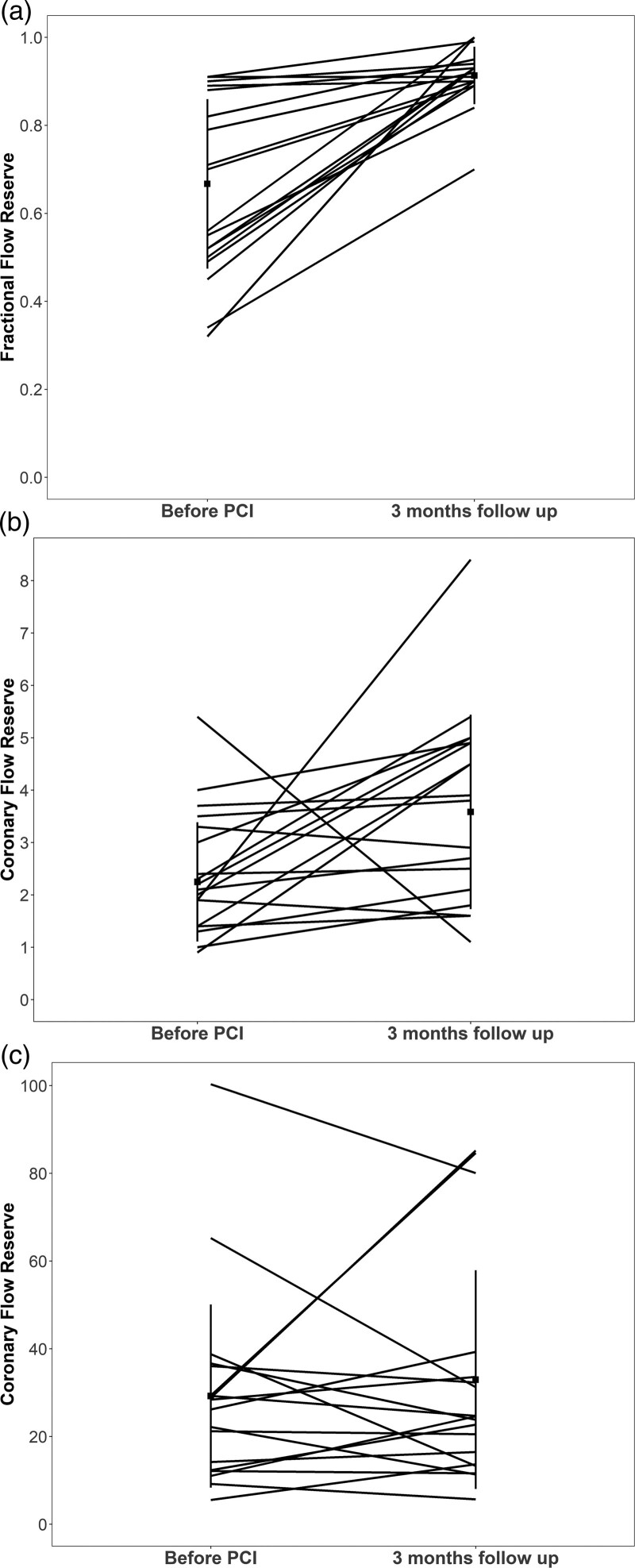
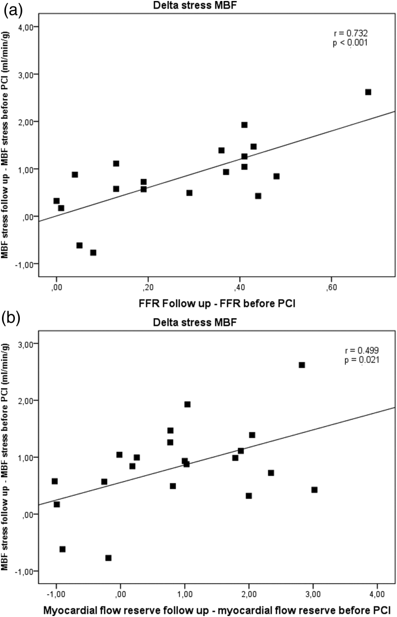
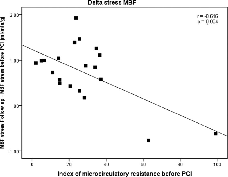
Results: In the affected myocardium, stress MBF and MFR increased significantly from before revascularization to 3 months after revascularization: stress MBF 2.4 ± 0.8 vs. 3.2 ± 0.8; P < 0.001 and MFR 2.5 ± 0.8 vs. 3.4 ± 1.1; P = 0.004. FFR and CFR increased significantly from baseline to after revascularization and remained stable from after revascularization to 3-month follow-up: FFR 0.64 ± 0.20 vs. 0.91 ± 0.06 vs. 0.91 ± 0.07; P < 0.001; CFR 2.4 ± 1.2 vs. 3.6 ± 1.9 vs. 3.6 ± 1.9; P < 0.001, whereas IMR did not change significantly: 30.3 ± 22.9 vs. 30.1 ± 25.3 vs. 31.9 ± 25.2; P = ns. After revascularization, an increase in stress MBF was associated with an increase in FFR ( r = 0.732; P < 0.001) and an increase in MFR ( r = 0.499; P = 0.021). IMR measured before PCI was inversely associated with improvement in stress MBF, ( r = -0.616; P = 0.004).
Conclusion: Recovery of myocardial perfusion after PCI was associated with an increase in FFR 3 months after revascularization. Microcirculatory dysfunction was associated with less improvement in myocardial perfusion.
Copyright © 2023 The Author(s). Published by Wolters Kluwer Health, Inc.
Conflict of interest statement
L.O.J. has received research grants from Biotronik, Biosensors, Abbott Vascular and Terumo to her institution and honoraria from Biotronik. For the remaining authors, there are no conflicts of interest.
Figures
References
-
- Tousoulis D, Androulakis E, Kontogeorgou A, Papageorgiou N, Charakida M, Siama K, et al. Insight to the pathophysiology of stable angina pectoris. Curr Pharm Des 2013; 19:1593–1600. - PubMed
-
- Schwartz L, Bourassa MG. Evaluation of patients with chest pain and normal coronary angiograms. Arch Intern Med 2001; 161:1825–1833. - PubMed
-
- Marzilli M, Merz CN, Boden WE, Bonow RO, Capozza PG, Chilian WM, et al. Obstructive coronary atherosclerosis and ischemic heart disease: an elusive link! J Am Coll Cardiol 2012; 60:951–956. - PubMed
-
- Pries AR, Badimon L, Bugiardini R, Camici PG, Dorobantu M, Duncker DJ, et al. Coronary vascular regulation, remodelling, and collateralization: mechanisms and clinical implications on behalf of the working group on coronary pathophysiology and microcirculation. Eur Heart J 2015; 36:3134–3146. - PubMed
-
- Lee JM, Jung JH, Hwang D, Park J, Fan Y, Na SH, et al. Coronary flow reserve and microcirculatory resistance in patients with intermediate coronary stenosis. J Am Coll Cardiol 2016; 67:1158–1169. - PubMed
MeSH terms
Substances
LinkOut - more resources
Full Text Sources
Medical
Miscellaneous
