Review
. 2024 Feb;11(5):e2302165.
doi: 10.1002/advs.202302165. Epub 2023 Nov 27.

Listen to Your Gut: Key Concepts for Bioengineering Advanced Models of the Intestine

Affiliations
Review

Listen to Your Gut: Key Concepts for Bioengineering Advanced Models of the Intestine

Oliver Cameron et al. Adv Sci (Weinh). 2024 Feb.

Abstract

The intestine performs functions central to human health by breaking down food and absorbing nutrients while maintaining a selective barrier against the intestinal microbiome. Key to this barrier function are the combined efforts of lumen-lining specialized intestinal epithelial cells, and the supportive underlying immune cell-rich stromal tissue. The discovery that the intestinal epithelium can be reproduced in vitro as intestinal organoids introduced a new way to understand intestinal development, homeostasis, and disease. However, organoids reflect the intestinal epithelium in isolation whereas the underlying tissue also contains myriad cell types and impressive chemical and structural complexity. This review dissects the cellular and matrix components of the intestine and discusses strategies to replicate them in vitro using principles drawing from bottom-up biological self-organization and top-down bioengineering. It also covers the cellular, biochemical and biophysical features of the intestinal microenvironment and how these can be replicated in vitro by combining strategies from organoid biology with materials science. Particularly accessible chemistries that mimic the native extracellular matrix are discussed, and bioengineering approaches that aim to overcome limitations in modelling the intestine are critically evaluated. Finally, the review considers how further advances may extend the applications of intestinal models and their suitability for clinical therapies.

Keywords: biomaterials; disease modelling; intestine; organ-on-chip; organoids.


Conflict of interest statement

The authors declare no conflict of interest.

Figures
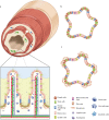
Figure 1
Figure 1
The intestinal epithelium and intestinal organoids. a) Cross section of the small intestine with a zoomed schematic of the intestinal epithelium. b) Adult stem cell‐derived intestinal organoid containing differentiated epithelial cells with both secretory and absorptive phenotypes. c) Embryonic or pluripotent stem cell‐derived intestinal organoid containing differentiated epithelial cells and surrounding mesenchymal cells.
Figure 2
Figure 2
Mechanical cues experienced by intestinal cells. a) Intrinsic mechanical cues. Top: Tensile forces generated by actomyosin contraction apply tension on neighboring cells and the extracellular matrix via focal adhesions. Middle; Stiffness describes the degree to which a material resists an applied force. Bottom: Cell density and shape provide mechanical cues to cells as a result of cell crowding or ECM confirmations. b) Extrinsic mechanical cues. Top: Applied tensile and compressive forces can directly impact cells, for example, by luminal contents applying normal forces to the gut wall as they pass. Middle; Hydrostatic pressures result from fluid itself pushing on a cell. Bottom: Shear forces act parallel to the gut wall and may cause lateral deformation of cells, for example, as luminal contents pass. c) Intestinal tissues are viscoelastic and thus possess properties of both elastic solids and viscous fluids.
Figure 3
Figure 3
Mechanical forces influencing the static and dynamic intestine. a) Longitudinal cross section portraying the layered organization of the gut wall. The cells of the epithelium experience a balance of tensile and compressive forces. The mucosal and submucosal tissues exist under constant compression both axially from luminal contents and laterally from nearby crypts. Layers farther from the lumen are under constant tension. b) Sections of the intestine can be cut to investigate the residual stress. The opening angle will indicate the balance between compression and tension between layers. c) The dynamic intestine is subject multiple macro‐mechanical processes during peristaltic and inter‐peristaltic periods. Left; Distension of the gut by digested food or microbial metabolism applies outward radial and longitudinal forces. Middle; Contraction of the muscle layers leads to inward radial and longitudinal forces. Right: Shear forces due to the passage of luminal contents act unidirectionally. Adapted with permission.[ 283 ] 2022, Springer Nature.
Figure 4
Figure 4
Keys factors for biomimicry of the intestine. The intestine contains diverse cell types that coordinate together to maintain intestinal function. Various chemical factors are critical for maintaining viability and function of intestinal cells. Components of the extracellular matrix provide a physical environment suited to each cell type and their local milieu. The intestine responds to and generates mechanical cues that maintain homeostasis while performing physical tasks. Diverse gradients are established and maintained within the intestine, including along the crypt‐villis axis and across the epithelium, that assist with intestinal homeostasis and defence. Together, interactions between intestinal cells and the chemical, physical, and mechanical features of the intestine is bidirectional and reciprocal, and can be recapitulated using advanced bioengineering technologies including hydrogel chemistries, microfluidics, and light‐based technologies. Enteroendocrine cell (EEC), smooth muscle cell (SMC), intestinal stem cell (ISC).
Figure 5
Figure 5
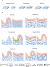
The effects of extracellular matrix cues on intestinal cells. a) Left; Cells with excess space form more focal adhesions, with reinforced cytoskeletal tension whereas cells with limited adhesive space form fewer focal adhesions and adopt a more rounded shape with less cytoskeletal tension. Middle; The more textured the ECM topography, the more focal adhesions cells form. Thus, cells spread on highly textured ECM while fewer topographical features promote rounded cells. Right: A highly crosslinked ECM with a higher stiffness prompts cells to adopt spread morphologies, while softer ECM promotes rounded cells with fewer focal adhesions and lower levels of cytoskeletal tension. b) Top left; Matrices created with lower stiffnesses enable intestinal monolayers to develop crypt/villus‐like morphologies, whereas at higher stiffnesses this morphology cannot develop. Top right: More adhesive motifs may improve intestinal monolayer viability. Middle left: Culture of IOs on matrices fabricated with crypt/villus‐like topographical features promote physiological cellular heterogeneity and localization of progenitor cells. Middle right: Matrices with degradability enable the development of crypt/villus‐like morphology. Bottom left; Matrix features, like hydrogel mesh size, can influence the diffusion of molecules of biologically relevant size. Mesh size can be modulated by altering polymer concentration, molecular weight, and crosslinking density. Bottom right: Matrices can be formed with fibrous structures, mimicking the native ECM. Pathological conditions such as fibrosis may be mimicked this way by altering the size and number of fibers.
Figure 6
Figure 6
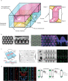
Examples of bioengineered intestinal models. a) Design of an intestine chip containing two parallel culture channels separated by a porous membrane lined on both sides by cells. Inlet and outlet channels allow for the addition of drugs or microbes, enabling independent analyses on each monolayer. Vacuum chambers on either side stretch the flexible membrane and adherent cells, mimicking peristalsis. b) (left) SEM images of PDMS stamp with crypt‐villus structures. (right) A hydrogel formed with the stamp and seeded with Lgr5‐GFP mIO and stained for E‐cadherin and AldoB. Lgr5+ ISCs localize to the crypts, while AldoB‐expressing enterocytes cover the villus. c) Design of cylindrical intestine chip. (left) The design consists of a central chamber containing a hydrogel flanked by inlet and outlet channels for luminal perfusion, and two lateral reservoirs supplying media and growth factors. (middle) Prior to cell loading, the hydrogel is laser ablated to construct a cylindrical channel with micro‐cavities resembling crypts. (right) The tubular scaffold is populated with cells to establish a confluent epithelium. d) (left) mIO cultured in pill‐shaped cavities and stained for E‐cadherin, YAP, and notch ligand (DLL) show differential expression in elongated versus restricted cells along the sides versus bases of the pill shape. (right) Proposed mechanism of geometry‐driven crypt specification. Panels (b) and (d) are reproduced with permission.[ 269 ] 2022, The American Association for the Advancement of Science. Panel (c) is reproduced with permission.[ 204 ] 2020, Springer Nature.
Figure 7
Figure 7
Schematic showing the potential of two photon microscopy to engineer an intestinal ECM. a) (left) One‐photon UV excitation and emission spectra. (right) Cross‐section of a synthetic hydrogel containing encapsulated cells designed to depolymerize in response to UV irradiation. The laser carves a topography through the hydrogel to create a 3D architecture reminiscent of the intestine. In this situation, encapsulated cells in the path of the laser light are negatively impacted. b) (left) Two‐photon infrared excitation and emission spectra. Two photon microscopy combines the energy of two colliding photons to equal that of UV irradiation. (right) Cross‐section of a synthetic hydrogel containing encapsulated cells designed to depolymerize in response to UV irradiation. Two‐photon laser depolymerization can carve an intestinal‐like topography, but here, encapsulated cells in the path of the laser are viable and the resolution of ablation is limited only by the optics.

Similar articles

Cited by

References

    1. Wang Y., Kasper L. H., Brain. Behav. Immun. 2014, 38, 1. - PMC - PubMed
    1. Roda G., Nat. Rev. Dis. Prim. 2020, 6, 22. - PubMed
    1. Kobayashi T., Nat. Rev. Dis. Prim. 2020, 6, 1. - PubMed
    1. Hallberg K., Grzegorczyk A., Larson G., Strandvik B., J. Pediatr. Gastroenterol. Nutr. 1997, 25, 290. - PubMed
    1. Soler A. P., Miller R. D., Laughlin K. V., Carp N. Z., Klurfeld D. M., Mullin J. M., Carcinogenesis 1999, 20, 1425. - PubMed

Publication types

LinkOut - more resources