. 2024 Apr;20(15):e2307066.
doi: 10.1002/smll.202307066. Epub 2023 Nov 27.

Pneumonic Plague Protection Induced by a Monophosphoryl Lipid A Decorated Yersinia Outer-Membrane-Vesicle Vaccine

Affiliations

Pneumonic Plague Protection Induced by a Monophosphoryl Lipid A Decorated Yersinia Outer-Membrane-Vesicle Vaccine

Saugata Majumder et al. Small. 2024 Apr.

Abstract

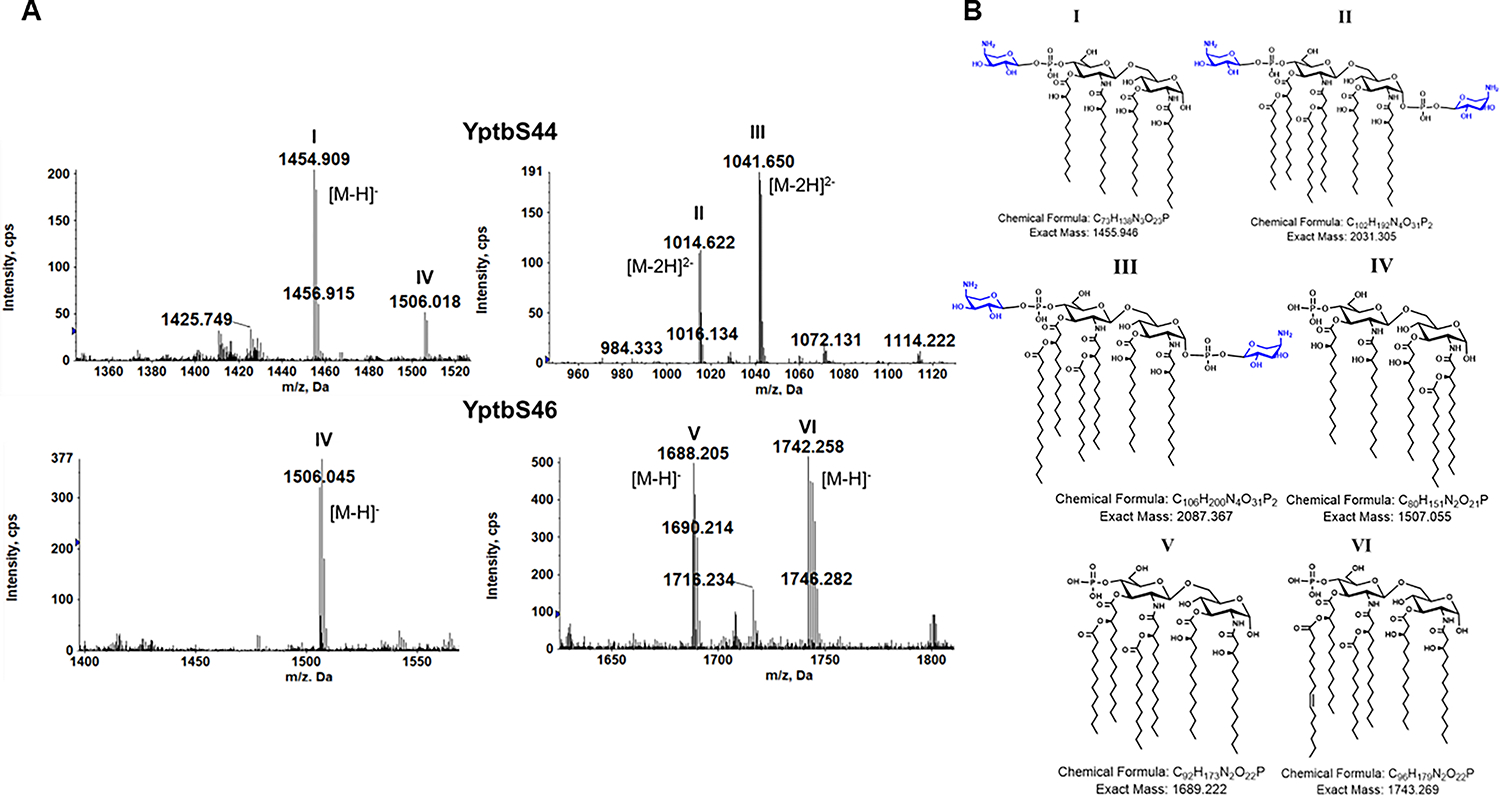
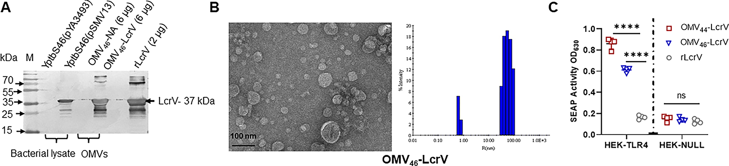
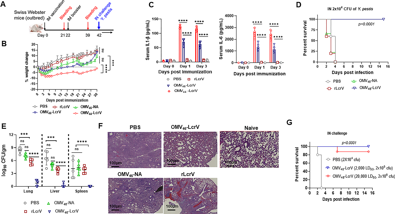
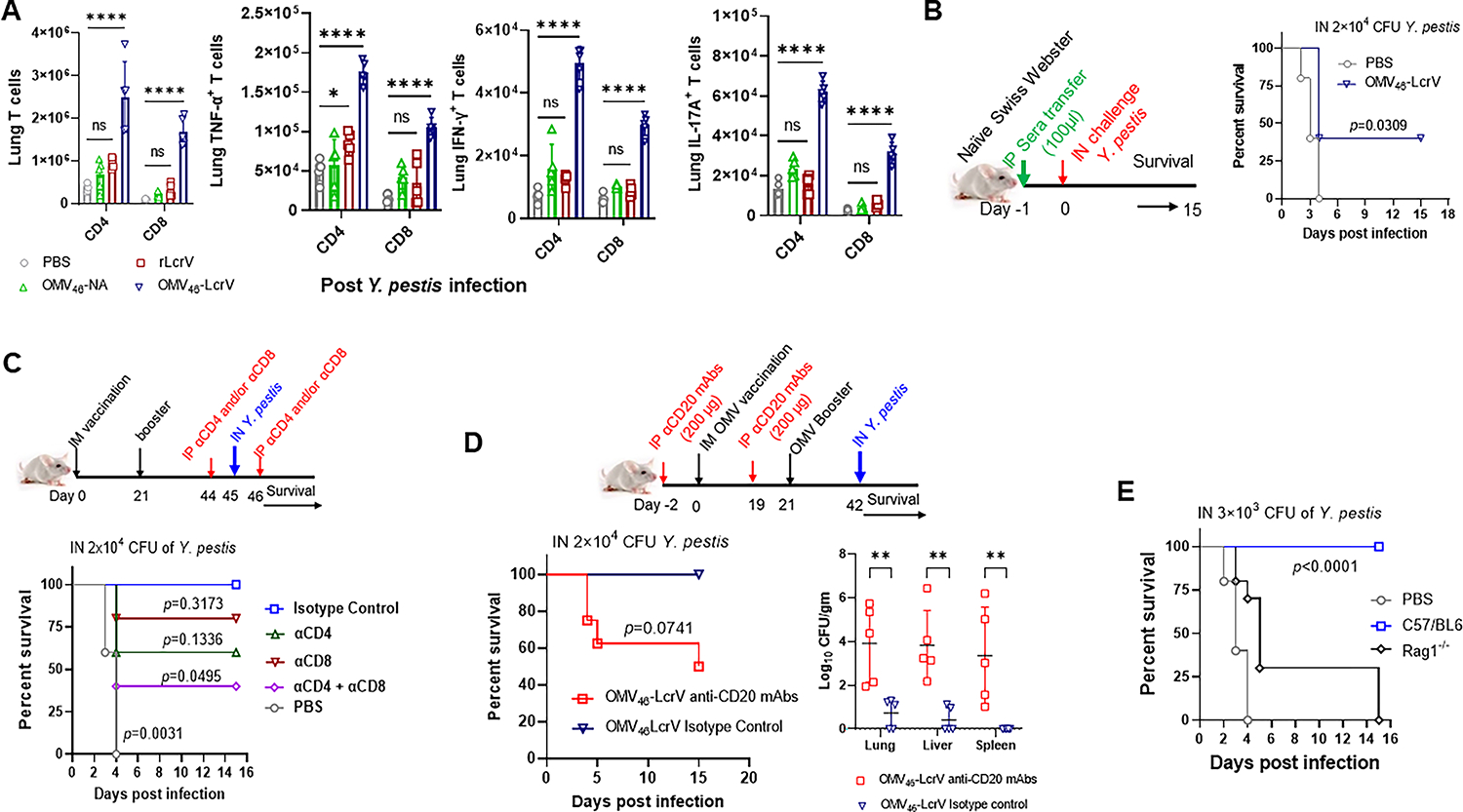
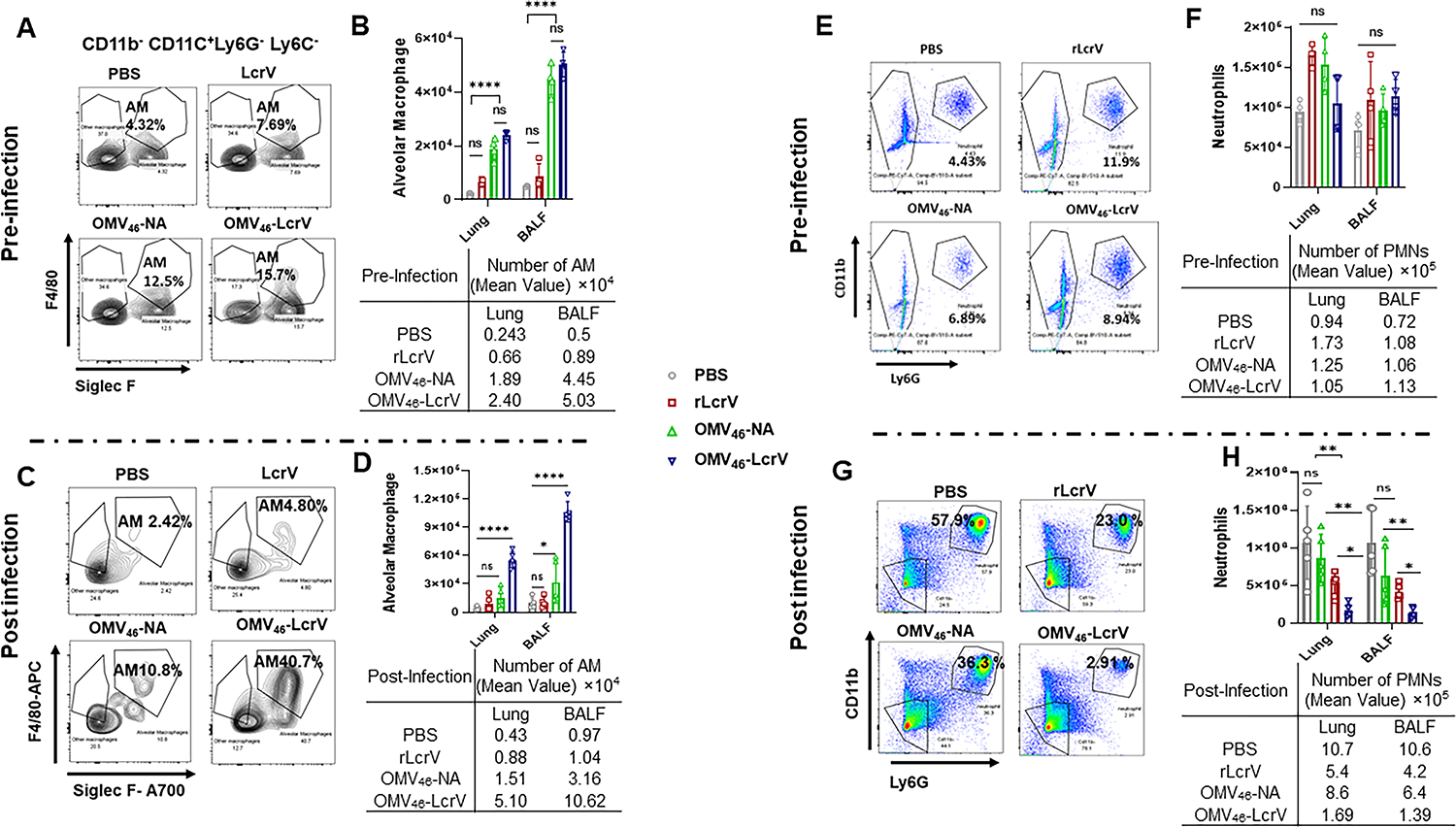
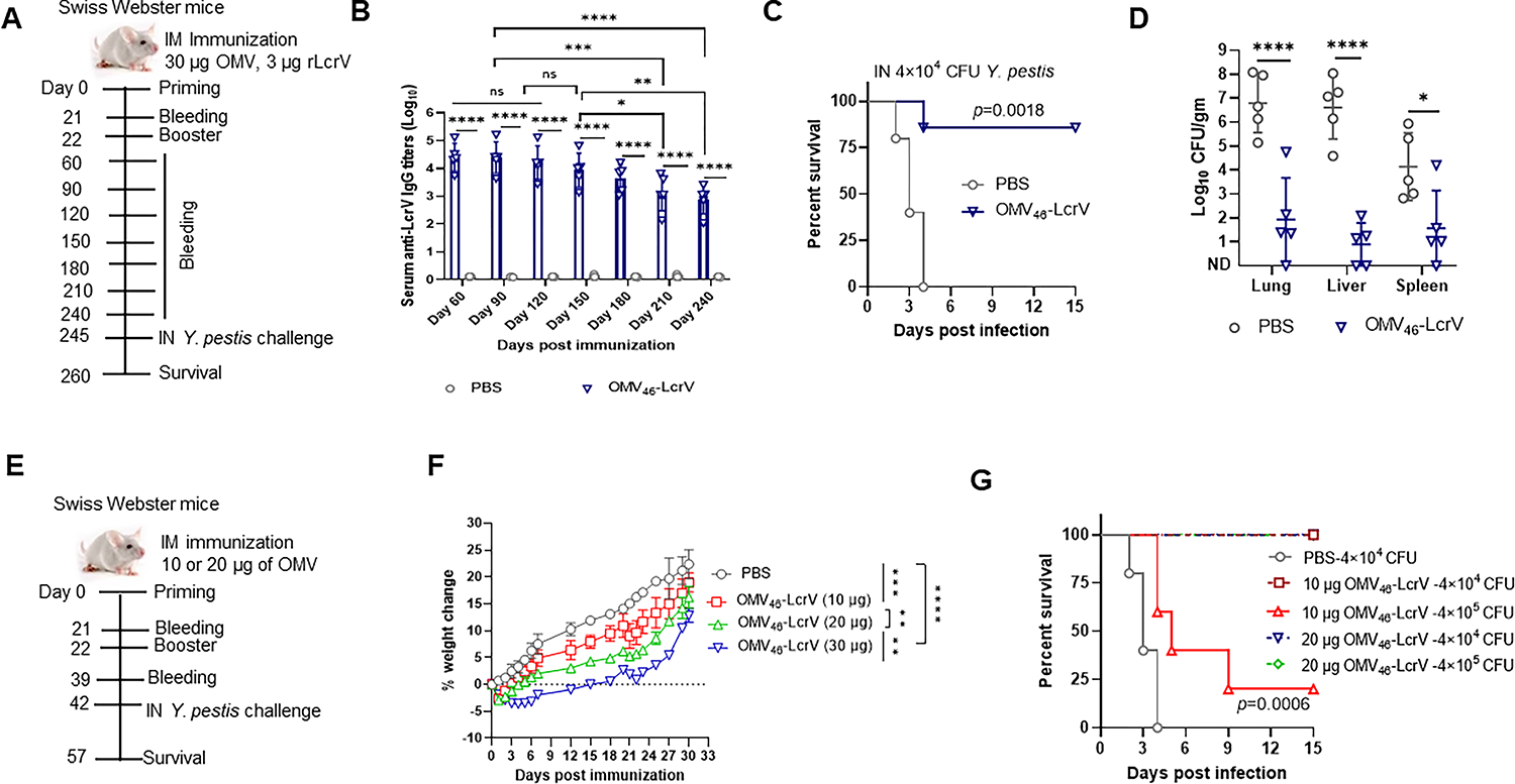
A new Yersinia pseudotuberculosis mutant strain, YptbS46, carrying the lpxE insertion and pmrF-J deletion is constructed and shown to exclusively produce monophosphoryl lipid A (MPLA) having adjuvant properties. Outer membrane vesicles (OMVs) isolated from YptbS46 harboring an lcrV expression plasmid, pSMV13, are designated OMV46-LcrV, which contained MPLA and high amounts of LcrV (Low Calcium response V) and displayed low activation of Toll-like receptor 4 (TLR4). Intramuscular prime-boost immunization with 30 µg of of OMV46-LcrV exhibited substantially reduced reactogenicity than the parent OMV44-LcrV and conferred complete protection to mice against a high-dose of respiratory Y. pestis challenge. OMV46-LcrV immunization induced robust adaptive responses in both lung mucosal and systemic compartments and orchestrated innate immunity in the lung, which are correlated with rapid bacterial clearance and unremarkable lung damage during Y. pestis challenge. Additionally, OMV46-LcrV immunization conferred long-term protection. Moreover, immunization with reduced doses of OMV46-LcrV exhibited further lower reactogenicity and still provided great protection against pneumonic plague. The studies strongly demonstrate the feasibility of OMV46-LcrV as a new type of plague vaccine candidate.

Keywords: OUTER membrane vesicles; Y. pestis; monophosphoryl lipid A; plague vaccine; protective immunity.


Conflict of interest statement

Conflict of interest: All other authors declare that they have no conflicts of interest.

Figures

Figure. 1.
Figure. 1.. Lipid A profiles of recombinant Yptb strains.
(A) Mass spectrometry analysis of lipid A species in the YptbS44 and YptbS46 strains. Shown are the negative ion electropray ionization (ESI) mass spectra of the [M-H] ions of lipid A species and their chemical structures. (B) Structure of corresponding lipid A species.
Figure. 2.
Figure. 2.. Analysis of OMVs and the amount of LcrV antigen from recombinant Yptb strains.
(A) Analysis of LcrV antigen by immunoblotting. YptbS46(pYA3493): YptbS46 harboring an empty Asd+ plasmid; YptbS46(pSMV13): YptbS46 harboring an Asd+ plasmid for lcrV expression; OMV46-NA: 6 μg of OMVs isolated from YptbS46(pYA3493); OMV46-LcrV: 6 μg of OMVs isolated from YptbS46(pSMV13); 2 μg of purified rLcrV used as a control; M: 10–250 kDa protein marker (Thermo Fischer Scientific). (B) Transmission electron microscopy (TEM) image of OMV-LcrV (left panel) and dynamic light scattering (DLS) of OMV-LcrV (right panel). (C) Comparison of embryonic alkaline phosphate (SEAP) activities in HEK-blue cells with or without mTLR4. HEK-blue mTLR4 cells were cocultured with 40 μg/mL OMVs for 8 hours. OMV44-LcrV from YptbS44(pSMV13) and rLcrV were used as controls. Data are shown as the mean ± SD. The statistical significance of differences among groups was analyzed by two-way ANOVA with Tukey’s post hoc test: ns, no significance; **** p<0.0001.
Figure 3:
Figure 3:. Protective efficacy of intramuscular immunization (IM) with OMVs against pneumonic plague.
Swiss Webster mice (n=10, equal males and females, 7 weeks old) were immunized IM with 30 μg of OMV44-LcrV, OMV46-LcrV, OMV46-NA, 3 μg of rLcrV/Alhydrogel, or Alhydrogel alone in 50 μL of PBS and then boosted on day 22 after priming. (A) Immunization schema for the animal study. (B) Weight change rates of animals post-immunization. (C) Analysis of serum cytokine levels in immunized mice. Cytokines (IL-1β and IL-6) in the sera collected on 0, 1, and 3 DPV were analyzed using corresponding ELISA kits (Invitrogen). (D) On 42 DPV, mice were intranasally challenged with 2×10⁴ CFU (200 LD50) of Y. pestis KIM6+(pCD1Ap). (E) At 2 DPI, the bacterial burden was determined in the lung, liver, and spleen. The experiments were performed twice, and the data were combined for analysis. (F) Lung histopathological analysis of representative mice from each group at 2 DPI. The lungs were microscopically examined and imaged using a Nanozoomer 2.0 RS Hamamatsu slide scanner (scale bar, 100 nm). (G) Swiss Webster mice immunized with OMV46-LcrV were intranasally challenged with 2×10⁵ CFU (2,000 LD50) and 2×10⁶ CFU (20,000 LD50) of Y. pestis KIM6+(pCD1Ap). After infection, animal morbidity and mortality were monitored for 15 days. Statistical significance was analyzed by the log-rank (Mantel-Cox) test for survival analysis. Data were analyzed and presented as the mean ± SD. The statistical significance of differences among groups was analyzed by two-way ANOVA with Tukey’s post hoc test: ns, no significance; *, P< 0.05; **, P< 0.01; ****, P< 0.0001.
Figure 4:
Figure 4:. Antibody and T-cell responses in the immunized mice.
Swiss Webster mice (n=5, equal males and females, 7 weeks old) were immunized IM with 30 μg of OMV46-LcrV, OMV46-NA, 3 μg of rLcrV/Alhydrogel or Alhydrogel alone in 50 μL of PBS and boosted on day 22 after priming. (A) Total serum IgG titers to LcrV in Swiss Webster mice at 21 and 39 DPV. (B) The ratio of IgG2a/IgG1 and IgG2b/IgG1 for antibodies specific to LcrV antigen on 21 and 39 DPV. (C) On 42 DPV, lymphocytes were aseptically isolated from the lungs and stimulated in vitro with 20 μg/mL rLcrV protein for 48 h to identify antigen-specific CD4+ and CD8+ T cells. Lung cells from sham mice were used as a control. Flow plots (left) and quantitative analysis (right) of total lung CD4+ and CD8+ T cells in the postimmunization. (D) Flow plots and (E) quantitative analysis of total CD4+ and CD8+ T cells producing IFN-γ, TNF-α, or IL-17A cytokines from the lungs. Each symbol was obtained from an individual mouse, and data are represented as the mean ± SD. The statistical significance of differences among groups was analyzed by two-way ANOVA with Tukey’s post hoc test: ns, no significance; * p<0.05; ** p<0.01; *** p<0.001; **** p<0.0001.
Figure 5:
Figure 5:. The role of adaptive immune responses in protection against pneumonic plague.
(A) Quantitative analysis of lung CD4+ and CD8+ T cells and their corresponding IFN-γ-, TNF-α-, or IL-17A-producing cells in immunized mice after pulmonary Y. pestis infection. On 42 DPV, mice were intranasally challenged with 2×10⁴ CFU (200 LD50) of Y. pestis KIM6+(pCD1Ap). At 2 DPI, lung cells were harvested from euthanized Swiss Webster mice (n=5/group females) and stained for T cells and specific cytokines (IFN-γ, TNF-α, or IL-17A) for flow cytometry. (B) Protection of serum transfer. (Left) The schema of serum transfer and (Right) naïve Swiss Webster mice (n=5/group females) were IP injected with 100 μl of sera from sham and OMV46-LcrV-immunized mice collected on 35 DPV. Twenty-four hours postinjection, recipient mice were intranasally challenged with 200 LD50 of Y. pestis KIM6+(pCD1Ap). (C) Protection of T-cell depletion. (Up) The schema of T-cell depletion and OMV46-LcrV-immunized Swiss Webster mice (n=5/group females) were IP administered either with anti-CD4, anti-CD8, or anti-CD4 plus anti-CD8 mAbs (200 μg/each mouse in 200 μl PBS) at a two-day interval for the depletion of CD4+ and/or CD8+ T cells, mice were injected with the isotype control mAbs as controls. The treated mice were IN challenged with 200 LD50 of Y. pestis KIM6+(pCD1Ap). (Down) Animal survival was monitored for 15 days. (D) Protection of B-cell depletion. (Top) Schema for B-cell depletion before OMV46-LcrV immunization. Swiss Webster mice (n=5–7/group, females) were IP administered anti-CD20 mAbs on day 2 before immunization and again treated with the same antibody on day 19 (two days before the booster at day 21) for the depletion of B cells. Mice were injected with isotype control mAbs as controls. Treated mice were IN challenged with 2.0×10⁴ CFU of Y. pestis KIM6+(pCD1Ap). (Lower left) Animal survival was monitored for 15 days. (Lower right) Bacterial burden was determined in the lung, liver, and spleen for the B-cell-depleted and isotype antibody-treated control mice at 2 DPI. The experiments were performed twice, and the data were combined for analysis. (E) Protection in B and T-cell-deficient mice against pneumonic plague. C57/BL6 and Rag1−/− mice (n=5, mixed gender) were intramuscularly immunized with 20 μg of OMV46-LcrV. Mice administered PBS were used as a negative control group. On 42 DPV, animals were intranasally challenged with 30 LD50 of Y. pestis. The mortality and morbidity of animals were monitored for 15 days. Statistical significance was analyzed by the log-rank (Mantel-Cox) test for survival analysis. Data were analyzed and presented as the mean ± SD. The statistical significance of differences among groups was analyzed by two-way ANOVA with Tukey’s post hoc test: ns, no significance; *, P< 0.05; **, P< 0.01; ****, P< 0.0001.
Figure 6:
Figure 6:. Pattern of alveolar macrophages (AMs) and neutrophils in the lung and BALF with or without infection.
Swiss Webster mice (n=5 females) were immunized with OMV46-LcrV, OMV46-NA, rLcrV, or PBS as described above. Single cells were collected from the lung and BALF at 42 DPV and 2 DPI and stained with fluorescent markers to characterize AMs and neutrophils using flow cytometry. (A) Representative flow plots showing the percentage of AMs and (B) quantitative analysis of the number of AMs in the lung (per lung) and BALF (per/mL) at 42 DPV. (C) Representative flow plot showing the percentage of AMs in the lung (per lung) and BALF (per/mL) at 2 DPI. (D) Quantitative analysis of the number of AMs in the lung (per lung) and BALF (per/mL) at 2 DPI. (E) Representative flow plot showing the percentage of neutrophils in the lung (per lung) and BALF (per/mL) at 42 DPV. (F) Quantitative analysis of the number of neutrophils in the lung (per lung) and BALF (per/mL) at 42 DPV. (G) Representative flow plot showing the percentage of neutrophils in the lung (per lung) and BALF (per/mL) at 2 DPI. (H) Quantitative analysis of the number of neutrophils in the lung (per lung) and BALF (per/mL) at 2 DPI. Data were analyzed and presented as the mean ± SD. The statistical significance of differences among groups was analyzed by two-way ANOVA with Tukey’s post hoc test: ns, no significance; *, P< 0.05; **, P< 0.01; ****, P< 0.0001.
Figure 7:
Figure 7:. Pneumonic plague protection in long-term and dose-reduced immunized mice.
Swiss Webster mice (n=10/group, equal males and females) were immunized intramuscularly with OMV46-LcrV or Alhydrogel alone in 50 μL of PBS (negative control) and then boosted on day 22 after the priming immunization as mentioned previously. (A) Schema for long-term protection in mice immunized with 30 μg of OMV46-LcrV. (B) Total anti-LcrV IgG titers in sera. Blood was collected from the immunized mice at 60, 90, 120, 150, 180, 210, and 240 DPV. (C) On 245 DPV, the immunized mice were intranasally challenged with 4.0×10⁴ CFU (400 LD50) of Y. pestis KIM6+(pCD1Ap), and survival was monitored for 15 days. (D) At 2 DPI (i.e., 247 DPV), the bacterial burden was determined in the lung, liver, and spleen. (E) Schema for evaluation of dose-reduction immunization with 20 or 10 μg of OMV46-LcrV. (F) Weight change rates of immunized mice. (G) At 42 DPV, the immunized mice were challenged with 400 LD50 and 4,000 LD50 of Y. pestis KIM6+(pCD1Ap). Survival was monitored for 15 days. Statistical significance was analyzed by the log-rank (Mantel-Cox) test for survival analysis. Data were analyzed and presented as the mean ± SD. The statistical significance of differences among groups was analyzed by two-way ANOVA with Tukey’s post hoc test: ns, no significance; *, P< 0.05; **, P< 0.01; ****, P< 0.0001.

Update of

Similar articles

Cited by

References

    1. Barbieri R; Signoli M; Cheve D; Costedoat C; Tzortzis S; Aboudharam G; Raoult D; Drancourt M, Clin Microbiol Rev 2020, 34 (1). DOI 10.1128/CMR.00044-19. - DOI - PMC - PubMed
    1. Hinnebusch BJ, Plague in the 21st Century: Global Public Health Challenges and Goals. Georgiev VS, Ed. Humana Press, Totowa, NJ: 2010.
    1. Riedel S, Proc (Bayl Univ Med Cent) 2005, 18 (2), 116–24. DOI 10.1080/08998280.2005.11928049. - DOI - PMC - PubMed
    1. Rosenzweig JA; Hendrix EK; Chopra AK, Appl Microbiol Biotechnol 2021, 105 (12), 4931–4941. DOI 10.1007/s00253-021-11389-6. - DOI - PMC - PubMed
    1. Perry RD; Fetherston JD, Clin Microbiol Rev 1997, 10 (1), 35–66. - PMC - PubMed

Publication types

LinkOut - more resources