Review
. 2024 Feb;46(2):e2300150.
doi: 10.1002/bies.202300150. Epub 2023 Nov 27.

Origin and development of primary animal epithelia

Affiliations
Review

Origin and development of primary animal epithelia

Sophia Doerr et al. Bioessays. 2024 Feb.

Abstract

Epithelia are the first organized tissues that appear during development. In many animal embryos, early divisions give rise to a polarized monolayer, the primary epithelium, rather than a random aggregate of cells. Here, we review the mechanisms by which cells organize into primary epithelia in various developmental contexts. We discuss how cells acquire polarity while undergoing early divisions. We describe cases where oriented divisions constrain cell arrangement to monolayers including organization on top of yolk surfaces. We finally discuss how epithelia emerge in embryos from animals that branched early during evolution and provide examples of epithelia-like arrangements encountered in single-celled eukaryotes. Although divergent and context-dependent mechanisms give rise to primary epithelia, here we trace the unifying principles underlying their formation.

Keywords: blastocoel; epithelialization; lumen; monolayer; polarity; self-organization.


Conflict of interest statement

CONFLICT OF INTEREST STATEMENT

The authors declare no conflicts of interest.

Figures
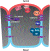
FIGURE 1
FIGURE 1
Conserved features of a typical animal epithelial cell. The apical cortical domain (red) is enriched with proteins that mutually antagonize proteins of the basolateral cortical domain (blue). Intercellular adhesion is mediated by homophilic E-Cadherins across the lateral domain. The Cadherin-Catenin complex directly interacts with the actin cytoskeleton. Additional adhesion complexes, not shown, are occluding junctions in the lateral domain between cells, and integrins that connect the basal plasma membrane to the extracellular matrix. Detailed reviews on the molecular characterization of epithelial cells and insights into the evolutionary origin of epithelial proteins have been previously published.[2,4,6,175]
FIGURE 2
FIGURE 2
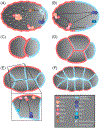
Apical-basal polarity establishment in the C. elegans embryo. (A) Anterior-posterior polarity is established upon fertilization of the oocyte. Sperm-associated microtubules establish the posterior domain which is enriched with the Par-2/Par-1 complex. Posterior inhibition of Ect-2 and RhoA triggers actomyosin flows that mediate the movement of Par-3/Par-6/aPKC to the anterior cortex. (B) Maintenance of anterior-posterior polarity. Chin-1 inhibits the posterior cortical binding of Cdc-42, and Par-1 inhibits Par-3. The Par-6/aPKC complex cycles between an inactive complex with Par-3 and a complex with Cdc-42, in which aPKC inhibits the cortical binding of posterior Par proteins. (C) An asymmetric cell division leads to the 2-cell stage. (D) During the early 4-cell stage and before the polarity shift discussed in text, Par-2 is enriched at all cell contacts and around the cortex of the germline precursor cell (blue). Par-3 is found uniformly on the cortices of the three somatic cells (red). (E) By the 8-cell-stage, cell-contact-enriched Pac-1 inhibits Cdc-42, and aPKC inhibits Par-1/−2, resulting in distinct apical-basal polarity. (F) During subsequent cell divisions, the basal surfaces of cells separate, forming a small blastocoel cavity which reaches its greatest volume by the 26-cell stage. Left is anterior and right is posterior.
FIGURE 3
FIGURE 3
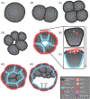
Lumen formation after polarity establishment in the mouse embryo. (A-D) Cells are round and unpolarized up to the early 8-cell stage. (E-F) During the 8-cell stage, compaction occurs, and apical-basal polarity is established. (E) RhoA is activated by the Phospholipase C - PtdIns (4,5) P2 - PKC pathway and drives actomyosin organization at distinct apical cups in the contact-free cortical domains. (F) Par-3 localizes at the apical domain along with Cdc-42, which recruits Par-6 and aPKC. Once Par-3, Par-6 and aPKC proteins localize at the apical cap, they exclude actomyosin. This leads to formation of an actin ring encompassing the Par-3/Par-6/aPKC domain. Basolateral cell contacts are enriched with E-Cadherin, Par-1, Scribble, and Lgl-1. (G) In the 16-cell embryo, apical actin rings expand toward cell contacts. Apical proteins Par-3, Par-6, and aPKC expand toward newly forming tight junctions at the apical-lateral domain of the outer cells (only tight junctions are shown). The pluripotent inner cell mass becomes apparent. (H) During the 32-cell stage, the lumen begins to form. Mature tight junctions seal the epithelium and basal ATPase ion pumps generate a gradient to osmotically draw water into the blastocoel cavity.
FIGURE 4
FIGURE 4
Xenopus embryonic cells polarize during the first cleavage. (A) The fertilized egg exhibits uniform cortical localization of aPKC prior to cleavage. (B) During cleavage, the new membrane is enriched with basolateral proteins Lgl and Par-1. The contact-free cortical surfaces are inherited from the egg cortex and are enriched with aPKC and Par-6. Antagonistic relationships between Lgl and aPKC keep cortical domains separate. Tight junctions begin to assemble during the first cleavage. (C) A small, expanding blastocoel appears. (D) The 32-cell stage embryo is a polarized monolayer.
FIGURE 5
FIGURE 5
Cell-cycle coupled epithelial organization in the Nematostella vectensis embryo. (A) Par-6 is enriched in the cortical domain and microvilli are apparent on the surface of fertilized oocytes suggesting early organization of the apical domain. (B) In the 4-cell embryo, Par-6 is restricted to the apical cortex. (C) By the 16-cell stage cells organize into a monolayer with distinct apical and basal adherens junctions. In the compacted embryo, polarity proteins Par-6 and aPKC occupy the apical domain, Par-3 is enriched at the apical junctions together with Cadherin3 and the α- and β-Catenins, while Lgl and Par-1 occupy the basolateral domain. Cadherin3 Is also present at the basal junctions. (D) During synchronous cell divisions, the embryo loses its compact shape, and some polarity proteins (Par-6 and Par-3) are no longer enriched in the apical domain and the apical junctions. The basolateral protein Lgl and the apical junctional proteins Cadherin3 and α-Catenin remain polarized in dividing cells (only Lgl is shown).
FIGURE 6
FIGURE 6
Gradual epithelialization from cell clusters in the hydrozoan Dynamena pumila embryo. (A) Cells in early embryos orient their mitotic spindles (white arrows) to give rise to external and internal cells after division. Cells may also shift toward the interior (magenta arrow). Both lead to an embryo of a solid cell mass with no obvious internal cavity. (B) Epithelialization gradually expands when several epithelial sheets (artificially colored in green, magenta, and cyan) begin to merge. Non-epithelialized cells (red) are still present. (C) Nuclei localize at the apical domain in epithelial cells (artificially colored in magenta and green). Yellow arrowhead points to where two independently epithelializing sheets meet. Reproduced with permission.[106]
FIGURE 7
FIGURE 7
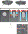
Cell contact-independent epithelialization in the Drosophila embryo. (A) After fertilization, nuclei undergo rapid divisions in the same cytoplasm giving rise to a syncytium. During the 8th and 9th divisions, nuclei migrate toward the cortex. (B) Interphase of the 14th cycle marks the onset of cellularization and the development of four distinguishable cortical domains. Insertion of new membrane starts from the embryo surface (apical), then apical-lateral, such that old membrane is transferred basally. The apical domain develops above the nucleus and the lateral domains attract basolateral and adherens junction proteins. (C) An epithelial monolayer emerges once the ingressing membrane fully envelops each nucleus and cells separate from the underlying yolk. Activation of RhoA triggers actomyosin contractility which is necessary to stabilize the basal furrow and to trigger the contractions that will drive basal closure. Lgl, Dlg-1, Scribble, and Par-1 are recruited to the lateral domain. The subapical domain is defined by the localization of Rap-1, Afadin, Par-3 and the E-Cadherin-Catenin complex at the adherens junctions. The apical domain is populated by Cdc-42, Par-6, and aPKC. Lateral Par-1 excludes subapical Par-3, lateral Lgl antagonizes apical Par-6, and apical aPKC excludes Lgl.
FIGURE 8
FIGURE 8
Monolayer emergence on the yolk surface of the chicken embryo. (A) The first cell cleavage is incomplete. (B) After a series of complete cell cleavages in the center, the five to six cell-thick layer is separated from the yolk by a fluid-filled cavity. (C) The thick layer thins out to the upper 1-cell-thick epiblast. Groups of cells ingress from the upper layer and fuse with cells from the periphery to form the lower epithelium of the hypoblast. Variations of hypoblast emergence exist across animals. Redrawn from[136]
FIGURE 9
FIGURE 9
Layer inversion and cell egression modes of epithelia formation in sponges. (A-F) Inversion gives rise to a flagellated monolayer in embryos of the calcarean sponge Sycon ciliatum. (A-C) The oocyte is fertilized and undergoes cleavages between the outer pinacocyte and the inner choanocyte epithelia of the parent. (D) During the 8-cell stage, cells are arranged into a hollow monolayered sphere with flagella pointing toward the interior. (E-F) Exposure of the flagella on the exterior occurs during the inversion of the monolayer as it moves from one maternal compartment into the neighboring lumen. Redrawn with modification from[153] (G-I) Egression of internal cells transforms a solid embryo into a flagellated monolayer in Oscarella. Enclosed within the blastula are bacterial symbionts. Redrawn with modification from.[152]
FIGURE 10
FIGURE 10
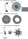
Transient epithelia-like structures formed by unicellular eukaryotes. (A) Aggregation of Dictyostelium discoideum cells into a fruiting body. A polarized monolayer develops at the tip of the fruiting body which contains the spores. Conserved α- and β-Catenins localize at the lateral cell contacts (blue) and recruit IQGAP1-Cortexillin which block myosin II and restrict its localization to the apical domain (red). (B) Cellularization of the Ichthyosporean Sphaeroforma arctica. Sequential nuclear divisions give rise to a syncytium. Contractile actomyosin drives synchronous plasma membrane invaginations around individual peripheral nuclei and the emergence of a transient polarized monolayer before cell dispersal. (C) Dividing cells of the choanoflagellate Salpingoeca rosetta remain attached and organize into a polarized rosette colony. Intercellular bridges keep cells connected. Additionally, extracellular matrix is secreted basally (blue) and facilitates colony organization.

Similar articles

Cited by

References

    1. Brusca GJ, Brusca RC, & Gilbert SF (1997). Characteristics of metazoan development. In: (Gilbert AM, S.F. and Raunio Ed.), Embryology: Constructing the Organism (pp. 3–19).
    1. Rodriguez-Boulan E, & Macara IG (2014). Organization and execution of the epithelial polarity programme. Nature Reviews Molecular Cell Biology, 15(4), 225–242. 10.1038/nrm3775 - DOI - PMC - PubMed
    1. Hay ED (2005). The mesenchymal cell, its role in the embryo, and the remarkable signaling mechanisms that create it. Developmental Dynamics, 233(3), 706–720. 10.1002/dvdy.20345 - DOI - PubMed
    1. Buckley CE, & St Johnston D (2022). Apical–basal polarity and the control of epithelial form and function. Nature Reviews Molecular Cell Biology, 23(8), 559–577. 10.1038/s41580-022-00465-y - DOI - PubMed
    1. Leys SP, & Riesgo A (2012). Epithelia, an evolutionary novelty of Metazoans. Journal of Experimental Zoology Part B: Molecular and Developmental Evolution, 318(6), 438–447. 10.1002/jez.b.21442 - DOI - PubMed

Publication types

LinkOut - more resources