. 2024 May;20(5):1098-1113.
doi: 10.1080/15548627.2023.2287925. Epub 2023 Dec 7.

TRIM22 facilitates autophagosome-lysosome fusion by mediating the association of GABARAPs and PLEKHM1

Affiliations

TRIM22 facilitates autophagosome-lysosome fusion by mediating the association of GABARAPs and PLEKHM1

Hansol Heo et al. Autophagy. 2024 May.

Abstract

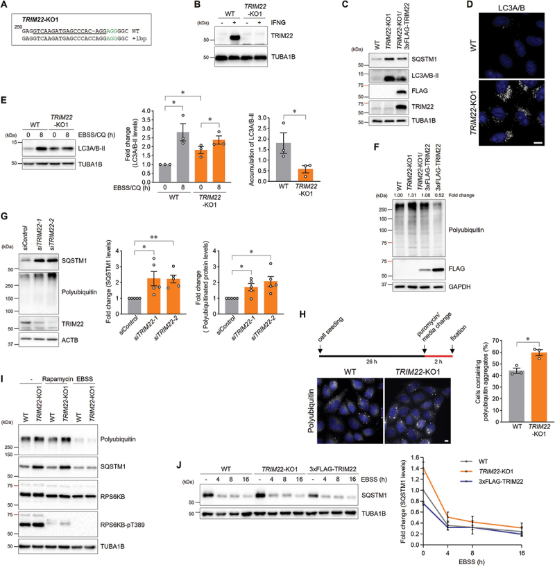
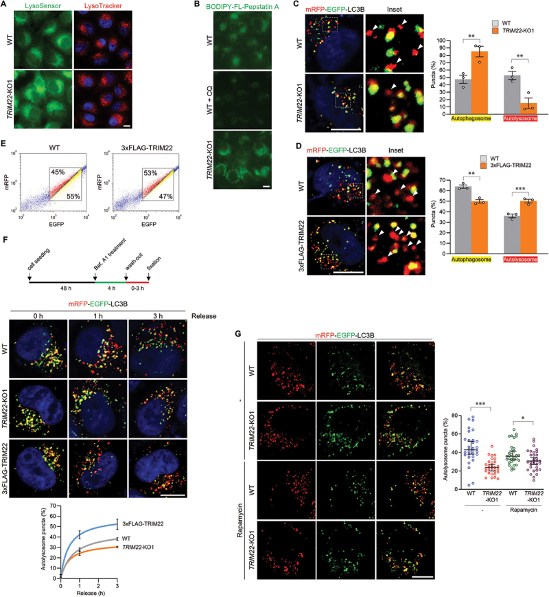
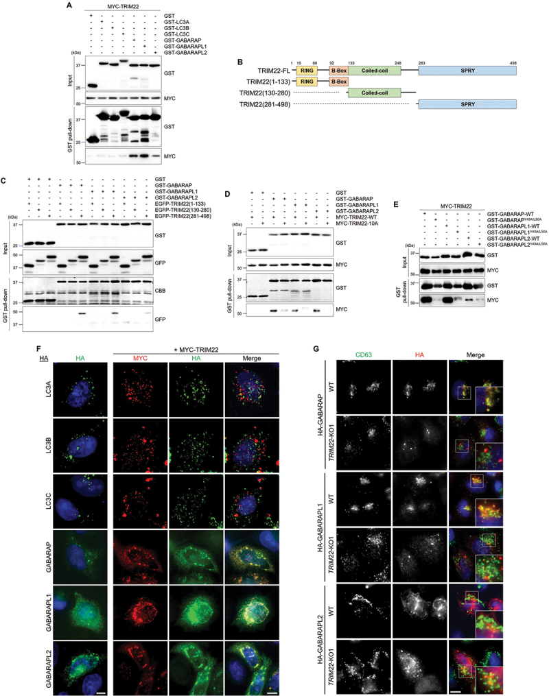
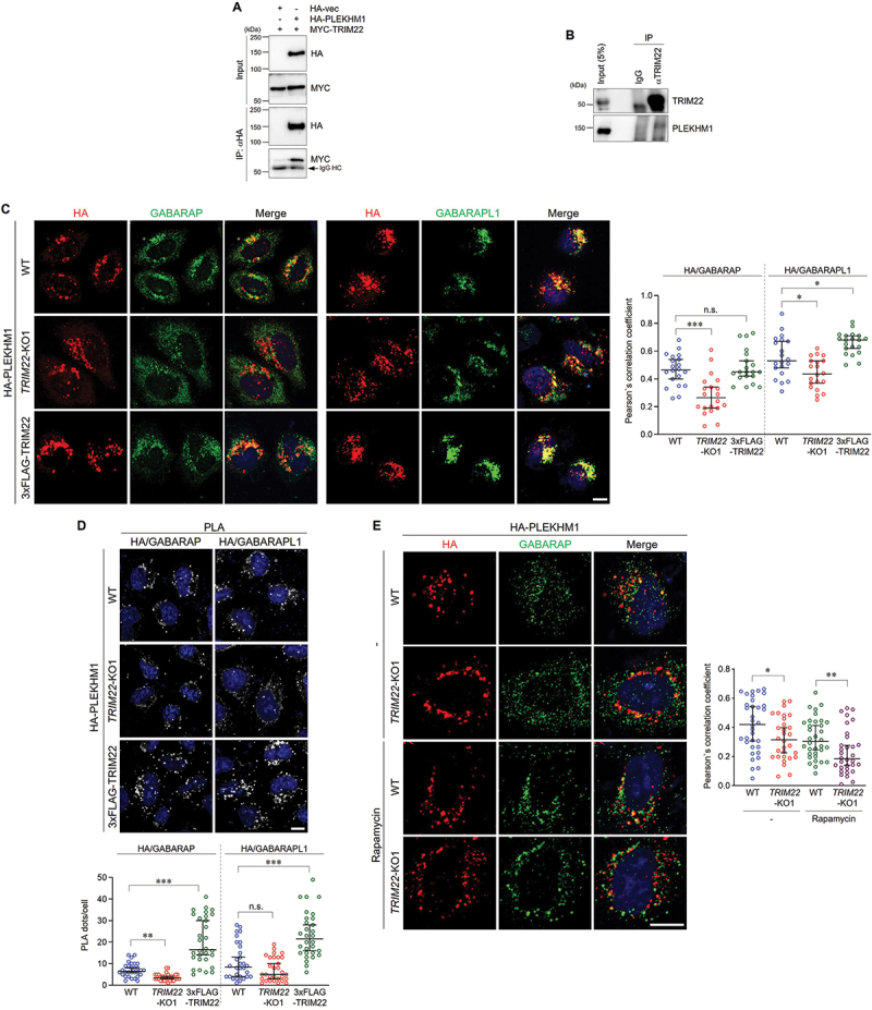
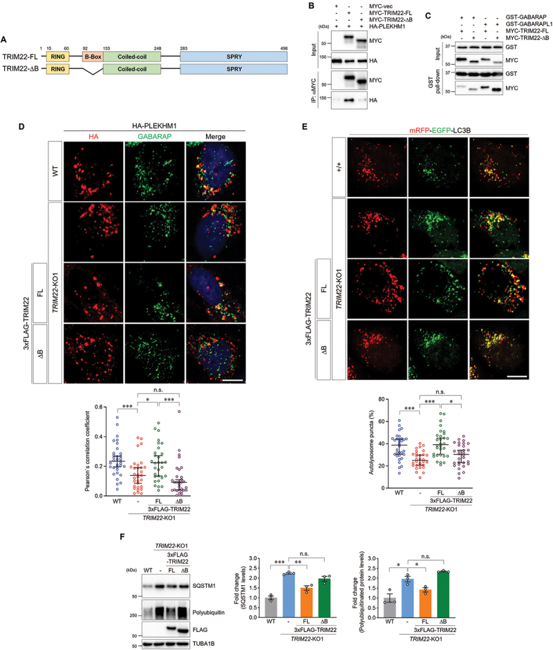
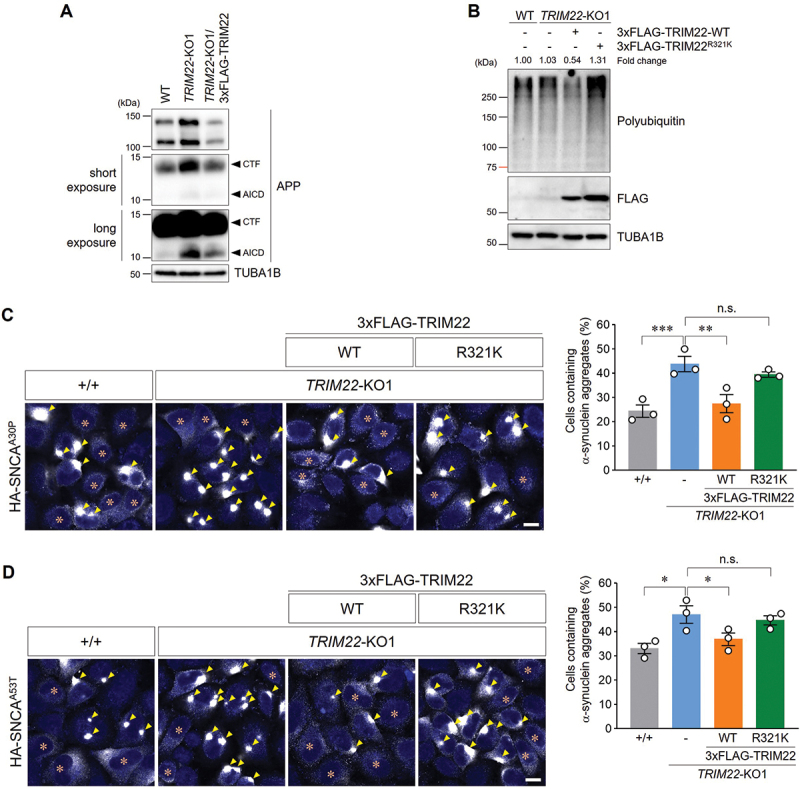
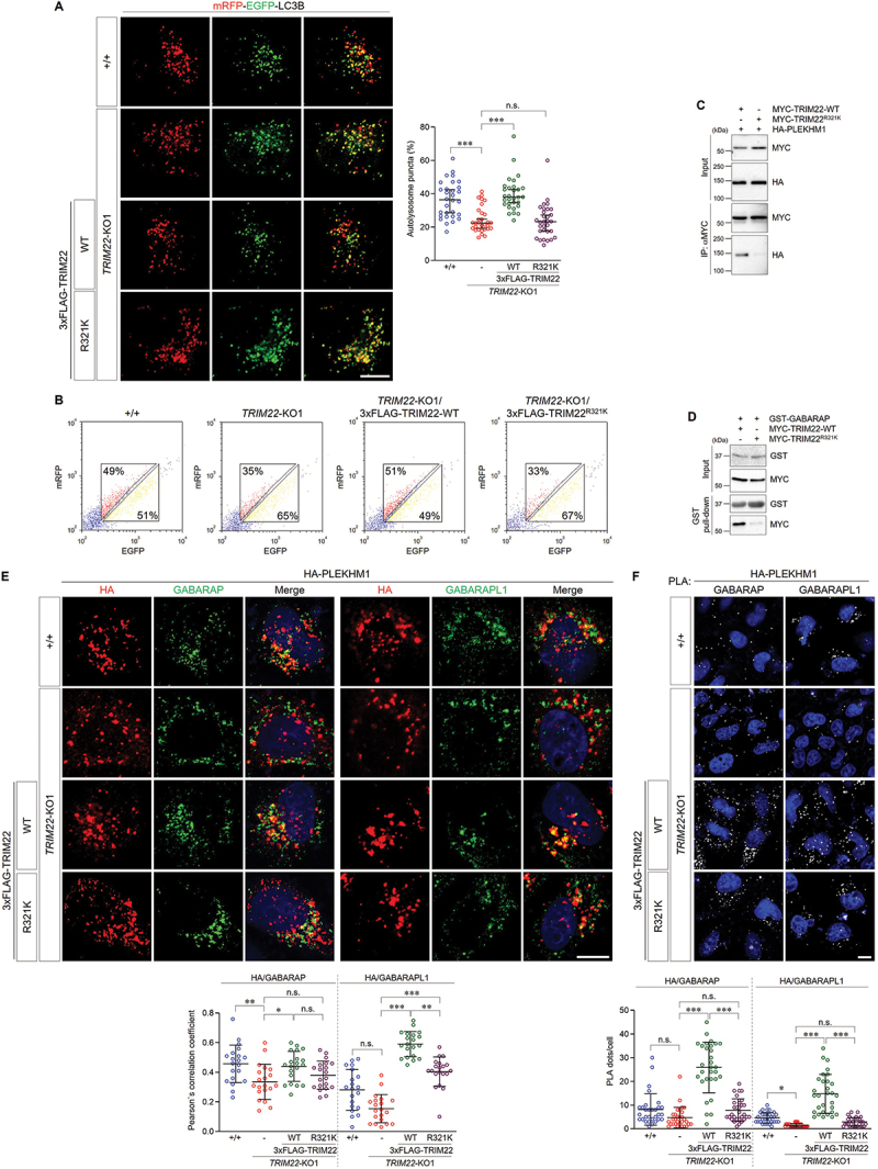
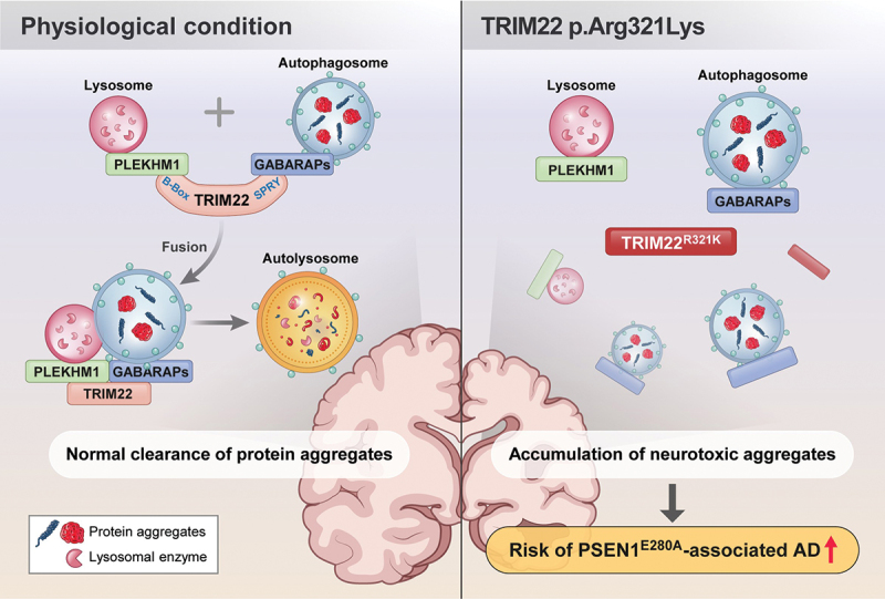
Tripartite motif (TRIM) proteins are a large family of E3 ubiquitin ligases implicated in antiviral defense systems, tumorigenesis, and protein quality control. TRIM proteins contribute to protein quality control by regulating the ubiquitin-proteasome system, endoplasmic reticulum-associated degradation, and macroautophagy/autophagy. However, the detailed mechanisms through which various TRIM proteins regulate downstream events have not yet been fully elucidated. Herein, we identified a novel function of TRIM22 in the regulation of autophagy. TRIM22 promotes autophagosome-lysosome fusion by mediating the association of GABARAP family proteins with PLEKHM1, thereby inducing the autophagic clearance of protein aggregates, independent of its E3 ubiquitin ligase activity. Furthermore, a TRIM22 variant associated with early-onset familial Alzheimer disease interferes with autophagosome-lysosome fusion and autophagic clearance. These findings suggest TRIM22 as a critical autophagic regulator that orchestrates autophagosome-lysosome fusion by scaffolding autophagy-related proteins, thus representing a potential therapeutic target in neurodegenerative diseases.Abbreviations: AD: Alzheimer disease; ADAOO: AD age of onset; AICD: APP intracellular domain; APP: amyloid beta precursor protein; BSA: bovine serum albumin; cDNAs: complementary DNAs; CQ: chloroquine; CTF: carboxyl-terminal fragment; EBSS: Earle's balanced salt solution; GABARAP: GABA type A receptor-associated protein; GST: glutathione S-transferase; HA: hemagglutinin; HOPS: homotypic fusion and protein sorting; IFN: interferon; IL1A/IL-1α: interleukin 1 alpha; KO: knockout; MTORC1: mechanistic target of rapamycin kinase complex 1; NFKBIA/IκBα: NFKB inhibitor alpha; NFE2L2/NRF2: NFE2 like bZIP transcription factor; PBS: phosphate-buffered saline; PI3K: class I phosphoinositide 3-kinase; PLA: proximity ligation assay; PLEKHM1: pleckstrin homology and RUN domain containing M1; PSEN1: presenilin 1; SEM: standard errors of the means; SNAREs: soluble N-ethylmaleimide-sensitive factor attachment protein receptors; SNCA: synuclein alpha; SNP: single nucleotide polymorphism; TBS: tris-buffered saline; TNF/TNF-α: tumor necrosis factor; TRIM: tripartite motif; ULK1: unc-51 like autophagy activating kinase 1; WT: wild-type.

Keywords: Alzheimer disease; PLEKHM1; TRIM22; autophagosome-lysosome fusion; autophagy.


Conflict of interest statement

No potential conflict of interest was reported by the author(s).

Figures

Figure 1.
Figure 1.
TRIM22 deficiency interferes with intracellular clearance. (A,B) The generation of a TRIM22-knockout cell line was validated via genomic DNA sequencing (A) and immunoblotting (B). (A) Sequence alignment of the allele from wild-type HeLa cells and TRIM22-KO1 cells identified through sequencing. Nucleotide numbers are expressed relative to the initiation codon. Sequences corresponding to the 19-nt sgRNA target are underlined and the 3-nt PAM is highlighted in green. (B) Wild-type HeLa cells or TRIM22-KO1 cells were grown in the absence or presence of 50 ng/mL of IFNG for 24 h and then analyzed via immunoblotting with the indicated antibodies. (C) Wild-type HeLa cells, TRIM22-KO1 cells, or TRIM22-KO1 cells stably expressing 3×FLAG-TRIM22 cells were subjected to immunoblotting with the indicated antibodies. (D) Wild-type HeLa cells or TRIM22-KO1 cells were immunostained for LC3A/B. (E) Wild-type HeLa cells or TRIM22-KO1 cells were incubated with EBSS containing 20 μM of CQ for 8 h and then analyzed via immunoblotting with the indicated antibodies. Band intensities for LC3A/B-II were measured and normalized to the mean intensity of the untreated wild-type cell group. The accumulation of LC3A/B-II was calculated by subtracting the intensity of the untreated group from that of the EBSS/CQ-treated group. Graphs show means ± SEM of three independent experiments. (F) Wild-type HeLa cells, TRIM22-KO1 cells, TRIM22-KO1 cells stably expressing 3×FLAG-TRIM22 cells, or 3×FLAG-TRIM22-expressing cells were subjected to immunoblotting with the indicated antibodies. Band intensities of polyubiquitinated proteins were measured and normalized to the intensity of wild-type cells. (G) hTERT-RPE1 cells were transfected with either control or TRIM22 siRNAs, and subsequently subjected to immunoblotting with the indicated antibodies. Band intensities of SQSTM1 and polyubiquitinated proteins were measured and normalized to the mean intensity of siControl-transfected cells. Graphs show means ± SEM of five independent experiments. (H) Wild-type HeLa cells or TRIM22-KO1 cells were treated with 5 μg/mL of puromycin for 2 h and immunostained with polyubiquitin-specific antibodies. Cells containing more than five polyubiquitin aggregates were quantified from more than 200 cells per group. Graphs show means ± SEM of three independent experiments. (I) Wild-type HeLa cells or TRIM22-KO1 cells were incubated with either 100 nM of rapamycin or with EBSS for 8 h. Cell lysates were subjected to immunoblotting with indicated antibodies. (J) Wild-type HeLa cells, TRIM22-KO1 cells, or 3×FLAG-TRIM22-expressing cells were incubated with EBSS for the indicated times, and cell lysates were subjected to immunoblotting with the indicated antibodies. Band intensities of SQSTM1 were measured and normalized to the mean intensity of untreated wild-type cell group. Graphs show means ± SEM of three independent experiments. *p < 0.05; **p < 0.01. Scale bars: 10 μm.
Figure 2.
Figure 2.
TRIM22 positively regulates autophagosome-lysosome fusion. (A) Wild-type HeLa cells or TRIM22-KO1 cells were treated with LysoSensor green DND-189 or LysoTracker red DND-99 for 30 min before fixation. (B) Wild-type HeLa cells or TRIM22-KO1 cells were treated with BODIPY-FL-Pepstatin A for 30 min. As a control, wild-type HeLa cells were cultured with CQ for 4 h before BODIPY-FL-Pepstatin A treatment. (C) mRFP-EGFP-LC3B was stably expressed in wild-type HeLa cells or TRIM22-KO1 cells, and fluorescent LC3B puncta were then analyzed in more than 30 cells per group. Puncta with mRFP+/EGFP+ indicate autophagosomes, and puncta with mRFP+/EGFP (arrowheads) indicate autolysosomes. Graphs show means ± SEM of three independent experiments. (D,E) mRFP-EGFP-LC3B was stably expressed in wild-type HeLa cells or cells expressing 3×FLAG-TRIM22. (D) Fluorescent LC3B puncta were analyzed in more than 30 cells per group. Arrowheads indicate autolysosomes. Graphs show means ± SEM of three independent experiments. (E) Cells were analyzed using flow cytometry. The intensities of mRFP and EGFP signals from more than 10,000 cells were analyzed, and the percentage of cells in corresponding gates is shown. (F) mRFP-EGFP-LC3B was stably expressed in wild-type HeLa cells, TRIM22-KO1 cells, or 3×FLAG-TRIM22-expressing cells. The cells were treated with bafilomycin A1 (40 nM), followed by washing out and fixation as indicated, and the ratio of autolysosomes was then analyzed. Graphs show means ± SEM of more than 20 cells per group. (G) mRFP-EGFP-LC3B was stably expressed in wild-type HeLa cells or TRIM22-KO1 cells. The cells were treated with 100 nM of rapamycin for 8 h, and then the cells with fluorescent LC3B puncta were analyzed. Graphs show median with 95% confidence interval of more than 30 cells per group. *p < 0.05; **p < 0.01; *** p < 0.001. Scale bars: 10 μm.
Figure 3.
Figure 3.
TRIM22 interacts with GABARAPs and mediates their lysosomal localization. (A) HEK293T cells were transfected with MYC-TRIM22, and lysates were incubated with the indicated GST-tagged proteins and precipitated with glutathione sepharose 4B beads. Precipitates were then immunoblotted as shown. (B) Schematic domain structure of TRIM22 and its deletion mutants. (C–E) HEK293T cells were transfected as indicated, and lysates were incubated with the indicated GST-tagged proteins and precipitated with glutathione sepharose 4B beads. Precipitates were then immunoblotted as shown. (F) HeLa cells were transfected with HA-tagged LC3A, LC3B, LC3C, GABARAP, GABARAPL1, or GABARAPL2 along with MYC-TRIM22. The cells were immunostained for HA (green) and MYC (red). (G) Wild-type HeLa cells or TRIM22-KO1 cells were transfected as indicated, and the cells were immunostained for CD63 (green) and HA (red). Scale bars: 10 μm.
Figure 4.
Figure 4.
TRIM22 positively regulates autophagosome-lysosome fusion by mediating the association of GABARAPs with PLEKHM1. (A) HEK293T cells were transfected as indicated, and lysates were immunoprecipitated with antibodies against HA. Precipitates were then immunoblotted as shown. An arrow denotes IgG heavy chain. (B) HeLa cells were treated with 50 ng/mL of IFNG for 24 h, followed by starvation with EBSS for 2 h. Lysates were immunoprecipitated with rabbit IgG or antibodies against TRIM22. Precipitates were then immunoblotted as shown. (C,D) Wild-type HeLa cells, TRIM22-KO1 cells, or 3×FLAG-TRIM22-expressing cells were transfected with HA-PLEKHM1. (C) The cells were immunostained for HA (red) and GABARAP (left, green) or GABARAPL1 (right, green). Colocalization of HA-PLEKHM1 with GABARAP or GABARAPL1 was analyzed by calculation of Pearson’s correlation coefficients. Graphs show median with 95% confidence interval of more than 20 cells per group. (D) The cells were subjected to PLA with anti-HA antibodies along with antibodies against GABARAP (left) or GABARAPL1 (right). The number of PLA dots was quantified. Graphs show median with 95% confidence interval of more than 30 cells per group. (E) Wild-type HeLa cells or TRIM22-KO1 cells were transfected with HA-PLEKHM1 and immunostained for HA (red) and GABARAP (green). Colocalization of HA-PLEKHM1 with GABARAP was analyzed by calculation of Pearson’s correlation coefficients. Graphs show median with 95% confidence interval of more than 30 cells per group. *p < 0.05; **p < 0.01; ***p < 0.001; n.s., not significant. Scale bars: 10 μm.
Figure 5.
Figure 5.
TRIM22 facilitates autophagosome-lysosome fusion by interacting with PLEKHM1. (A) Schematic domain structure of TRIM22 and the B-box deletion mutant. (B) HEK293T cells were transfected as indicated, and lysates were immunoprecipitated with antibodies against MYC. Precipitates were then immunoblotted as shown. (C) HEK293T cells were transfected with MYC-TRIM22-FL or ΔB, and lysates were incubated with the indicated GST-tagged proteins and precipitated with glutathione sepharose 4B beads. Precipitates were then immunoblotted as shown. (D) Wild-type HeLa cells, TRIM22-KO1 cells, and TRIM22-KO1 cells expressing 3×FLAG-TRIM22-FL or ΔB were transfected with HA-PLEKHM1. The cells were immunostained as indicated. Colocalization of HA-PLEKHM1 with GABARAP was analyzed by calculation of Pearson’s correlation coefficient. Graphs show median with 95% confidence interval of more than 30 cells per group. (E) mRFP-EGFP-LC3B was stably expressed in wild-type HeLa cells, TRIM22-KO1 cells, and TRIM22-KO1 cells expressing 3×FLAG-TRIM22-FL or ΔB. The cells with fluorescent LC3B puncta were analyzed. mRFP+/EGFP puncta were counted as autolysosomes. Graphs show median with 95% confidence interval of more than 30 cells per group. (F) Wild-type HeLa cells, TRIM22-KO1 cells, and TRIM22-KO1 cells expressing 3×FLAG-TRIM22-FL or ΔB were subjected to immunoblotting with the indicated antibodies. Band intensities of SQSTM1 or polyubiquitinated proteins were measured and normalized to the mean intensity of wild-type cells. Graphs show means ± SEM of three independent experiments. FL, full-length. *p < 0.05; **p < 0.01; ***p < 0.001; n.s., not significant. Scale bars: 10 μm.
Figure 6.
Figure 6.
An AD-related TRIM22 variant interferes with intracellular clearance. (A) Wild-type HeLa cells, TRIM22-KO1 cells, or TRIM22-KO1 cells stably expressing 3×FLAG-TRIM22 were subjected to immunoblotting with the indicated antibodies. Arrowheads denote the CTF of APP or the AICD. (B) Wild-type HeLa cells, TRIM22-KO1 cells, or TRIM22-KO1 cells stably expressing 3×FLAG-TRIM22-WT or R321K were subjected to immunoblotting with the indicated antibodies. Band intensities for polyubiquitinated proteins were measured and normalized to the intensity of wild-type cells. (C,D) Wild-type HeLa cells, TRIM22-KO1 cells, and TRIM22-KO1 cells expressing 3×FLAG-TRIM22-WT or R321K were transfected with HA-SNCAA30P (C) or HA-SNCAA53T (D). Cells were immunostained for HA, and cells containing HA-SNCA aggregates were then analyzed as described (more than 500 cells per group). Arrowheads indicate aggregated α-synuclein, and asterisks indicate the cells with no aggregates. Graphs show means ± SEM of three independent experiments. Representative images are shown. *p < 0.05; **p < 0.01; ***p < 0.001; n.s., not significant. Scale bars: 10 μm.
Figure 7.
Figure 7.
The R321K variant inhibits the role of TRIM22 in autophagosome-lysosome fusion. (A,B) mRFP-EGFP-LC3B was stably expressed in wild-type HeLa cells, TRIM22-KO1 cells, and TRIM22-KO1 cells expressing 3×FLAG-TRIM22-WT or R321K. (A) The cells with fluorescent LC3B puncta were analyzed. Puncta with mRFP+/EGFP+ indicate autophagosomes, and puncta with mRFP+/EGFP indicate autolysosomes. Graphs show median with 95% confidence interval of more than 30 cells per group. (B) Cells were analyzed using flow cytometry. The intensities of mRFP and EGFP signals from more than 2,000 cells were analyzed, and the percentages of cells in corresponding gates are indicated. (C) HEK293T cells were transfected as indicated, and lysates were immunoprecipitated with antibodies against MYC. Precipitates were then immunoblotted as shown. (D) HEK293T cells were transfected as indicated, and lysates were incubated with GST-GABARAP proteins and precipitated with glutathione sepharose 4B beads. Precipitates were then immunoblotted as shown. (E,F) Wild-type HeLa cells, TRIM22-KO1 cells, and TRIM22-KO1 cells expressing 3×FLAG-TRIM22-WT or R321K were transfected with HA-PLEKHM1. (E) The cells were immunostained as indicated. Colocalization of HA-PLEKHM1 with GABARAP (left) or GABARAPL1 (right) was analyzed via calculation of Pearson’s correlation coefficient. Graphs show median with 95% confidence interval of more than 20 cells per group. (F) Cells were subjected to PLA with anti-HA antibodies along with antibodies against GABARAP (left) or GABARAPL1 (right). The number of PLA dots was quantified. Graphs show median with 95% confidence interval of more than 30 cells per group. *p < 0.05; **p < 0.01; *** p < 0.001; n.s., not significant. Scale bars: 10 μm.
Figure 8.
Figure 8.
Roles of TRIM22 in intracellular clearance. Under physiological conditions, TRIM22 facilitates the association of GABARAPs and PLEKHM1, thereby inducing autophagosome-lysosome fusion. The autolysosomes effectively remove protein aggregates. However, the R321K SNP in TRIM22 fails to mediate the association between GABARAPs and PLEKHM1. As a result, clearance of neurotoxic aggregates is inhibited, which finally potentially contributes to increasing the risk of neurodegenerative diseases, including PSEN1E280A-associated AD.

References

    1. Gatica D, Lahiri V, Klionsky DJ.. Cargo recognition and degradation by selective autophagy. Nat Cell Biol. 2018. Mar;20(3):233–242. doi: 10.1038/s41556-018-0037-z - DOI - PMC - PubMed
    1. Rabanal-Ruiz Y, Otten EG, Korolchuk VI., et al. mTORC1 as the main gateway to autophagy. Essays Biochem. 2017 Dec 12;61(6):565–584. doi: 10.1042/EBC20170027 - DOI - PMC - PubMed
    1. Kim J, Kundu M, Viollet B, et al. AMPK and mTOR regulate autophagy through direct phosphorylation of Ulk1. Nat Cell Biol. 2011. Feb;13(2):132–141. doi: 10.1038/ncb2152 - DOI - PMC - PubMed
    1. Turco E, Fracchiolla D, Martens S. Recruitment and activation of the ULK1/Atg1 kinase complex in selective autophagy. J Mol Biol. 2020 Jan 3;432(1):123–134. doi: 10.1016/j.jmb.2019.07.027 - DOI - PMC - PubMed
    1. Antonioli M, Di Rienzo M, Piacentini M, et al. Emerging mechanisms in initiating and terminating autophagy. Trends In Biochemical Sciences. 2017. Jan;42(1):28–41. doi: 10.1016/j.tibs.2016.09.008 - DOI - PubMed

Publication types

MeSH terms

Substances

LinkOut - more resources