Review
. 2023 Dec 20;87(4):e0018422.
doi: 10.1128/mmbr.00184-22. Epub 2023 Nov 27.

Programmed chromosome fragmentation in ciliated protozoa: multiple means to chromosome ends

Affiliations
Review

Programmed chromosome fragmentation in ciliated protozoa: multiple means to chromosome ends

Mireille Bétermier et al. Microbiol Mol Biol Rev. .

Abstract

SUMMARYCiliated protozoa undergo large-scale developmental rearrangement of their somatic genomes when forming a new transcriptionally active macronucleus during conjugation. This process includes the fragmentation of chromosomes derived from the germline, coupled with the efficient healing of the broken ends by de novo telomere addition. Here, we review what is known of developmental chromosome fragmentation in ciliates that have been well-studied at the molecular level (Tetrahymena, Paramecium, Euplotes, Stylonychia, and Oxytricha). These organisms differ substantially in the fidelity and precision of their fragmentation systems, as well as in the presence or absence of well-defined sequence elements that direct excision, suggesting that chromosome fragmentation systems have evolved multiple times and/or have been significantly altered during ciliate evolution. We propose a two-stage model for the evolution of the current ciliate systems, with both stages involving repetitive or transposable elements in the genome. The ancestral form of chromosome fragmentation is proposed to have been derived from the ciliate small RNA/chromatin modification process that removes transposons and other repetitive elements from the macronuclear genome during development. The evolution of this ancestral system is suggested to have potentiated its replacement in some ciliate lineages by subsequent fragmentation systems derived from mobile genetic elements.

Keywords: IESs; Paramecium; Tetrahymena; chromosome fragmentation; ciliated protozoa; spirotrich; telomeres; transposons.


Conflict of interest statement

The authors declare no conflict of interest.

Figures
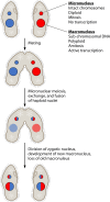
Fig 1
Fig 1
Nuclear dimorphism in ciliated protozoa. Idealized ciliate cells are shown at the top containing a MIC and a macronucleus (MAC). Both nuclei replicate and divide during asexual or vegetative reproduction. In response to starvation, cells of different mating types pair, the MIC undergoes meiosis, and exchanged haploid meiotic products fuse to form a diploid zygotic nucleus. The zygotic nucleus divides, with one product becoming the MIC, while the other product undergoes developmental genome rearrangement to form a new MAC. The old MAC (hatched circle) is degraded.
Fig 2
Fig 2
Two forms of DNA rearrangement occur in the developing MAC. A segment of MIC DNA is shown at the top that includes two segments of DNA that will form MAC chromosomes (green rectangles; C1 and C2). MAC development begins with multiple rounds of DNA replication without karyokinesis or cell division. Two types of DNA rearrangement then occur: (i) internal eliminated sequences (IESs; red rectangles) and other repetitive elements are excised from internal regions of the chromosomes, and (ii) the chromosomes are fragmented and telomeric repeats (“T”) are synthesized de novo onto the DNA ends. Following rearrangement, additional cycles of DNA replication give rise to the mature polyploid MAC.
Fig 3
Fig 3
Genome-scanning model for epigenetic control of DNA elimination in Paramecium. Maternal MAC transcripts (wavy blue lines in top and middle panels) are produced constitutively from the whole rearranged genome. During meiosis, the MIC genome is transcribed on both strands, yielding double-stranded RNAs (dsRNA) that cover the entire non-rearranged genome. After dsRNA processing by Dicer-like proteins Dcl2 and Dcl3, 25-nt scnRNAs (wavy black and red lines, middle panel) are loaded onto Piwi proteins Ptiwi01 and Ptiwi09. Following comparison with maternal MAC transcripts, the scnRNA population is enriched in MIC-specific scnRNAs (wavy red lines in middle and bottom panels, red boxes and double-headed arrow represent IESs and an imprecisely eliminated TE, respectively). The latter scnRNAs are imported into the new developing MACs, where they direct Ptiwi01/09 to homologous sequences by pairing to nascent transcripts (in blue, bottom panel). Ptiwi01/09 recruits the PRC2 polycomb complex, which contains the Ezl1 histone methyl transferase responsible for H3K9 and H3K27 trimethylation (represented by orange and white circles). RNA-mediated heterochromatin formation is thought to drive the elimination complex to MIC-specific DNA. See text for references.
Fig 4
Fig 4
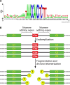
The Tetrahymena Cbs. (A) DNA sequence logo showing sequence conservation among the 225 Cbss in the T. thermophila MIC genome. The Cbs AC-rich strand is illustrated. The Cbs, comprising the central 15 nucleotides (positions 16–30), is shown along with 15 adjacent nucleotide positions on each side of the Cbs. Only Cbs positions 1, 11, 13, 14, and 15 show any diversity [figure taken from reference (18)]. (B) Schematic of chromosome breakage and telomere addition events in Tetrahymena. A segment of a MIC chromosome containing a Cbs and two flanking MAC chromosome precursors (“C1” and “C2”) is shown at the top. During MAC development, this MIC chromosome region is first endoreplicated and then fragmented at or near the Cbs. Telomeric repeats (5′-CCCCAA-3′/3′-GGGGTT-5′; yellow “T” circles) are added to the ends at various positions within the TAR. Note that the Cbs DNA is lost along with a variable length of sequence on either side of the Cbs.
Fig 5
Fig 5
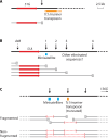
Molecular analysis of three Paramecium chromosome fragmentation regions. (A) MIC chromosome (bold line) including the telomere addition region (vertical arrow) downstream of the P. tetraurelia 51G surface antigen gene is shown, along with the corresponding MAC chromosome (thin line). A Tc1/mariner transposon (orange) is adjacent to the TAR on the MIC chromosome. Adapted from reference (28). (B) Multiple TARs in the vicinity of the P. tetraurelia 51A surface antigen gene. Vertical arrows mark the position of the TARs that were mapped upstream and downstream of the 51A gene. Position 1 accounts for 50% of MAC telomeric ends in wild-type strains and overlaps the 3′ end of a minisatellite (blue box). Other as yet uncharacterized germline eliminated sequences were proposed to locate downstream of position 2. The dashed arrow is the major TAR used in the d48 MAC deletion mutant. [See references (103, 104).] (C) Fragmentation region of the P. primaurelia chromosome harboring the 156G surface antigen gene. The three confirmed TARs are marked by vertical arrows. The locus harbors a truncated copy of a Tc1/mariner transposon (orange box) and two minisatellites (blue boxes). [See reference (36).] New MAC chromosomes are represented at the bottom of each panel, with the telomeres shown as hatched boxes to account for the microheterogeneity of telomere addition positions (within 500–800 bp regions). In panel C, internal deletions of germline DNA repeats (minisatellites and transposons) are represented by red dotted lines between brackets on individual chromosomes. The same scale bar applies to all panels.
Fig 6
Fig 6
Fragmentation of a germline chromosome during development generates multiple somatic chromosomes. The JBrowse image of the MIC scaffold that bears the P. tetraurelia 51G and 51C surface antigen (SAg) genes is shown (top), with tracks for annotations showing genes (107), IESs (28), MAC Illumina read coverage, preliminary TE annotations, curated retro- (LINE) and Tc1/mariner (TIR) transposons (29), and remapped MAC telomere repeats found on individual reads. Note that the arrows representing the orientation of the SAg genes and TEs (TIR and LINE) are not drawn to scale. The true extent of the hypothetical MIC chromosome, beyond the MAC-destined region shown, has not been determined (dotted lines at left and right ends). Sites of fragmentation (light blue shading) are identified from the positions of TEs and remapped MAC telomere repeats combined with the MAC read coverage. The lower part of the figure shows hypothetical MAC chromosomes predicted to result from programmed genome rearrangement. The six molecules carrying the 51C SAg gene (a to f, in black) are compatible with the sizes of the P. tetraurelia MAC chromosomes that were observed on southern blots, following pulsed-field gel electrophoresis and hybridization with a 51C SAg gene probe (108). Note that the region displayed here is homologous to the P. primaurelia region shown in Fig. 5C. (Linda Sperling contributed to the design of this figure.)
Fig 7
Fig 7
Model of chromosome fragmentation and de novo telomere addition in Euplotes. A segment of MIC DNA is shown at top, which includes two MDSs (green ovals C1 and C2) as well as flanking DNA eliminated during MAC development (thin blue line). An enlargement of the segments of DNA that will be fragmented is shown below the top line, with individual base pairs indicated (N = A, G, C, or T). Note that the right end of MDS “C1” and the left end of MDS “C2” overlap by 6 bp. During MAC development, a fragmentation enzyme (red object) interacts with the E-CBS and cleaves the 5′-strand 11 bases upstream and the 3′-strand 17 bases upstream to generate 6 bp 3′ overhangs. Telomerase then adds telomeric repeats (highlighted in yellow) to the 3′ ends. The 5′ strands of the telomeres are then synthesized, along with the complements of the original six base overhangs (bases in red). Sequences which are not retained as MAC nanochromosomes are degraded, as indicated by the large red “X.”
Fig 8
Fig 8
Chromosome fragmentation in Oxytricha and Stylonychia. Alternative processing and microheterogeneity of chromosome fragmentation/telomerization are illustrated for a segment of MIC DNA containing two coding regions (green ovals C1 and C2) and three chromosome fragmentation/telomerization regions (F/T), including one that is used in an alternative manner (Alt. F/T). Endoreplication during MAC development generates multiple DNA strands that can be alternatively fragmented to yield a two-nanochromosome family. Chromosome fragmentation and/or de novo telomere addition occurs with variability at all ends, leading to microheterogeneity in the position of telomeres (“T”).
Fig 9
Fig 9
Telomere-to-telomere RNA aids in unscrambling and telomerization. A MAC DNA molecule formed from three MDSs (green rectangles) in the MIC (1, 2, and 3), two of which are scrambled (2 and 3), produces a telomere-to-telomere transcript (red line) in the maternal MAC. The transcript is transferred to the developing MAC where it provides a template for unscrambling the MDSs and for IES (orange lines) removal. The RNA may also play a role in defining chromosome fragmentation and providing a template for the de novo synthesis of telomeres (T).
Fig 10
Fig 10
Summary of proposed two-stage model for the evolution of ciliate chromosome fragmentation systems. See text for details. Relative evolutionary relationships of species are shown, but evolutionary distances are not drawn to scale.
Fig 11
Fig 11
Transposon-based model for the origin of ”CBS”-based ciliate chromosome fragmentation. The process initiates with the invasion of the MIC genome (blue line with green ovals representing genes/coding regions) by transposon X with terminal inverted repeats (TIRs), which proliferates by transposition. Developmental activation of the transposase would produce breaks at the transposon ends in the genome during MAC development. The transposase gene (hatched rectangle) ultimately is domesticated as the developmental nuclease catalyzing breaks. Further transposition of decayed elements populates the genome with TIRs that are the current ciliate ”CBSs”.

Similar articles

Cited by

References

    1. Chapman JR, Taylor MRG, Boulton SJ. 2012. Playing the end game: DNA double-strand break repair pathway choice. Mol Cell 47:497–510. doi:10.1016/j.molcel.2012.07.029 - DOI - PubMed
    1. Putnam CD, Pennaneach V, Kolodner RD. 2004. Chromosome healing through terminal deletions generated by de novo telomere additions in Saccharomyces cerevisiae. Proc Natl Acad Sci U S A 101:13262–13267. doi:10.1073/pnas.0405443101 - DOI - PMC - PubMed
    1. Estrem B, Wang J. 2023. Programmed DNA elimination in the parasitic nematode Ascaris. PLoS Pathog 19:e1011087. doi:10.1371/journal.ppat.1011087 - DOI - PMC - PubMed
    1. Parfrey LW, Lahr DJG, Knoll AH, Katz LA. 2011. Estimating the timing of early eukaryotic diversification with multigene molecular clocks. Proc Natl Acad Sci U S A 108:13624–13629. doi:10.1073/pnas.1110633108 - DOI - PMC - PubMed
    1. McGrath CL, Zufall RA, Katz LA. 2007. Variation in macronuclear genome content of three ciliates with extensive chromosomal fragmentation: a preliminary analysis. J Eukaryot Microbiol 54:242–246. doi:10.1111/j.1550-7408.2007.00257.x - DOI - PubMed

LinkOut - more resources