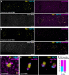
. 2023 Dec 15;150(24):dev202284.
doi: 10.1242/dev.202284. Epub 2023 Dec 11.

Caenorhabditis elegans germ granules are present in distinct configurations and assemble in a hierarchical manner

Affiliations

Caenorhabditis elegans germ granules are present in distinct configurations and assemble in a hierarchical manner

Celja J Uebel et al. Development. .

Abstract

RNA silencing pathways are complex, highly conserved, and perform crucial regulatory roles. In Caenorhabditis elegans germlines, RNA surveillance occurs through a series of perinuclear germ granule compartments - P granules, Z granules, SIMR foci, and Mutator foci - multiple of which form via phase separation. Although the functions of individual germ granule proteins have been extensively studied, the relationships between germ granule compartments (collectively, 'nuage') are less understood. We find that key germ granule proteins assemble into separate but adjacent condensates, and that boundaries between germ granule compartments re-establish after perturbation. We discover a toroidal P granule morphology, which encircles the other germ granule compartments in a consistent exterior-to-interior spatial organization, providing broad implications for the trajectory of an RNA as it exits the nucleus. Moreover, we quantify the stoichiometric relationships between germ granule compartments and RNA to reveal discrete populations of nuage that assemble in a hierarchical manner and differentially associate with RNAi-targeted transcripts, possibly suggesting functional differences between nuage configurations. Our work creates a more accurate model of C. elegans nuage and informs the conceptualization of RNA silencing through the germ granule compartments.

Keywords: Caenorhabditis elegans; Gene regulation; Germ granules; Nuage; RNAi; Small RNAs.


Conflict of interest statement

Competing interests The authors declare no competing or financial interests.

Figures
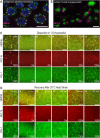
Fig. 1.
Fig. 1.
Mutator foci and P granule separation is independent of nuclear association and can be re-established after perturbation. (A) Widefield immunofluorescence of mut-16::mCherry; pgl-1::gfp germlines with endogenous Mutator foci (MUT-16, magenta) and P granules (PGL-1, green) as adjacent yet distinct compartments. (B) Ectopically expressed MUT-16::mCherry (magenta) and PGL-1::GFP (green) driven by the myo-3 muscle-specific promoter create condensates that maintain separation in the cytoplasm of a muscle cell. (C) Top: Live images of the transition zone of mut-16::mCherry; pgl-1::gfp gonads dissected in M9 buffer (Ci) or varying concentrations of 1,6-hexanediol (Cii-Cvi). Middle: MUT-16 foci (red) are dispersed in all concentrations of 1,6-hexanediol except 0.625%. Numbers in the bottom right of each panel indicate the percentage of gonads displaying Mutator foci. Bottom: PGL-1 foci (green) are disrupted in only 10% and 5% 1,6-hexanediol, indicating different sensitivities to perturbation of weak hydrophobic interactions. Numbers in the bottom right of each panel indicate the percentage of gonads displaying P granules. (D) mut-16::mCherry; pgl-1::gfp animals were subjected to heat stress at 32°C for 2 h and allowed to recover at room temperature (21°C) for 2 h. Top: Representative live images of the pachytene region collected before heat stress (Di, no H.S.), immediately after heat stress (Dii, 0 min recovery), and for every 30 min during recovery (Diii-Dvi). Middle: MUT-16 (red) weakly colocalizes with P granules immediately after heat shock in 80% of samples. At 30- and 60-min room temperature recovery, MUT-16 loses colocalization with PGL-1 but remains dispersed in the cytoplasm. By 90- and 120-min room temperature recovery, MUT-16 reappears as separate, punctate foci adjacent to PGL-1, indicating the interaction is able to be re-established after perturbation. The percentage of gonads displaying punctate Mutator foci is indicated in the bottom-right corner of each panel (n=10 gonads). Bottom: PGL-1 does not completely disperse after heat stress. Percentage of gonads displaying P granules indicated in the bottom-right corner of each panel. Scale bars: 5 µm.
Fig. 2.
Fig. 2.
P granules form unique pocket morphologies in the mid and late pachytene. (A-C) 3D-SIM images of immunostained mut-16::mCherry; pgl-1::gfp germlines display P granules (PGL-1, green) and Mutator foci (MUT-16, magenta). (A) P granule and Mutator foci are adjacent in the transition zone as previously described. (B) Some P granules appear to form an arc or circular morphology (arrows) around Mutator foci in the mid pachytene region. (C) Toroidal, ‘donut-like’ P granule morphologies are prevalent in the late pachytene region (arrow and insets). Insets (i-iii) highlight this unique morphology, termed ‘P granule pocket’. Images in A and B comprise ten maximum projection z-stacks (0.125 µm z-step). Image in C comprises 55 maximum projection z-stacks (0.125 µm z-step). Scale bars: 5 µm.
Fig. 3.
Fig. 3.
Nuage compartments exhibit a hierarchical stoichiometry. (A) Violin plot of fluorescently tagged germ granules surrounding nuclei in the late pachytene, with each dot corresponding to one nucleus (n=30). Asterisks indicate average foci per nucleus. **P≤0.01, ****P≤0.0001. Significance was determined with a two-tailed, equal variance Student's t-test. (B) Manual adjacency quantification from either mut-16::gfp; rfp::znfx-1; pgl-1::bfp to determine P granule populations (left) or mut-16::gfp simr-1::mCherry; pgl-1::bfp to determine SIMR foci populations (right). Overlapping pie charts illustrate distinct populations of granule association. (C) Summary of combined granule stoichiometry indicating the percentage that any one compartment (rows) is adjacent to a second compartment (columns). ND, no data collected. (D) Representative widefield image of a fixed mut-16::gfp; rfp::znfx-1; pgl-1::bfp late pachytene nucleus displaying the different P granule populations: P granule only (P, asterisks), P granule associated with Z granule (PZ, arrowheads), P granule associated with both Z granule and Mutator focus (PZM, arrows). Scale bar: 1 µm.
Fig. 4.
Fig. 4.
P granule pockets exhibit an exterior-to-interior organization. (A) Structured illumination of immunostained germlines with endogenously tagged SIMR-1 to detect SIMR foci (cyan), and ZNFX-1 to detect Z granules (magenta). P granules (yellow) are visualized with anti-PGL-1. Insets (magnifications of the boxed area) show a Z granule occupying the entire interior of the P granule pocket and a SIMR focus innermost still. (B) Confocal image of fixed pachytene nuclei from the mut-16::gfp; tagRFP::znfx-1; pgl-1::bfp germline. Insets show a Z granule (magenta) encompassing a Mutator focus (cyan). (C) Structured illumination of immunostained germlines with endogenously tagged ZNFX-1 (magenta) and MUT-16 (green). Nuclear pore complexes (cyan) are visualized with anti-Nup 107 (mAb414). Insets show a Z granule occupying the interior of the nuclear pore pocket and a Mutator focus localized in the center. (Di) Endogenous fluorescence in a confocal image of the late pachytene region of fixed gfp::znfx-1, tagRFP::npp-9; pgl-1::bfp germlines demonstrate nuclear pore interaction with germ granules. Arrows indicate P granules associated with nuclear pores but not Z granules. Asterisk indicates solitary nuclear pores. (Dii) Violin plot of pixel distance between nuclear pores and P granules (yellow), and between nuclear pores and Z granules (magenta). ****P≤0.0001. Significance was determined with a two-tailed Mann–Whitney test. (Diii) Example of a line profile used to generate the pixel distance in Dii, displaying the fluorescence intensity along a line bisecting a single P and Z granule and the associated nuclear pores. The line used to generate this graph is indicated by a dotted white line in Di. Scale bars: 1 µm.
Fig. 5.
Fig. 5.
Silenced RNA associates preferentially with specific germ granule populations. (A,B) Confocal image of three oocytes in the diakinesis region of a mut-16::gfp; pgl-1::bfp germline following 6 h on control (L4440) RNAi (A) or mex-6 RNAi (B). Proximal oocytes are oriented to the right. (B) smFISH for both oma-1 (control) and mex-6 RNA shows that mex-6 RNA, and not oma-1 RNA, associates with germ granules after mex-6 RNAi. Scale bars: 5 µm. (C,D) Insets (magnifications of the boxed areas in A and B) show mex-6 RNA association with individual germ granules. For mex-6 RNAi-treated animals, the mex-6 RNA signal is located between the signals for PGL-1 and MUT-16. (E) Quantification of granules associated with RNA from mut-16::gfp; pgl-1::bfp following 6 h on mex-6 RNAi to determine frequency of P and PM interactions with mex-6 RNA.
Fig. 6.
Fig. 6.
Working model of P granule pocket organization and nuage assembly. Top: Model of a P granule exhibiting toroidal ‘pocket’ morphology at the periphery of a C. elegans germ cell nucleus (gray). Nuclear pores (dark gray) interact primarily with the P granule pocket (teal), enabling P granules to capture nascent RNA (black). The P granule pocket encircles a Z granule (red), which balances secondary siRNA synthesis across transcripts and is required for siRNA inheritance. The Z granule further encompasses a SIMR focus (purple), which acts as an intermediate between primary and secondary siRNA pathways, and a Mutator focus, which is required for secondary siRNA synthesis (orange). Bottom: Assembly hierarchy of P granule populations proceeding from P to PZSM (left to right). P granules associated with Mutator foci are associated with all known nuage compartments.

Update of

References

    1. Alcazar, R. M., Lin, R. and Fire, A. Z. (2008). Transmission dynamics of heritable silencing induced by double-stranded RNA in Caenorhabditis elegans. Genetics 180, 1275-1288. 10.1534/genetics.108.089433 - DOI - PMC - PubMed
    1. Ambros, V. and Ruvkun, G. (2018). Recent molecular genetic explorations of Caenorhabditis elegans MicroRNAs. Genetics 209, 651-673. - PMC - PubMed
    1. Andralojc, K. M., Campbell, A. C., Kelly, A. L., Terrey, M., Tanner, P. C., Gans, I. M., Senter-Zapata, M. J., Khokhar, E. S. and Updike, D. L. (2017). ELLI-1, a novel germline protein, modulates RNAi activity and P-granule accumulation in Caenorhabditis elegans. PLoS Genet. 13, e1006611. 10.1371/journal.pgen.1006611 - DOI - PMC - PubMed
    1. Aoki, K., Moriguchi, H., Yoshioka, T., Okawa, K. and Tabara, H. (2007). In vitro analyses of the production and activity of secondary small interfering RNAs in C. elegans. EMBO J. 26, 5007-5019. 10.1038/sj.emboj.7601910 - DOI - PMC - PubMed
    1. Ashe, A., Sapetschnig, A., Weick, E.-M., Mitchell, J., Bagijn, M. P., Cording, A. C., Doebley, A.-L., Goldstein, L. D., Lehrbach, N. J., Le Pen, J.et al. (2012). piRNAs can trigger a multigenerational epigenetic memory in the germline of C. elegans. Cell 150, 88-99. 10.1016/j.cell.2012.06.018 - DOI - PMC - PubMed

Publication types

MeSH terms

LinkOut - more resources