. 2023 Dec 20;8(6):e0040323.
doi: 10.1128/msphere.00403-23. Epub 2023 Nov 27.

Characterization of HcaA, a novel autotransporter protein in Helicobacter cinaedi, and its role in host cell adhesion

Affiliations

Characterization of HcaA, a novel autotransporter protein in Helicobacter cinaedi, and its role in host cell adhesion

Sae Aoki et al. mSphere. .

Abstract

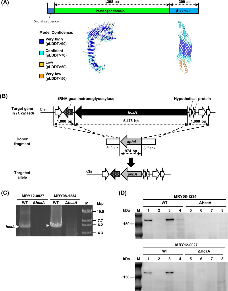
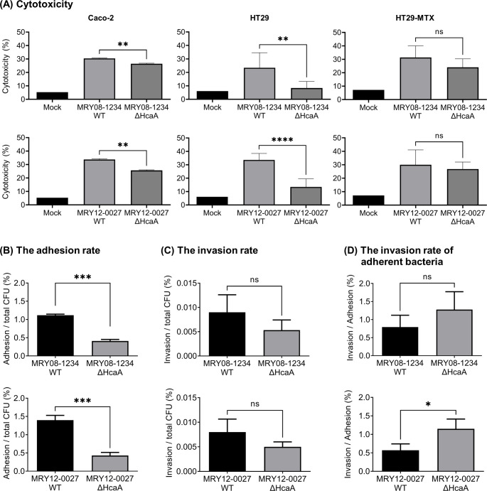
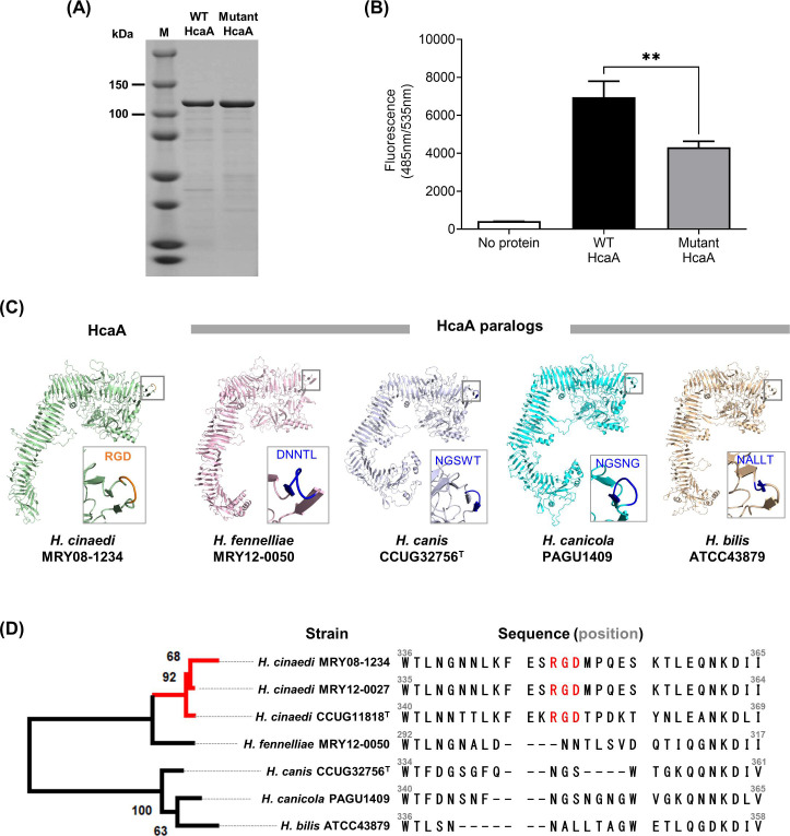
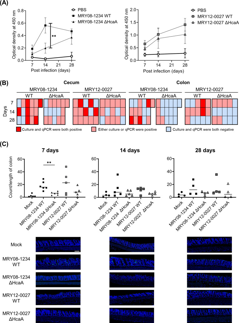
Helicobacter species are classified as gastric or enterohepatic according to their habitat. Among enterohepatic Helicobacter species, which inhabit the intestine, colon, and liver, Helicobacter cinaedi has been most frequently isolated from humans. H. cinaedi often causes bacteremia and cellulitis in immunocompromised hosts. Here, we focused on the H. cinaedi autotransporter protein A (HcaA), a novel virulence factor in H. cinaedi. We discovered that HcaA contributes to cell adhesion via its Arg-Gly-Asp motif. Furthermore, in animal experiments, bacterial colonization was reduced in mice infected with HcaA-knockout strains, supporting the hypothesis that HcaA contributes to H. cinaedi adhesion to host cells. Our study provides a novel mechanism for the establishment of H. cinaedi infections and provides new insights into the role of autotransporter proteins in the establishment of Helicobacter infection.

Keywords: Helicobacter cinaedi; autotransporter protein; cell adhesion; non-Helicobacter pylori Helicobacter species; type V secretion system.


Conflict of interest statement

The authors declare no conflict of interest.

Figures

Fig 1
Fig 1
Construction of hcaA-knockout strains. (A) HcaA contains features of putative autotransporter proteins. The three-dimensional structure of HcaA predicted by AlphaFold2 is color-coded by pLDDT values, which indicate the confidence level of the prediction. (B) Schematic diagram of the knockout strategy for the hcaA gene. The hcaA locus on H. cinaedi MRY12-0027 and MRY08-1234 chromosomes, the donor fragment used to disrupt the hcaA gene locus, and the predicted hcaA gene locus after homologous recombination are shown. (C and D) Confirmation of hcaA gene knockout. (C) PCR using genomic DNA from wild-type (MRY08-1234 WT and MRY12-0027 WT) and HcaA-knockout strains (MRY08-1234 ΔhcaA and MRY12-0027 ΔhcaA) as templates. Lane 1, MRY12-0027 WT; lane 2, MRY12-0027 ΔhcaA; lane 3, MRY08-1234 WT; lane 4, MRY08-1234 ΔhcaA; and lane 5, OneSTEP Marker 6 (λ/Sty I digest) (Nippon Gene Co., Ltd.). White arrows indicate amplicons of the hcaA gene (5,478 bp). Absence of the amplicons in HcaA-knockout strains was confirmed. (D) Immunoblot of HcaA. The concentration of the extracted protein was measured using the bicinchoninic acid (BCA) protein assay. The samples were adjusted to a concentration of 0.5 mg/mL with phosphate-buffered saline (PBS) and separated by 5%–20% sodium dodecyl sulfate-polyacrylamide gel electrophoresis. The top and bottom panels show results for MRY08-1234 and MRY12-0027, respectively. Lane M, Precision Plus Protein Dual Color Standards (Bio-Rad); lanes 1 and 5, whole cell lysate; lanes 2 and 6, culture supernatant fraction; lanes 3 and 7, membrane fraction; and lanes 4 and 8, soluble fraction. The expected size of HcaA was 150 kDa, and a band corresponding to 150 kDa was observed in the WT (lanes 1 and 3) but not in the ΔHcaA strain (lanes 5 and 7), confirming the knockout and localization of HcaA.
Fig 2
Fig 2
Knockout of HcaA reduced H. cinaedi cytotoxicity and adherence to cells. (A) LDH assay to evaluate cytotoxicity. Each cell was infected with wild-type (MRY08-1234 WT or MRY12-0027 WT) and HcaA-knockout (MRY08-1234 ΔHcaA or MRY12-0027 ΔHcaA) strains for 24 h (multiplicity of infection [MOI], 100). Cytotoxicity (%) was calculated using the method described in the instructions. (B–D) Adhesion and invasion assays against Caco-2 cells. Caco-2 cells were infected with wild-type (MRY08-1234 WT or MRY12-0027 WT) and HcaA knockout (MRY08-1234 ΔHcaA or MRY12-0027 ΔHcaA) strains for 4 h (MOI 100). The adhesion rate (B) and invasion rate (C) were calculated by dividing the number of adherent bacterial cells and invading bacterial cells, respectively, by the total number of infected bacterial cells. The invasion rate of adherent bacteria (D) was calculated by dividing the number of invading bacteria cells by the number of adherent bacteria cells. Results are presented as the mean ± standard deviation (SD) of six independent experiments. *P  <  0.05, **P < 0.01, ***P < 0.001, ****P  <  0.0001, and ns: not specified.
Fig 3
Fig 3
RGD motif on HcaA contributes to the adhesion of H. cinaedi. (A) SDS-PAGE of the purified WT HcaA and mutant HcaA, stained with Coomassie brilliant blue. HcaA (120 kDa) with the RGD motif (WT HcaA) and HcaA with glycine replaced with alanine in the RGD motif (mutant HcaA) were expressed in Escherichia coli and purified. Proteins were adjusted to 1 mg/mL and applied. (B) Adhesion of purified WT HcaA (with the RGD motif) and mutant HcaA (with the RAD motif) to U937 cells. Fluorescence-labeled U937 cells attached to HcaA were compared by measuring the fluorescence at 490 nm. Means and SD reflect values of six per group. **P  <  0.01. (C) Three-dimensional structure of HcaA and HcaA homologs predicted by AlphaFold2. Of the five models generated, the top-ranked model was used in this study. The predicted structure was visualized using PyMOL Molecular Graphics System version 2.2.5. HcaA homologs from H. bilis, H. fennelliae, H. canicola, and H. canis were analyzed and compared. The RGD motif is located at the protrusion in HcaA (within the square). HcaA homologs had a structure protruding from the surface, like HcaA; however, the protruding structure did not contain the RGD motif. (D) Phylogenetic tree based on the alignment of the passenger domain of HcaA and HcaA homologs. The alignment was generated using ClustalW version 2.1. The maximum-likelihood tree was constructed using RAxML (https://github.com/amkozlov/raxml-ng) and visualized using MEGA-X v 10.2.6. A comparison of the amino acid sequence of HcaA showed that the RGD motif was present only in HcaA from H. cinaedi (red text).
Fig 4
Fig 4
Knockout of HcaA reduced H. cinaedi colonization in mice. (A) Reactivity to H. cinaedi in infected mice, as determined by enzyme-linked immunosorbent assay. Sera from mice at 7, 14, and 28 days after inoculation were used. Squares and triangles indicate serum from the group of infected wild-type (black square, MRY08-1234 WT; gray square, MRY12-0027 WT) or HcaA knockout (black triangle, MRY08-1234 ΔHcaA; gray triangle, MRY12-0027 ΔHcaA) strains, respectively. Means and SD reflect values from five of six mice per group. **P  <  0.01. (B) Heatmap combining the results of the culture method and qPCR assays. Columns indicate each mouse and rows indicate the duration of infection. Positive results for both the culture method and qPCR assays are shown in red, positive results for either are shown in pink, and negative results for both are shown in light blue. (C) Quantitative bacteria count per length of the colon and representative images of the colon of H. cinaedi-infected mice. Adhesion of H. cinaedi to the colon was quantified using ImageJ version 1.53t, by counting the number of H. cinaedi in images taken with a 20× objective lens. Results are expressed as the number of H. cinaedi per length of colon. Means and SD reflect the values of five or six mice per group. **P  <  0.01. Green, H. cinaedi and blue, nucleus. Scale bar 100 μm.

Similar articles

Cited by

References

    1. Polk DB, Peek RM. 2010. Helicobacter pylori: gastric cancer and beyond. Nat Rev Cancer 10:403–414. doi:10.1038/nrc2857 - DOI - PMC - PubMed
    1. Matsuo T, Mori N, Mizuno A, Sakurai A, Kawai F, Starkey J, Ohkushi D, Abe K, Yamasaki M, Ito J, Yoshino K, Mikami Y, Uehara Y, Furukawa K. 2020. Infected aortic aneurysm caused by Helicobacter cinaedi: case series and systematic review of the literature. BMC Infect Dis 20:854. doi:10.1186/s12879-020-05582-7 - DOI - PMC - PubMed
    1. Akiyama T, Imamura H, Fukui N, Sakai N. 2021. Helicobacter cinaedi-infected chronic subdural hematoma mimicking an expanding hematoma: a case report. Surg Neurol Int 12:288. doi:10.25259/SNI_387_2021 - DOI - PMC - PubMed
    1. Kawamura Y, Tomida J, Morita Y, Fujii S, Okamoto T, Akaike T. 2014. Clinical and bacteriological characteristics of Helicobacter cinaedi infection. J Infect Chemother 20:517–526. doi:10.1016/j.jiac.2014.06.007 - DOI - PubMed
    1. Suzuki T, Kutsuna S, Tsuboi M, Ota M, Hayakawa K, Ohmagari N. 2019. Helicobacter cinaedi hepatic cyst infection with bacteremia. Emerg Infect Dis 25:603–604. doi:10.3201/eid2503.180936 - DOI - PMC - PubMed

Substances

Supplementary concepts