

A proposed structural connectivity matrices approach for the superior fronto-occipital fascicle in the Harvard-Oxford Atlas comparative framework following the Pandya comparative extrapolation principle
- PMID: 38010231
- PMCID: PMC11019921
- DOI: 10.1002/cne.25562
A proposed structural connectivity matrices approach for the superior fronto-occipital fascicle in the Harvard-Oxford Atlas comparative framework following the Pandya comparative extrapolation principle
Abstract
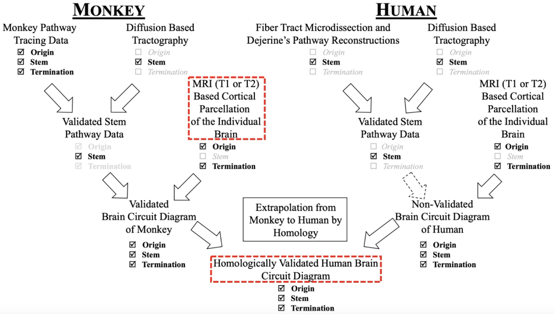
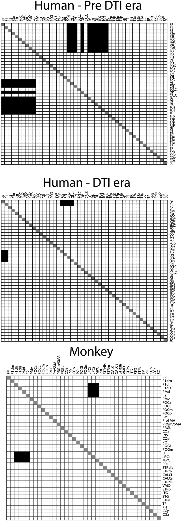
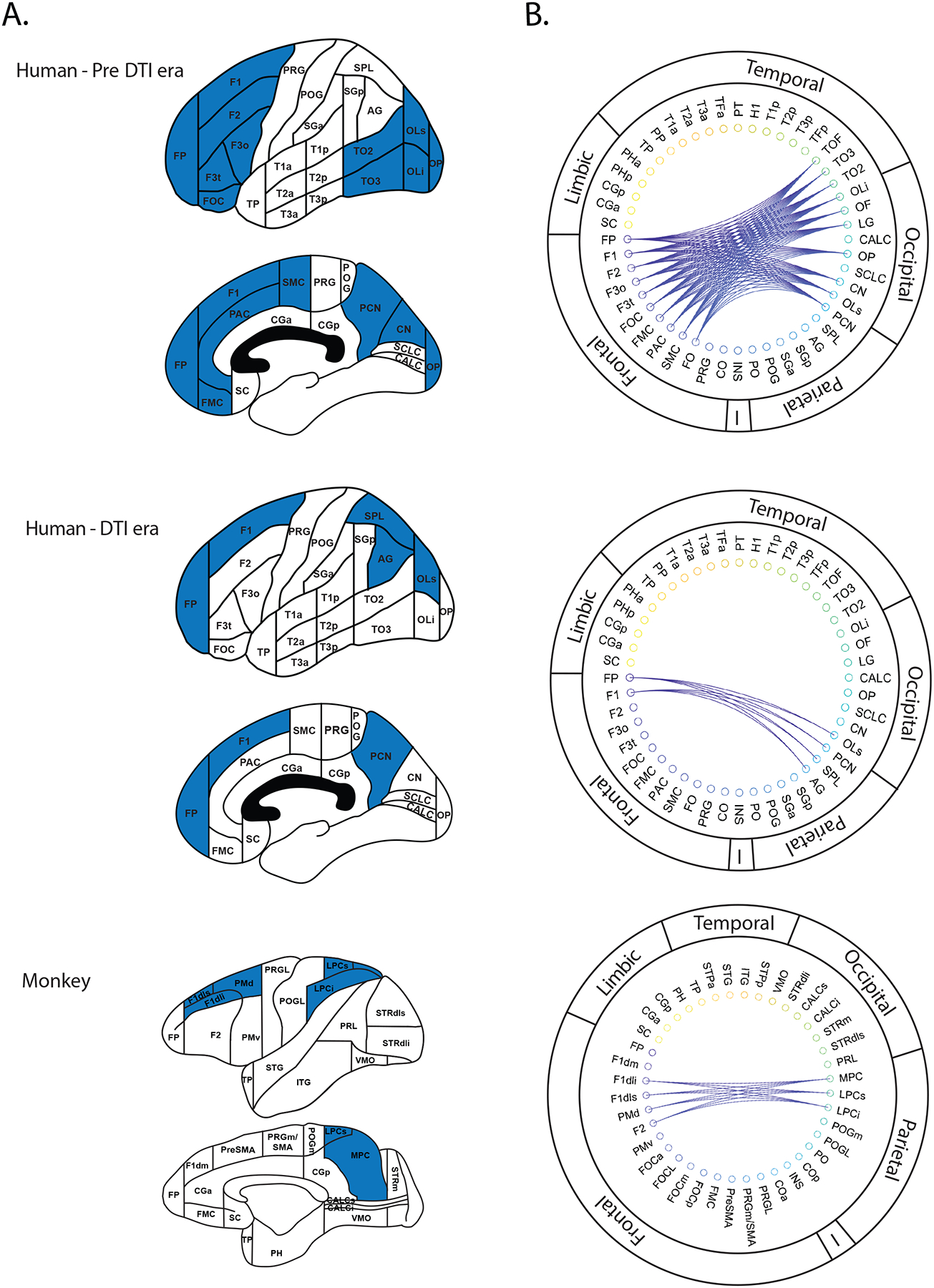
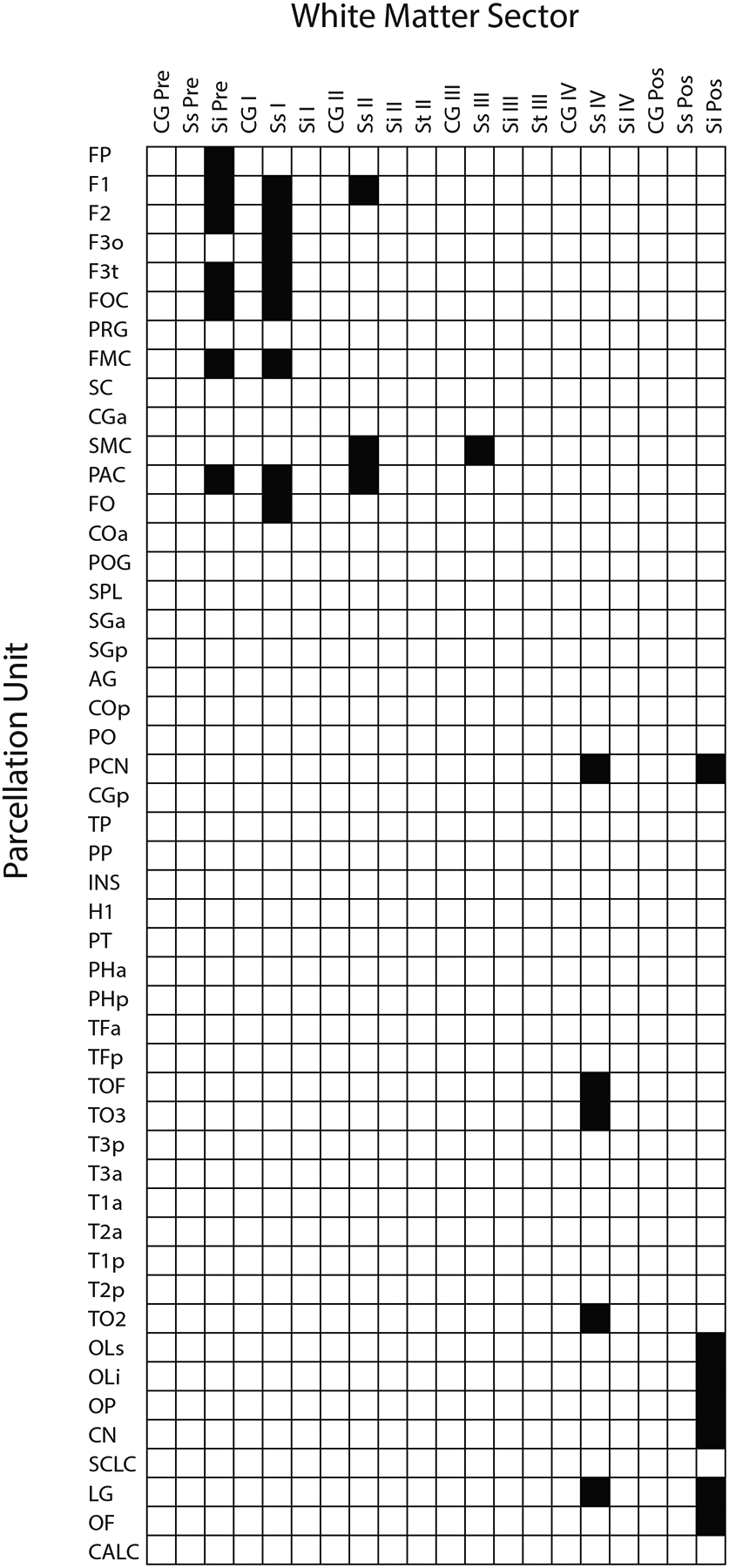
A key set of connections necessary for the most complex brain functions are the long association cortico-cortical fiber tracts. These pathways have been described by the Dejerines and others using post mortem histological or brain dissection techniques. Given methodological limitations, these fiber connections have not been delineated completely in humans. Although the stem portions of fiber tracts have been identified in humans, their precise origins and terminations remain to be determined. By contrast, the origins and terminations as well as the stems of long cortico-cortical association fiber pathways in monkeys have been detailed in the macaque monkey brain using experimental tract tracing methods. Deepak Pandya made major contributions to the delineation of fiber tracts in the monkey brain. In the early 1990s, he compared his observations in monkeys with the original descriptions in humans by the Dejerines. With the advent of diffusion-weighted imaging, Dr. Pandya extended this line of investigation to the human brain with Dr. Nikos Makris. In this translational analysis of long association cortico-cortical fiber tracts, they applied a principle of extrapolation from monkey to human. In the present study, we addressed the reasoning and the complex methodology in translating brain structural connectivity from monkey to human in one cortico-cortical fiber tract, namely the superior fronto-occipital fascicle, which was delineated in both species by Dr. Pandya and colleagues. Furthermore, we represented this information in the form of connectional matrices in the context of the HOA2.0-ComPaRe framework, a homological monkey-to-human translational system used in neuroimaging.
Keywords: DTI; Harvard-Oxford Atlas; dMRI; fiber bundle; superior fronto-occipital fascicle; tractography.
© 2023 Wiley Periodicals LLC.
Conflict of interest statement
Conflicts of Interest
The authors declare they have no conflicts of interest.
Figures
Similar articles
-
A Proposed Human Structural Brain Connectivity Matrix in the Center for Morphometric Analysis Harvard-Oxford Atlas Framework: A Historical Perspective and Future Direction for Enhancing the Precision of Human Structural Connectivity with a Novel Neuroanatomical Typology.Dev Neurosci. 2023;45(4):161-180. doi: 10.1159/000530358. Epub 2023 Mar 28. Dev Neurosci. 2023. PMID: 36977393 Free PMC article.
-
Mapping temporo-parietal and temporo-occipital cortico-cortical connections of the human middle longitudinal fascicle in subject-specific, probabilistic, and stereotaxic Talairach spaces.Brain Imaging Behav. 2017 Oct;11(5):1258-1277. doi: 10.1007/s11682-016-9589-3. Brain Imaging Behav. 2017. PMID: 27714552 Free PMC article.
-
Long association tracts of the human white matter: an analysis of 18 hemisphere dissections and in vivo HARDI-CSD tractography.Zh Vopr Neirokhir Im N N Burdenko. 2017;81(1):13-25. doi: 10.17116/neiro201780713-25. Zh Vopr Neirokhir Im N N Burdenko. 2017. PMID: 28291210 English, Russian.
-
The vertical superior longitudinal fascicle and the vertical occipital fascicle.J Neurosurg Sci. 2021 Dec;65(6):581-589. doi: 10.23736/S0390-5616.21.05368-6. J Neurosurg Sci. 2021. PMID: 35128919 Review.
-
Fiber anatomy of dorsal and ventral language streams.Brain Lang. 2013 Nov;127(2):192-204. doi: 10.1016/j.bandl.2012.04.015. Epub 2012 May 24. Brain Lang. 2013. PMID: 22632814 Review.
Cited by
-
Optimization of TMS target engagement: current state and future perspectives.Front Neurosci. 2025 Jan 29;19:1517228. doi: 10.3389/fnins.2025.1517228. eCollection 2025. Front Neurosci. 2025. PMID: 39944889 Free PMC article.
References
-
- Brodmann K 1999. Brodmann’s “Localisation in the Cerebral Cortex.” Springer.
-
- Bürgel U, Amunts K, Hoemke L, Mohlberg H, Gilsbach JM, Zilles K. 2006. White matter fiber tracts of the human brain: three-dimensional mapping at microscopic resolution, topography and intersubject variability. Neuroimage 29:1092–1105. - PubMed
-
- Bürgel U, Mecklenburg I, Blohm U, Zilles K. 1997. Histological visualization of long fiber tracts in the white matter of adult human brains. J Hirnforsch 38:397–404. - PubMed
-
- Catani M, Howard RJ, Pajevic S, Jones DK. 2002. Virtual in vivo interactive dissection of white matter fasciculi in the human brain. Neuroimage 17:77–94. - PubMed
Publication types
MeSH terms
Grants and funding
- K24 MH116366/MH/NIMH NIH HHS/United States
- R01 MH111917/NH/NIH HHS/United States
- R21 DA042271/NH/NIH HHS/United States
- R01 NS125307/NH/NIH HHS/United States
- R01 MH112748/MH/NIMH NIH HHS/United States
- R01 AG042512/NH/NIH HHS/United States
- R21 DA042271/DA/NIDA NIH HHS/United States
- R01 MH125860/NH/NIH HHS/United States
- R01 NS125307/NS/NINDS NIH HHS/United States
- R01 MH112748/NH/NIH HHS/United States
- K24 MH116366/NH/NIH HHS/United States
- R01 MH125860/MH/NIMH NIH HHS/United States
- R01 AG042512/AG/NIA NIH HHS/United States
- R01 MH111917/MH/NIMH NIH HHS/United States
LinkOut - more resources
Full Text Sources
