Review
. 2024 Mar;13(7):e2303161.
doi: 10.1002/adhm.202303161. Epub 2023 Dec 10.

Patient-Derived Microphysiological Systems for Precision Medicine

Affiliations
Review

Patient-Derived Microphysiological Systems for Precision Medicine

Jihoon Ko et al. Adv Healthc Mater. 2024 Mar.

Abstract

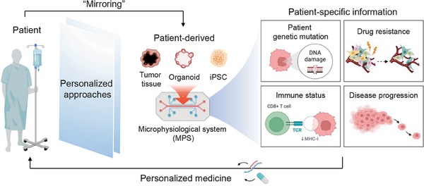
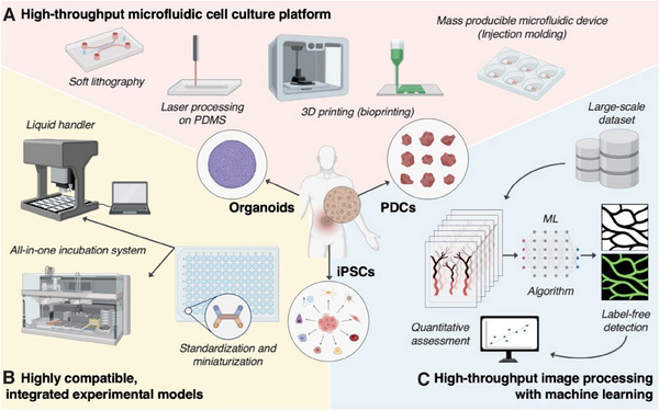
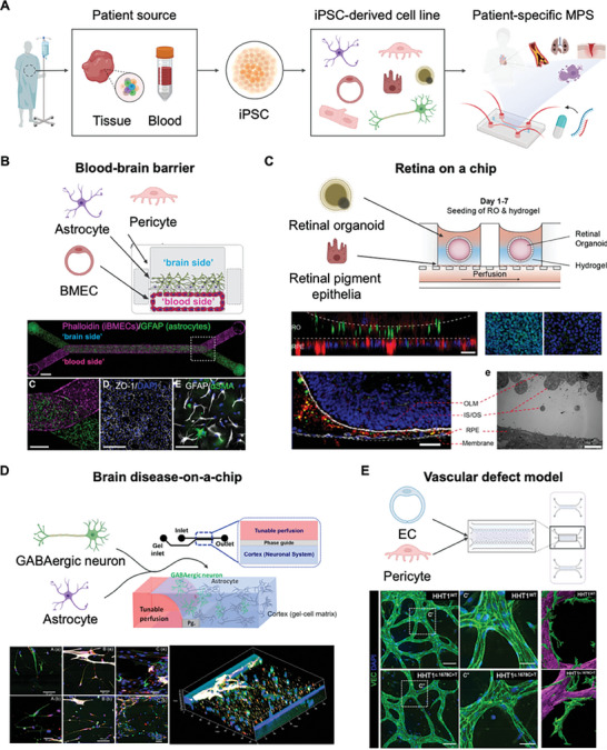
Patient-derived microphysiological systems (P-MPS) have emerged as powerful tools in precision medicine that provide valuable insight into individual patient characteristics. This review discusses the development of P-MPS as an integration of patient-derived samples, including patient-derived cells, organoids, and induced pluripotent stem cells, into well-defined MPSs. Emphasizing the necessity of P-MPS development, its significance as a nonclinical assessment approach that bridges the gap between traditional in vitro models and clinical outcomes is highlighted. Additionally, guidance is provided for engineering approaches to develop microfluidic devices and high-content analysis for P-MPSs, enabling high biological relevance and high-throughput experimentation. The practical implications of the P-MPS are further examined by exploring the clinically relevant outcomes obtained from various types of patient-derived samples. The construction and analysis of these diverse samples within the P-MPS have resulted in physiologically relevant data, paving the way for the development of personalized treatment strategies. This study describes the significance of the P-MPS in precision medicine, as well as its unique capacity to offer valuable insights into individual patient characteristics.

Keywords: microphysiological system; patient-derived; physiologically-relevant; precision medicine.


Conflict of interest statement

The authors declare no conflict of interest.

Figures

Figure 1
Figure 1
P‐MPS for precision medicine. The illustration describes the application of P‐MPS in precision medicine. P‐MPS enables the mirroring of patient‐specific characteristics by utilizing patient‐derived samples including iPSCs, organoids, and tumor tissues. This platform plays a critical role in accelerating precision medicine by providing valuable insights into an individual such as genetic variants, drug resistance, immune status, and disease progression. The illustration was created using BioRender.
Figure 2
Figure 2
Components for facilitating high‐throughput microfluidic cell culture experiments. A) Diverse efficient fabrication methods for microfluidics devices: soft lithography, laser‐based PDMS direct processing, 3D printing, and injection molding. B) Development of standardized and highly compatible microfluidic devices for integration with automation devices such as liquid handlers and incubation systems. C) Utilize machine learning for analysis of large‐scale data obtained from high‐throughput experiments. Constructing a system for quantitative analysis, including protein expression and morphological features. Streamlining experimental processes through label‐free virtual staining, eliminating the need for fluorescent staining. The illustration was created using BioRender.
Figure 3
Figure 3
Representative models of MPS for tissue modeling. A) A preferred blood vessel‐on‐a‐chip design that facilitates perfusion through multicellular tumor spheroids to enhance the efficacy of anti‐cancer drug delivery. Red: RFP‐HUVEC, Blue: Nucleus. Reproduced with permission from Park et al. (Lab on a chip 22, 22, (2022); Copyright 2022, Royal Society of Chemistry).[ 88 ] B) Skin‐on‐a‐chip with vascular integration for investigating vascular absorption. HE‐stained and immune‐stained epidermal layers. Reproduced with permission from Mori et al. (Biomaterials 116 (2017); Copyright 2021, Elsevier).[ 95 ] C) Blood‐retinal barrier‐on‐a‐chip for simulating pathological angiogenesis and assessing the effectiveness of treatments for choroidal neovascularization. Images show VEGF treatment with and without bevacizumab. Bevacizumab cotreatment inhibits angiogenic sprouting. Red: HUVEC (Anti‐human CD31 Alexa Fluor 647 conjugated). Reproduced with permission from Chung et al. (Advanced Healthcare Material 7, 2, (2018); Copyright 2018, Wiley).[ 92 ] D) Blood–brain barrier (BBB)‐on‐a‐chip platform for assessing the delivery of BBB‐opening agents through the microvasculature. Reproduced with permission from Seo et al. (Advanced Functional Materials 32(10) (2022); Copyright 2022, Wiley).[ 99 ] E) Real‐time monitoring of natural killer (NK) cell infiltration and cytotoxicity in a tumor vasculature‐on‐a‐chip. Red: HUVEC (lectin), Green: Colorectal cancer cell (EpCAM), White: NK cells (CellTrace Far Red), Blue: Dead cell (Cytox Blue). Reproduced with permission from Song et al. (Frontiers in Immunology 12,1 (2021); Copyright 2021 Frontiers).[ 101 ] F) Multi‐organ‐on‐a‐chip with tissue‐specific niche enabling cross‐talk between organs within the system through the vascular circulation (upper), and immunofluorescence or immunohistochemical images for liver, heart, bone, skin, and vasculature (lower). Reproduced with permission from Ronaldson‐Bouchard et al. (Nature Biomedical Engineering 64,4 (2022); Copyright 2022, Springer Nature).[ 105 ]
Figure 4
Figure 4
Representative examples of patient‐derived cell/organoid‐based MPS. A) A patient‐derived gastric cancer spheroid‐on‐a‐chip model for demonstrating blood vessel formation induced by PDC and evaluating the anti‐cancer drug efficacy. Green: HUVEC (lectin, 488), Red: Patient‐derived gastric cancer cell (EpCAM, 594). Reproduced with permission from Lee et al. (Cancer Medicine 10, 20 (2021); Copyright 2021 John Wiley & Sons Ltd.).[ 123 ] B) A patient‐derived pancreas‐on‐a‐chip for mimicking defective conductance regulator (CFTR) function associated with cystic fibrosis. Defective CFTR function in pancreatic ductal epithelial cells (PDECs) led to a significant reduction in insulin secretion within islet cells. Reproduced with permission from Mun et al. (Nature Communications 10, 1 (2019); Copyright 2019, Springer Nature).[ 139 ] C) A patient‐derived colorectal cancer organoid model on a nested array chip for high‐throughput drug screening. PDO model on the chip accurately predicted chemotherapy outcomes. Reproduced with permission from Cui et al. (Bio‐Design and Manufacturing 5 (2022); Copyright 2022 Springer).[ 140 ] D) A superhydrophobic microwell array chip for patient‐derived lung cancer organoid models that recapitulate the histological and genetic features of the parental tumors to produce clinically meaningful drug responses within a week. Reproduced with permission from Hu et al. (Nature Communications 12,1 (2021); Copyright 2021, Springer Nature).[ 141 ] Illustrations were created using BioRender.
Figure 5
Figure 5
MPS with iPSCs developed from patient‐derived samples. A) The workflow involves obtaining iPSCs from a patient‐derived source and differentiating them into specific cell lines for the model. The P‐MPS facilitates the 3D co‐culture of two or more cell types, enabling a more comprehensive representation of cellular interactions. B) Development and validation of a personalized BBB model within the MPS. The BBB model was established by differentiating key cell lines (astrocyte, brain microvascular endothelial cell (BMEC), and pericyte) derived from iPSCs obtained from the same donor. Key protein expression was observed in each cell line from both the brain and blood sides of the membrane, indicating the functionality and relevance of the BBB model. Reproduced with permission from Vatine et al. (Cell stem cell 24, 6 (2019); Copyright 2019 Cell press).[ 173 ] C) Microphysiological model of the human retina incorporating over seven essential retinal cell types derived from iPSCs. The retina on a chip cultures retinal pigment epithelia by introducing a downstream perfusion channel that can complement the avascularity of the retinal organoid. Reproduced with permission from Achberger et al. (Elife 8 (2019); Copyright 2019, eLife Sciences Publications Ltd.).[ 174 ] D) 3D brain disease on chip platform with iPSC‐derived GABAergic neurons and astrocytes for studying neurotoxicity and therapeutic intervention in organophosphate exposure. Neuron‐astrocyte interaction, synapse formation, and enhanced viability with Butyrylcholinesterase treatment were observed. Reproduced with permission from Liu et al. (PLoS One 15, 3 (2020); Copyright 2022, PLOS).[ 175 ] E) Patient‐specific hiPSCs were used to generate diseased and healthy endothelial cells reflecting haploinsufficiency. Characterization of the 3D vascular network in ECs with HHT gene mutations revealed abnormal morphological features, protein expression, and pericyte interaction. Reproduced with permission from Orlova et al. (Stem Cell Reports 17, 7 (2022); Copyright 2022, Cell Press).[ 176 ] Illustrations were created using BioRender.

Similar articles

Cited by

References

    1. Ma C., Peng Y., Li H., Chen W., Trends Pharmacol. Sci. 2021, 42, 119. - PMC - PubMed
    1. Roth A., Science 2021, 373, 1304. - PubMed
    1. Miller C. P., Shin W., Ahn E. H., Kim H. J., Kim D.‐H., Trends Biotechnol. 2020, 38, 857. - PMC - PubMed
    1. Seyhan A. A., Transl. Med. Commun. 2019, 4, 18.
    1. Crippa M., Bersini S., Gilardi M., Arrigoni C., Gamba S., Falanga A., Candrian C., Dubini G., Vanoni M., Moretti M., Lab Chip 2021, 21, 1061. - PubMed

Publication types

LinkOut - more resources