

Association of Male Sex and Microvascular Alterations on Optical Coherence Tomography Angiography in Diabetes
- PMID: 38010281
- PMCID: PMC10683768
- DOI: 10.1167/tvst.12.11.30
Association of Male Sex and Microvascular Alterations on Optical Coherence Tomography Angiography in Diabetes
Abstract
Purpose: Epidemiologically, men have a higher incidence, severity, and progression of diabetic retinopathy (DR) than women. We investigated microvascular differences between men and women with diabetes on optical coherence tomography angiography (OCTA).
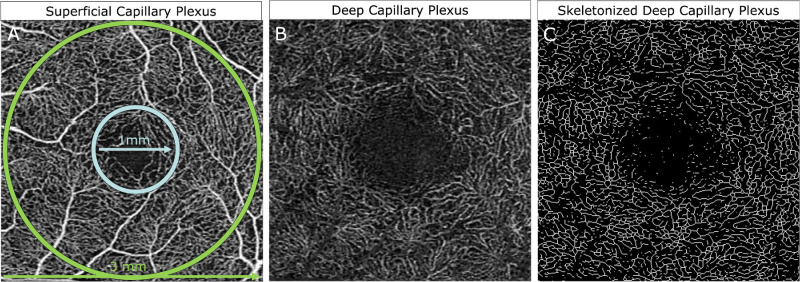
Methods: Three × 3 mm OCTA macula scans of non-diabetic and patients with diabetes were obtained. Vascular parameters included parafoveal vessel density (VD), vessel length density (VLD), and flow index (FI) of the superficial capillary plexus (SCP) and deep capillary plexus (DCP) as well as foveal avascular zone (FAZ) area and perimeter. Multivariable linear regression was used for statistical analysis.
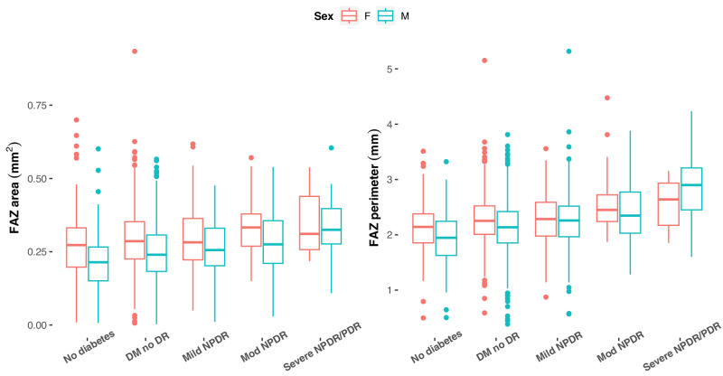
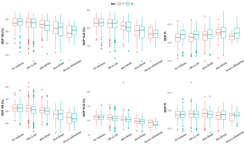
Results: There were 1809 patients with diabetes and 217 non-diabetic participants that were included in this study. Diabetic individuals included those with no DR (n = 1356), mild non-proliferative DR (NPDR; n = 286), moderate NPDR (n = 126), and severe NPDR/proliferative DR (PDR; n = 41). Male sex was significantly associated with smaller FAZ area/perimeter and lower DCP VLD in both non-diabetic subjects and patients with diabetes. Male sex in the diabetic group was additionally associated with lower SCP VD/VLD and DCP VD. Addition of an interaction between male sex and diabetes status in the interaction analysis showed that being male and diabetic conferred increased reduction in DCP VD and VLD compared to sex-based changes in non-diabetics. Larger FAZ perimeter, lower SCP VD/VLD, and lower DCP VLD were associated with poorer visual acuity in diabetics.
Conclusions: On OCTA, male patients with diabetes may have more severe microvascular disease especially in the DCP compared to women.
Translational evidence: Sex-based alterations in diabetic microvascular disease has the potential to influence future basic and clinical studies as well as the implementation of OCTA disease markers.
Conflict of interest statement
Disclosure:
Figures
Similar articles
-
Superficial capillary perfusion on optical coherence tomography angiography differentiates moderate and severe nonproliferative diabetic retinopathy.PLoS One. 2020 Oct 22;15(10):e0240064. doi: 10.1371/journal.pone.0240064. eCollection 2020. PLoS One. 2020. PMID: 33091032 Free PMC article.
-
Macular Capillary Perfusion in Chinese Patients With Diabetic Retinopathy Obtained With Optical Coherence Tomography Angiography.Ophthalmic Surg Lasers Imaging Retina. 2019 Apr 1;50(4):e88-e95. doi: 10.3928/23258160-20190401-12. Ophthalmic Surg Lasers Imaging Retina. 2019. PMID: 30998251
-
Optical Coherence Tomography Angiography Projection Artifact Removal: Impact on Capillary Density and Interaction with Diabetic Retinopathy Severity.Transl Vis Sci Technol. 2020 Jun 5;9(7):10. doi: 10.1167/tvst.9.7.10. eCollection 2020 Jun. Transl Vis Sci Technol. 2020. PMID: 32832217 Free PMC article.
-
Evaluation of Posterior Ocular Blood Flow in Diabetic Retinopathy Patients Without Macular Edema Using Optical Coherence Tomography Angiography.Photodiagnosis Photodyn Ther. 2023 Dec;44:103777. doi: 10.1016/j.pdpdt.2023.103777. Epub 2023 Sep 3. Photodiagnosis Photodyn Ther. 2023. PMID: 37669724 Review.
-
Optical coherence tomography angiography for the assessment of retinal microvasculature characteristics in preterm-born children: A systematic review and meta-analysis.Indian J Ophthalmol. 2024 May 1;72(Suppl 3):S372-S380. doi: 10.4103/IJO.IJO_2268_23. Epub 2024 Mar 8. Indian J Ophthalmol. 2024. PMID: 38454847 Free PMC article.
Cited by
-
Potential of Pterostilbene as an Antioxidant Therapy for Delaying Retinal Damage in Diabetic Retinopathy.Antioxidants (Basel). 2025 Feb 20;14(3):244. doi: 10.3390/antiox14030244. Antioxidants (Basel). 2025. PMID: 40227230 Free PMC article.
-
Diabetic Retinopathy (DR) nomogram construction based on optical coherence tomography angiography parameters: a preliminary exploration of DR prediction.Graefes Arch Clin Exp Ophthalmol. 2025 Jul;263(7):1867-1876. doi: 10.1007/s00417-025-06824-7. Epub 2025 Apr 8. Graefes Arch Clin Exp Ophthalmol. 2025. PMID: 40198363
References
-
- Anand SS, Islam S, Rosengren A, et al. .. Risk factors for myocardial infarction in women and men: insights from the INTERHEART study. Eur Heart J. 2008; 29(7): 932–940. - PubMed
-
- The DECODE Study Group, Hu G. Gender difference in all-cause and cardiovascular mortality related to hyperglycaemia and newly-diagnosed diabetes. Diabetologia. 2003; 46(5): 608–617. - PubMed
MeSH terms
LinkOut - more resources
Full Text Sources
Medical
Miscellaneous
