Clinical Trial
. 2024 Mar;93(3):177-189.
doi: 10.1007/s00280-023-04600-0. Epub 2023 Nov 27.

Pharmacodynamic effects of the PARP inhibitor talazoparib (MDV3800, BMN 673) in patients with BRCA-mutated advanced solid tumors

Affiliations
Clinical Trial

Pharmacodynamic effects of the PARP inhibitor talazoparib (MDV3800, BMN 673) in patients with BRCA-mutated advanced solid tumors

Arjun Mittra et al. Cancer Chemother Pharmacol. 2024 Mar.

Abstract

Purpose: Talazoparib is an inhibitor of the poly (ADP-ribose) polymerase (PARP) family of enzymes and is FDA-approved for patients with (suspected) deleterious germline BRCA1/2-mutated, HER2‑negative, locally advanced or metastatic breast cancer. Because knowledge of the pharmacodynamic (PD) effects of talazoparib in patients has been limited to studies of PARP enzymatic activity (PARylation) in peripheral blood mononuclear cells, we developed a study to assess tumoral PD response to talazoparib treatment (NCT01989546).

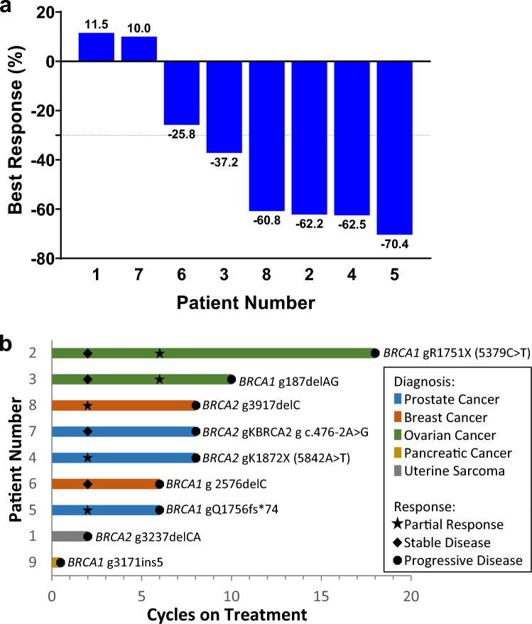
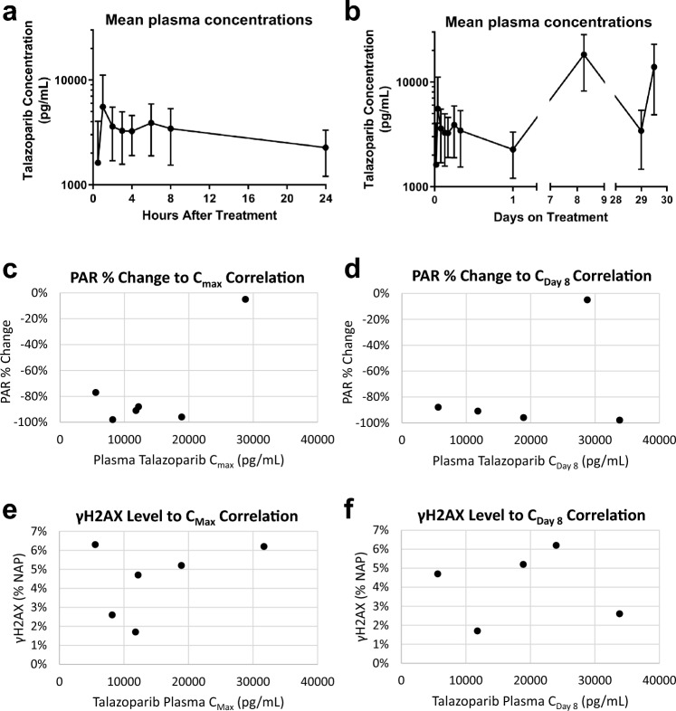
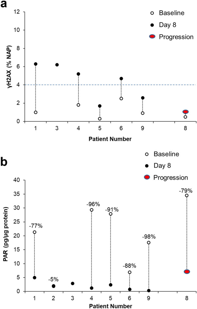
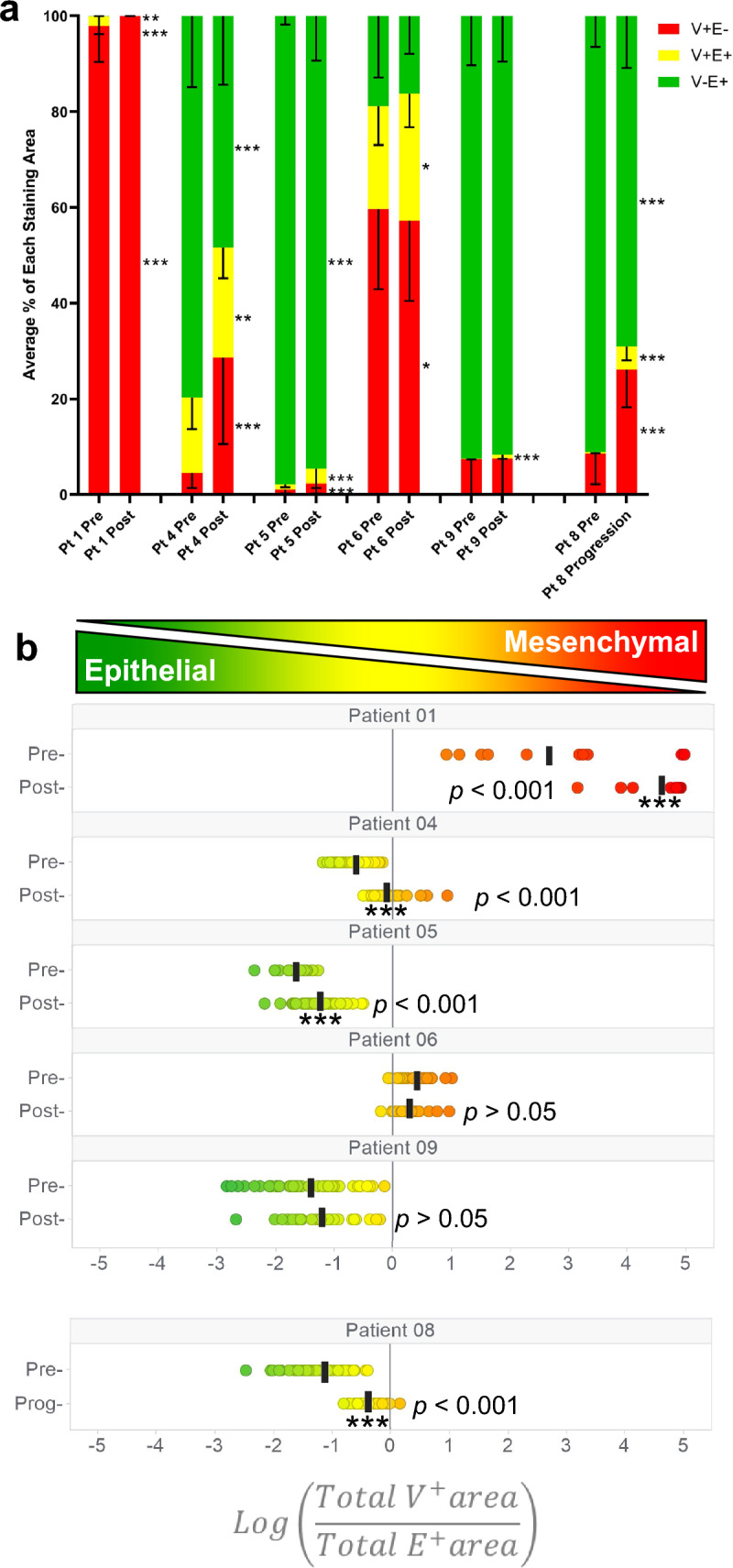
Methods: We administered single-agent talazoparib (1 mg/day) orally in 28-day cycles to adult patients with advanced solid tumors harboring (suspected) deleterious BRCA1 or BRCA2 mutations. The primary objective was to examine the PD effects of talazoparib; the secondary objective was to determine overall response rate (ORR). Tumor biopsies were mandatory at baseline and post-treatment on day 8 (optional at disease progression). Biopsies were analyzed for PARylation, DNA damage response (γH2AX), and epithelial‒mesenchymal transition.

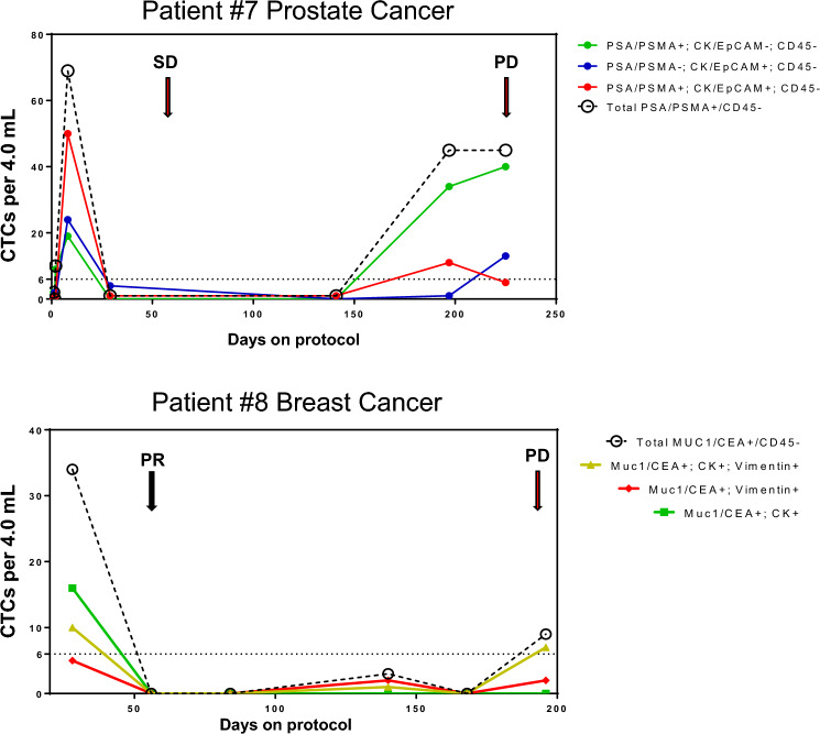
Results: Nine patients enrolled in this trial. Four of six patients (67%) evaluable for the primary PD endpoint exhibited a nuclear γH2AX response on day 8 of treatment, and five of six (83%) also exhibited strong suppression of PARylation. A transition towards a more mesenchymal phenotype was seen in 4 of 6 carcinoma patients, but this biological change did not affect γH2AX or PAR responses. The ORR was 55% with the five partial responses lasting a median of six cycles.

Conclusion: Intra-tumoral DNA damage response and inhibition of PARP enzymatic activity were confirmed in patients with advanced solid tumors harboring BRCA1/2 mutations after 8 days of talazoparib treatment.

Keywords: Clinical trial; DNA damage repair; Homologous recombination repair; Pharmacology; Poly-ADP-ribosylation; Targeted agent.


Conflict of interest statement

The authors declare no potential conflicts of interest.

Figures

Fig. 1
Fig. 1
Patient outcomes. a Best proportion change in RECIST measurements from baseline (listed below each bar) is plotted for all patients except for patient #9, who left the trial due to clinical progression before their first restaging. b Number of cycles on treatment for all patients colored by diagnosis. Response and BRCA1/2 eligibility mutation are shown for each patient
Fig. 2
Fig. 2
Pharmacokinetic analysis. Average talazoparib plasma levels (± standard deviation) for all 9 patients a during the 24 h following administration of the first dose and b during the entire collection period. Blood samples were collected prior to treatment; 0.5, 1, 2, 3, 4, 6, 8, and 24 h after the first dose; 3 to 6 h after the day 8 dose; and before and 3 to 6 h after the cycle 2, day 1 dose. c The most significant correlation between individual patient plasma exposure and PD effect was observed with the talazoparib maximal concentration (CMax) and the proportion of PAR inhibition observed at day 8 compared to baseline (0.76 correlation [0.25, 0.94 80% CI]). d No correlation between day 8 plasma talazoparib levels (CDay 8) and PAR inhibition was observed (0.35 correlation [− 0.5, 0.85 80% CI]), and neither did e CMax nor f CDay 8 talazoparib levels correlate with individual patient γH2AX levels (− 0.03 correlation [− 0.73, 0.7 80% CI] and 0.39 correlation [− 0.32, 0.82 80% CI], respectively)
Fig. 3
Fig. 3
Tumoral γH2AX and PAR changes. a Nuclear γH2AX levels (the proportion of nuclear area positive, or %NAP, for γH2AX) and b PAR levels in evaluable tumor biopsies collected at baseline and 3 to 6 h after talazoparib treatment on day 8, except for patient #8, whose on-treatment biopsy was taken at progression (after cycle 8, 24 h after last dose). The blue dotted line in (a) represents the γH2AX effect level threshold established in Wilsker et al.[29] The proportion change in PAR from before to after treatment is shown above each patient’s data points in (b). More tumor biopsy cores were evaluable with the validated PAR assay than the γH2AX assay because some biopsy cores for the γH2AX immunofluorescence assay were judged to have insufficient viable tumor cellularity for image analysis during the required pathologist’s review
Fig. 4
Fig. 4
Tumor epithelial-mesenchymal phenotypes. EMT-IFA results from tumor biopsies collected at baseline and on-treatment in patients that had suitable paired biopsy samples with sufficient viable tumor cellularity for analysis: a Proportion of tumor area staining positive for vimentin only (V + E −, red), E-cadherin only (V − E + , green), co-localized vimentin and E-cadherin (V + E + , yellow); b The log(V/E) ratio of vimentin and E-cadherin-stained areas within regions of interest (ROIs) in a tissue section. In each case, significance, as calculated by the Mann–Whitney test treating each ROI as a separate observation, is shown with asterisks (* p < 0.05; ** p < 0.01; *** p < 0.001). The on-treatment biopsy for patient #8 was taken at progression (after cycle 8, 24 h after last dose), not day 8 as for all other patients, and therefore is separately analyzed
Fig. 5
Fig. 5
Longitudinal CTC collections. Venous blood specimens collected from patients #7 and #8 were analyzed for CTC numbers and histology- and EMT-related phenotypic markers throughout the course of treatment and progression. The dotted line represents the lower limit of quantitation (LLQ), which CTC counts at most timepoints were below for both patients; however, CTC counts rose at (patient #8) or near (patient #7) the time of disease progression and the distribution of epithelial-mesenchymal-transitional cell phenotypes differ between enrollment and progression

References

    1. O'Sullivan CC, Moon DH, Kohn EC, Lee JM. Beyond breast and ovarian cancers: PARP inhibitors for BRCA mutation-associated and BRCA-like solid tumors. Front Oncol. 2014;4:42. doi: 10.3389/fonc.2014.00042. - DOI - PMC - PubMed
    1. Schreiber V, Ame JC, Dolle P, Schultz I, Rinaldi B, Fraulob V, Menissier-de Murcia J, de Murcia G. Poly(ADP-ribose) polymerase-2 (PARP-2) is required for efficient base excision DNA repair in association with PARP-1 and XRCC1. J Biol Chem. 2002;277(25):23028–23036. doi: 10.1074/jbc.M202390200. - DOI - PubMed
    1. Memisoglu A, Samson L. Base excision repair in yeast and mammals. Mutat Res. 2000;451(1–2):39–51. doi: 10.1016/S0027-5107(00)00039-7. - DOI - PubMed
    1. Bryant HE, Schultz N, Thomas HD, Parker KM, Flower D, Lopez E, Kyle S, Meuth M, Curtin NJ, Helleday T. Specific killing of BRCA2-deficient tumours with inhibitors of poly(ADP-ribose) polymerase. Nature. 2005;434(7035):913–917. doi: 10.1038/nature03443. - DOI - PubMed
    1. Farmer H, McCabe N, Lord CJ, Tutt AN, Johnson DA, Richardson TB, Santarosa M, Dillon KJ, Hickson I, Knights C, Martin NM, Jackson SP, Smith GC, Ashworth A. Targeting the DNA repair defect in BRCA mutant cells as a therapeutic strategy. Nature. 2005;434(7035):917–921. doi: 10.1038/nature03445. - DOI - PubMed

Publication types

Associated data