. 2024 Feb 16;190(3):402-414.
doi: 10.1093/bjd/ljad402.

Diverse macrophage populations contribute to distinct manifestations of human cutaneous graft-versus-host disease

Affiliations

Diverse macrophage populations contribute to distinct manifestations of human cutaneous graft-versus-host disease

Johanna Strobl et al. Br J Dermatol. .

Erratum in

  • Correction.
    [No authors listed] [No authors listed] Br J Dermatol. 2024 Jun 20;191(1):e1. doi: 10.1093/bjd/ljae157. Br J Dermatol. 2024. PMID: 38709154 No abstract available.

Abstract

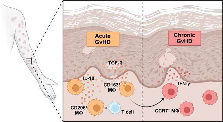
Background: Graft-versus-host disease (GvHD) is a major life-threatening complication of allogeneic haematopoietic stem cell transplantation (HSCT), limiting the broad application of HSCT for haematological malignancies. Cutaneous GvHD is described as a post-transplant inflammatory reaction by skin-infiltrating donor T cells and remaining recipient tissue-resident memory T cells. Despite the major influence of lymphocytes on GvHD pathogenesis, the complex role of mononuclear phagocytes (MNPs) in tissues affected by GvHD is increasingly appreciated.

Objectives: To characterize the identity, origin and functions of MNPs in patients with acute cutaneous GvHD.

Methods: Using single-cell RNA sequencing and multiplex tissue immunofluorescence, we identified an increased abundance of MNPs in skin and blood from 36 patients with acute cutaneous GvHD. In cases of sex-mismatched transplantation, we used expression of X-linked genes to detect rapid tissue adaptation of newly recruited donor MNPs resulting in similar transcriptional states of host- and donor-derived macrophages within GvHD skin lesions.

Results: We showed that cutaneous GvHD lesions harbour expanded CD163+ tissue-resident macrophage populations with anti-inflammatory and tissue-remodelling properties including interleukin-10 cytokine production. Cell-cell interaction analyses revealed putative signalling to strengthen regulatory T-cell responses. Notably, macrophage polarization in chronic cutaneous GvHD types was proinflammatory and drastically differed from acute GvHD, supporting the notion of distinct cellular players in different clinical GvHD subtypes.

Conclusions: Overall, our data reveal a surprisingly dynamic role of MNPs after HSCT. Specific and time-resolved targeting to repolarize this cell subset may present a promising therapeutic strategy in combatting GvHD skin inflammation.


Conflict of interest statement

Conflicts of interest The authors declare no conflicts of interest related to this study.

Figures

Graphical Abstract
Graphical Abstract
Figure 1
Figure 1
Cell populations in skin and blood of patients with acute graft-versus-host disease (aGvHD) and healthy donors. (a) Graphical overview of patient sampling. (b) UMAP clustering of aGvHD (n = 5) and healthy (n = 4) single-cell RNA-sequencing (scRNa-seq) datasets (left panel) and blood and skin cells according to cell barcode distribution (right panel). (c) UMAP showing Leiden clustering according to neighbourhood analysis in cells from (b). (d) Dot plot displaying cell type marker gene expression in Leiden clusters from (c). Data are shown as mean expression of respective genes in each group. (e) Leucocyte clusters identified by marker gene expression in (d). (f) Relative fraction of cell clusters identified in (e) derived from healthy (n = 4) and aGvHD (n = 5) datasets. Data are shown as mean cell fraction per cell type. HSC, haematopoietic stem cells; MNP, mononuclear phagocytes; NK, natural killer; UMAP, Uniform Manifold Approximation and Projection.
Figure 2
Figure 2
Expansion of macrophages (MΦ) in acute graft-versus-host disease (aGvHD) skin lesions. (a) UMAP showing Leiden subclusters from mononuclear phagocyte (MNP) cells from aGvHD (n = 5) and healthy control single-cell RNA-sequencing datasets (n = 4). (b) Distribution of skin and blood-derived cells in (a). (c) Dot plot showing marker genes used to identify MNP subclusters from (a). (d) Dataset origin in cells from (a). (e) Number of MΦ in aGvHD (n = 28) and healthy control skin (n = 10). Data are shown as CD11b/CD68 double-positive cells mm–2 ± SEM. Statistical analysis used an unpaired Student’s t-test. (f) Representative immunofluorescence staining of CD11b and CD68 in aGvHD and healthy control skin section. Dashed line indicates dermoepidermal border. Scale bar = 100 µm. DAPI, 4′,6-diamidino-2-phenylindole; DC, dendritic cells; Mono, monocytes; Neutro, neutrophils; p, plasmacytoid; UMAP, Uniform Manifold Approximation and Projection.
Figure 3
Figure 3
Acute graft-versus-host disease (aGvHD) skin lesions are marked by increased interleukin (IL)-10-producing CD163+ macrophages (MΦ). (a) UMAP clustering of aGvHD skin mononuclear phagocyte (MNP) cell states. (b) Dot plot of representative genes in cells from (a). (c) Representative image of CD68 and CD163 immunostaining in aGvHD skin. Scale bar = 20 µm. (d) CCR7+ and CD163+ MΦ in aGvHD skin (n = 15). Data are shown as percentage of MΦ (individual values and mean ± SEM). (e) Dot plot showing expression of profibrotic and proinflammatory cytokines and cytokine receptors in cells from (a). (f) interferon (IFN)-γ+ and IL-10+ MΦ in aGvHD skin (n = 18). Data are shown as mean percentage of cytokine-expressing MΦ (individual values and mean ± SEM). (g) Ratio of proinflammatory/profibrotic MΦs in aGvHD skin (n = 17) vs. healthy controls (n = 10). Data are shown as mean ratio of IFN-γ+ to IL-10+ MΦ. Bar represents mean ± SEM. Statistical analysis used paired (d, f) and unpaired (g) Student’s t-test. DC, dendritic cells; IFNG, IFN gamma; LC, Langerhans cells; TGFB, transforming growth factor beta; UMAP, Uniform Manifold Approximation and Projection.
Figure 4
Figure 4
Interaction of macrophages (MΦ) and T cells in acute graft-versus-host disease (aGvHD) skin. (a) UMAP clustering of aGvHD skin-derived cells used for cell–cell interaction predictions. (b) Dot plot displaying putative receptor-ligand signalling of mononuclear phagocytes (MNP) to T cells (left) and T cells to MNP (right). Interactions were considered for P-values < 0.01 and significant gene upregulation in > 15 cells of each cluster. Data are shown as relative communication probability. (c) Image showing immunofluorescence labelling of CD68 + CD206+ and CD3 + CD45+ cells in aGvHD skin. Left panel is an overview image: arrows show cell–cell contact of MΦ and T cells, and line indicates dermoepidermal border (scale bar = 100 µm). Right panel shows magnification of MΦ–T-cell contact (scale bar = 20 µm). (d) Graphical representation of putative cell–cell interaction proteins between MΦ and T cells in GvHD skin. IFN-γ, interferon gamma; TNF-α, tumour necrosis factor alpha; TNFR, TNF receptor; UMAP, Uniform Manifold Approximation and Projection.
Figure 5
Figure 5
X-linked gene expression reveals similar transcriptional profiles of host and donor macrophages (MΦ). (a) Graphical representation of mismatched haematopoietic stem cell transplantation (HSCT) and sampling. (b) UMAP showing XIST gene expression in acute graft-versus-host disease (aGvHD) lesional skin MΦ of patients after mismatched HSCT (n = 2). (c) UMAP clustering (left panel) and relative cell fraction (right panel) of host and donor skin MΦ identified by XIST expression (b). (d) Dot plot showing proinflammatory and tissue-remodelling gene expression in cells from (c). IFNG, interferon gamma; IL, interleukin; scRNA-seq, single-cell RNA sequencing; TGFB, transforming growth factor beta; UMAP, Uniform Manifold Approximation and Projection.
Figure 6
Figure 6
Distinct macrophage (MΦ) polarization profiles are found in acute graft-versus-host disease (aGvHD), chronic lichenoid (cl)GvHD and chronic sclerotic (cs)GvHD subtypes. (a) Graphical representation of GvHD patient sampling. (b) Quantification of CD11b + CD68+ cells in aGvHD (n = 15), clGvHD (n = 9) and csGvHD (n = 7) lesions vs. healthy skin (n = 10). Data are shown as number mm–2 ± SEM. (c, d) MΦ polarization in GvHD subtypes. Data are shown as percentage of MΦs positive for surface markers CCR7 (c) and CD163 (d). Bar indicates mean ± SEM. (e–h) Cytokine expression by CD68+ MΦ in samples from (b). Data are shown as mean ± SEM and individual percentage of interferon gamma (IFN-γ)+ (e) and interleukin (IL)-10+ cells among CD68+ MΦ positive for either cytokine. (f, h) Representative images of immunofluorescence staining for IFN-γ and IL-10 in MΦ clusters of the dermis of an aGvHD sample (scale bar = 20 µm). **P < 0.01, ***P < 0.001, ****P < 0.0001.

Comment in

References

    1. Zeiser R, Blazar BR. Acute graft-versus-host disease – biologic process, prevention, and therapy. N Engl J Med 2017; 377:2167–79. - PMC - PubMed
    1. Strobl J, Pandey RV, Krausgruber T. et al. Long-term skin-resident memory T cells proliferate in situ and are involved in human graft-versus-host disease. Sci Transl Med 2020; 12:eabb7028. - PMC - PubMed
    1. Matte CC, Liu J, Cormier J. et al. Donor APCs are required for maximal GVHD but not for GVL. Nat Med 2004; 10:987–92. - PubMed
    1. Tugues S, Amorim S, Spath S. et al. Graft-versus-host disease, but not graft-versus-leukemia immunity, is mediated by GM-CSF-licensed myeloid cells. Sci Transl Med 2018; 10:eaat8410. - PubMed
    1. Haniffa M, Ginhoux F, Wang XN. et al. Differential rates of replacement of human dermal dendritic cells and macrophages during hematopoietic stem cell transplantation. J Exp Med 2009; 206:371–85. - PMC - PubMed

MeSH terms