

Soft tissue prediction in orthognathic surgery: Improving accuracy by means of anatomical details
- PMID: 38011187
- PMCID: PMC10681161
- DOI: 10.1371/journal.pone.0294640
Soft tissue prediction in orthognathic surgery: Improving accuracy by means of anatomical details
Abstract
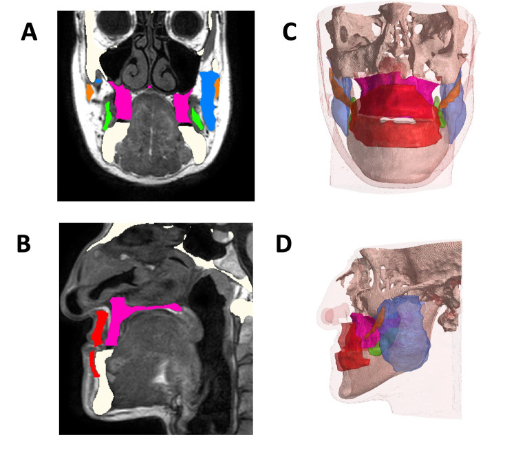
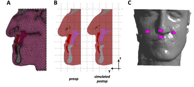
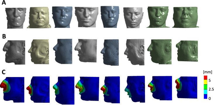
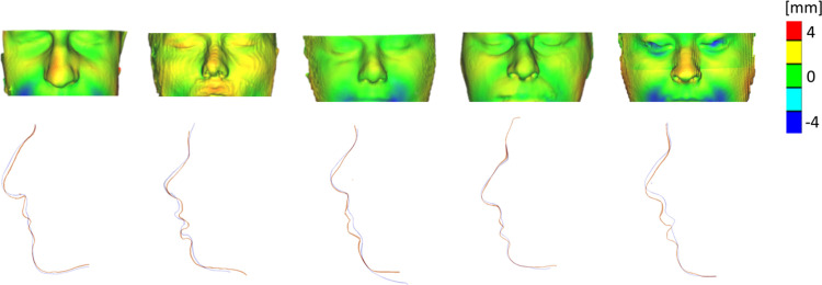
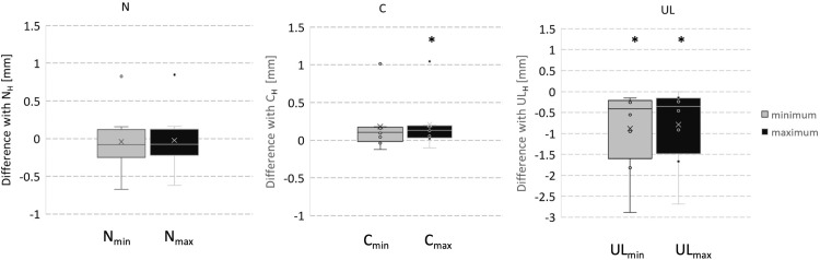
Three-dimensional virtual simulation of orthognathic surgery is now a well-established method in maxillo-facial surgery. The commercial software packages are still burdened by a consistent imprecision on soft tissue predictions. In this study, the authors produced an anatomically detailed patient specific numerical model for simulation of soft tissue changes in orthognathic surgery. Eight patients were prospectively enrolled. Each patient underwent CBCT and planar x-rays prior to surgery and in addition received an MRI scan. Postoperative soft-tissue change was simulated using Finite Element Modeling (FEM) relying on a patient-specific 3D models generated combining data from preoperative CBCT (hard tissue) scans and MRI scans (muscles and skin). An initial simulation was performed assuming that all the muscles and the other soft tissue had the same material properties (Homogeneous Model). This model was compared with the postoperative CBCT 3D simulation for validation purpose. Design of experiments (DoE) was used to assess the effect of the presence of the muscles considered and of their variation in stiffness. The effect of single muscles was evaluated in specific areas of the midface. The quantitative distance error between the homogeneous model and actual patient surfaces for the midface area was 0.55 mm, standard deviation 2.9 mm. In our experience, including muscles in the numerical simulation of orthognathic surgery, brought an improvement in the quality of the simulation obtained.
Copyright: © 2023 Ruggiero et al. This is an open access article distributed under the terms of the Creative Commons Attribution License, which permits unrestricted use, distribution, and reproduction in any medium, provided the original author and source are credited.
Conflict of interest statement
The authors have declared that no competing interests exist.
Figures
References
-
- Xia J. J., Gateno J., Teichgraeber J. F., Christensen A. M., Lasky R. E., Lemoine J. J. et al.. (2007) ‘Accuracy of the Computer-Aided Surgical Simulation (CASS) System in the Treatment of Patients With Complex Craniomaxillofacial Deformity: A Pilot Study’, Journal of Oral and Maxillofacial Surgery, 65(2), pp. 248–54. doi: 10.1016/j.joms.2006.10.005 - DOI - PubMed
-
- Aboul-Hosn Centenero S. and Hernández-Alfaro F. (2012) ‘3D planning in orthognathic surgery: CAD/CAM surgical splints and prediction of the soft and hard tissues results ‐ Our experience in 16 cases’, Journal of Cranio-Maxillofacial Surgery, 40(2), pp. 162–168. doi: 10.1016/j.jcms.2011.03.014 - DOI - PubMed
-
- Mazzoni S., Bianchi A., Schiariti G., Badiali G. and Marchetti C. (2015) ‘Computer-aided design and computer-aided manufacturing cutting guides and customized titanium plates are useful in upper maxilla waferless repositioning’, Journal of Oral and Maxillofacial Surgery, 73(4), pp. 701–707. doi: 10.1016/j.joms.2014.10.028 - DOI - PubMed
MeSH terms
LinkOut - more resources
Full Text Sources
