. 2024 Apr;8(4):397-414.
doi: 10.1038/s41551-023-01142-x. Epub 2023 Nov 27.

Genetically encoding multiple functionalities into extracellular vesicles for the targeted delivery of biologics to T cells

Affiliations

Genetically encoding multiple functionalities into extracellular vesicles for the targeted delivery of biologics to T cells

Devin M Stranford et al. Nat Biomed Eng. 2024 Apr.

Abstract

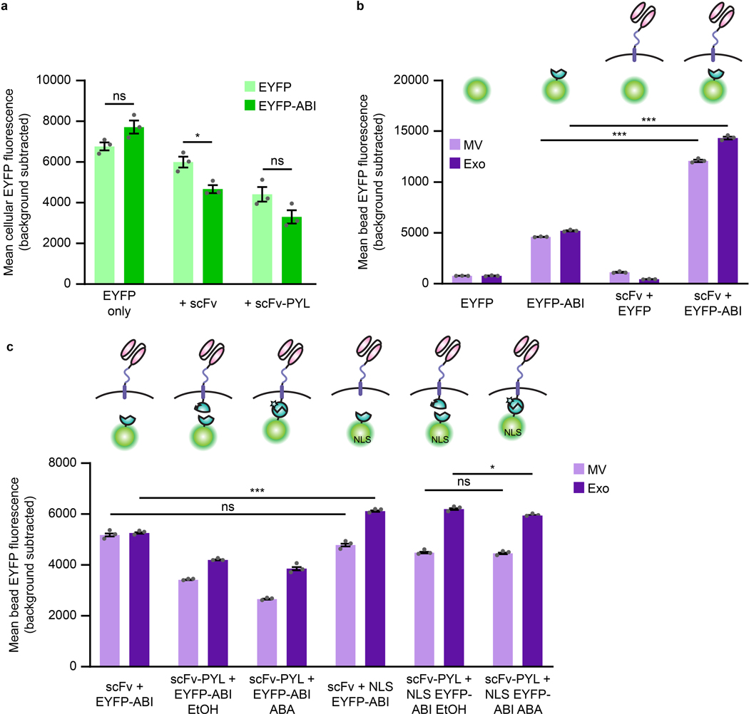
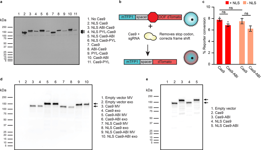
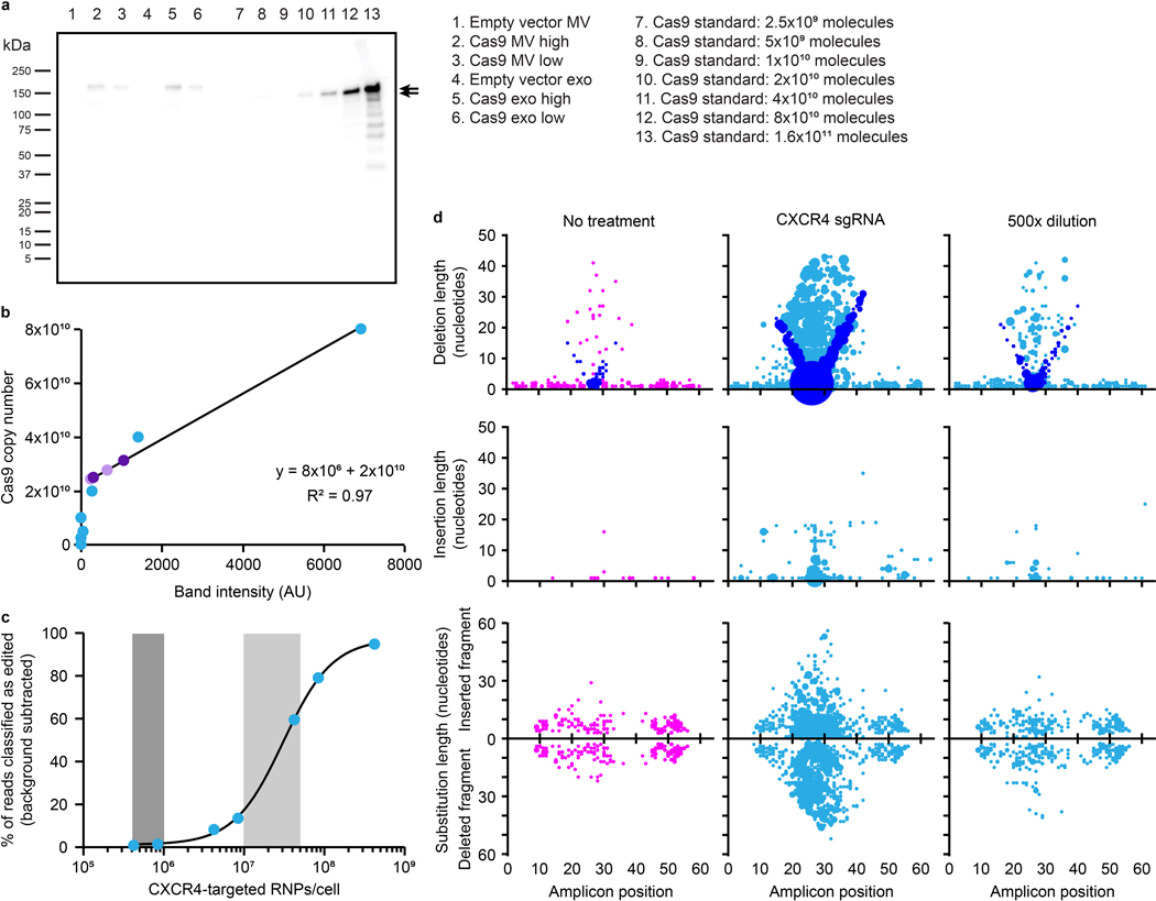
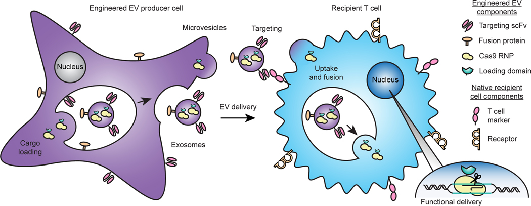
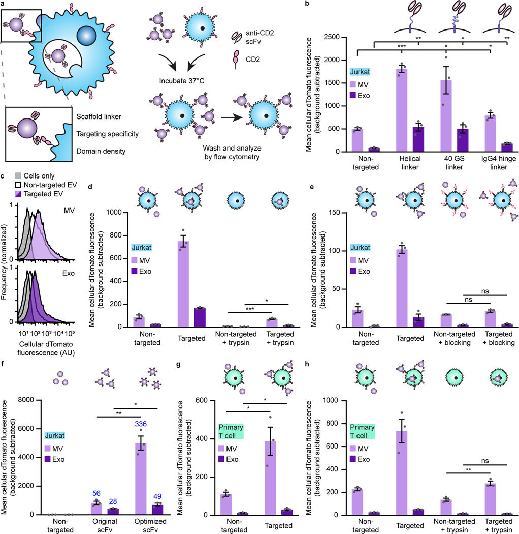
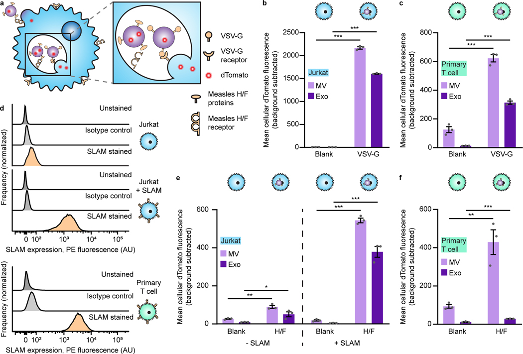
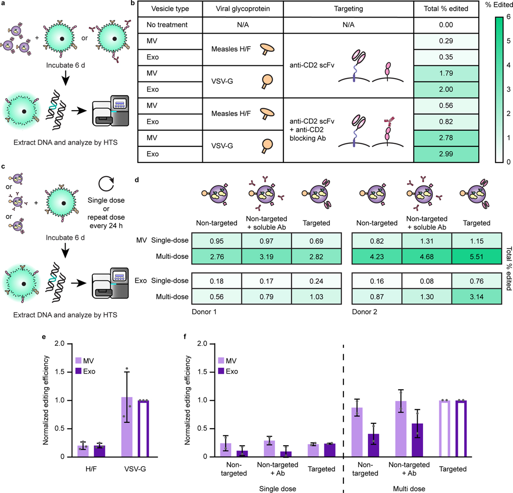
The genetic modification of T cells has advanced cellular immunotherapies, yet the delivery of biologics specifically to T cells remains challenging. Here we report a suite of methods for the genetic engineering of cells to produce extracellular vesicles (EVs)-which naturally encapsulate and transfer proteins and nucleic acids between cells-for the targeted delivery of biologics to T cells without the need for chemical modifications. Specifically, the engineered cells secreted EVs that actively loaded protein cargo via a protein tag and that displayed high-affinity T-cell-targeting domains and fusogenic glycoproteins. We validated the methods by engineering EVs that delivered Cas9-single-guide-RNA complexes to ablate the gene encoding the C-X-C chemokine co-receptor type 4 in primary human CD4+ T cells. The strategy is amenable to the targeted delivery of biologics to other cell types.


Conflict of interest statement

J.N.L. and D.M.S. are co-inventors on patent pending intellectual property that covers some technologies reported in this manuscript. J.N.L. and D.M.S. have financial interest in Syenex, which could potentially benefit from the outcomes of this research.

Figures

ED Fig. 1
ED Fig. 1. Different scFv display techniques result in different EV targeting properties.
a, Cartoon highlighting the structures of the PDGFR transmembrane domain scFv display and lactadherin C1C2 domain anchoring to phosphatidylserine. b, Expression of scFv constructs in EV producer cell lysates. 1 μg cell lysate was loaded per lane. Expected band sizes: ~40 kDa and ~75 kDa (black arrows). c, Loading of scFv constructs into EVs generated from cell lines in b. 5.0×10⁸ EVs were loaded per lane. d, Binding of targeted EVs to Jurkat T cells following a 2 h incubation. e, Representative histograms corresponding to the summary data reported in d. The subpopulation of cells showing a skewed, high degree of exosome binding is indicated by the red box. f, Recipient Jurkat T cells were incubated for 1 h in the presence or absence of anti-CD2 antibodies prior to a 2 h incubation with EVs. g, Representative histograms corresponding to the summary data reported in f. Flow cytometry experiments were performed in biological triplicate, and error bars (panels d, f) indicate standard error of the mean. EV dTomato loading evaluations are presented in Supplementary Fig. 5. Statistical tests comprise two-tailed Student’s t-tests using the Benjamini-Hochberg method to reduce the false discovery rate. (*p < 0.05, **p < 0.01, ***p < 0.001). Exact p-values are reported in Supplementary Table 1.
ED Fig. 2
ED Fig. 2. The ABI domain increases EV cargo loading independent of total protein expression.
a, Expression of EYFP and EYFP-ABI in the presence of anti-CD2 targeting constructs in transiently transfected HEK293FT cells analyzed by flow cytometry. A key observation is that addition of the ABI domain does not increase overall cargo protein expression in producer cells. b, Repeat of EYFP-ABI EV loading trends in the presence of an scFv shown in Fig. 3c. c, Comparison of EYFP loading into EVs with and without an NLS with ABA-binding constructs and under ABA-induced dimerization conditions. Addition of an NLS did not substantially impact EYFP loading, nor did ABA-induced dimerization substantially impact loading of nuclear-localized cargo. Experiments were performed in biological triplicate, and error bars indicate standard error of the mean. Statistical tests comprise two-tailed Student’s t-tests using the Benjamini-Hochberg method to reduce the false discovery rate. (*p < 0.05, **p < 0.01, ***p < 0.001). Exact p-values are reported in Supplementary Table 1.
ED Fig. 3
ED Fig. 3. The ABI domain increases Cas9 loading into EVs and Cas9-ABI retains function.
a, Expression of Cas9 fused to either the ABI or PYL domain in transiently transfected HEK293FT cells. 2 μg cell lysate was loaded per lane. Expected band sizes: ~160, 183, and 195 kDa (arrows). b, Cartoon illustrating the Cas9 reporter construct. Successful editing by Cas9 results in the deletion of a stop codon and (in some random fraction of cases) a repair-mediated frame shift induces express dTomato. c, Absence of an NLS or presence of the ABI domain does not meaningfully reduce Cas9 editing efficiency in transiently transfected Jurkat T cells. Cells were analyzed by flow cytometry 3 d post-transfection. Experiments were performed in biological triplicate, and error bars indicate standard error of the mean. Statistical tests comprise two-tailed Student’s t-tests using the Benjamini-Hochberg method to reduce the false discovery rate. (*p < 0.05, **p < 0.01, ***p < 0.001). Exact p-values are reported in Supplementary Table 1. Samples with high cellular autofluorescence were excluded from analysis. d, Full blot of Cas9 EV active loading data presented in Fig. 3e. e, Cellular expression of Cas9 with and without the ABI domain or an NLS. 2 μg cell lysate was loaded per lane.
ED Fig. 4
ED Fig. 4. Electroporation of recombinant Cas9-sgRNA ribonucleoprotein complexes into primary T cells confers dose-dependent editing of the genomic CXCR4 target locus.
a, Analysis of actively loaded Cas9 molecules per EV in vesicles displaying an anti-CD2 scFv and VSV-G. Lanes loaded with 4.0 × 10⁸ (“high”) or 2.0 × 10⁸ (“low”) EVs were compared to samples loaded with specified numbers of recombinant Cas9 molecules quantified based upon the manufacturer’s analysis (lanes 7–13). Expected band sizes (~160 or 195 kDa, arrows) correspond to Cas9 +/− the ABI domain. b, Quantification of Cas9 RNPs per EV. Band intensities from Cas9 standards in a were plotted against Cas9 molecules loaded (blue points), and loading of Cas9 into EVs was calculated using a line fit to the linear regime of the recombinant Cas9 standard curve (purple points; light: MV, dark: exo); this analysis indicates a loading of ~100 Cas9 molecules per EV. c, Frequency of indels detected at the Cas9-targeted CXCR4 locus after electroporation of CD4+ T cells with different doses of CXCR4-targeted RNPs. Doses of recombinant Cas9 that correspond to equivalent Cas9 molecules per cell as EV delivery and equivalent Cas9 editing efficiencies as EVs are highlighted in light and dark grey, respectively. Background subtraction was performed using an untreated control; treatment of T cells with RNPs complexed with a non-targeted sgRNA produces similar levels of apparent CXCR4 editing as did the untreated controls, likely indicating that these conditions both represent noise associated with this assay. d, Distributions of RNP-mediated edits, by type, as described in Fig. 5. The no treatment, non-diluted CXCR4 sgRNA treatment (maximum editing), and 500x sgRNA dilution (similar editing frequency as EV-mediated delivery of Cas9-sgRNA) conditions are shown.
Fig. 1:
Fig. 1:. Overview of the GEMINI strategy for genetically engineering multifunctional EVs.
EV cargo proteins are expressed in producer cells to facilitate incorporation into multiple vesicle populations: microvesicles, which bud from the cell surface, or exosomes, which are produced by endosomal invaginations into multivesicular bodies. Surface-displayed targeting and fusion proteins aid in binding to and uptake by recipient cells and subsequent cargo release via cell surface fusion or endosomal escape. In the proof-of-principle application explored in this study, the objective is to deliver a Cas9-sgRNA complex to T cells in order to knock out a gene, as described in subsequent sections.
Fig. 2:
Fig. 2:. Display of scFvs on EVs mediates receptor-specific, targeted binding and uptake to T cells.
a, Strategy for targeting EVs to T cells (left) and illustration of EV binding experiments (right). b, Targeted EVs binding to Jurkats (2 h incubation). To evaluate potential differences in dTomato loading, average EV fluorescence was analyzed separately (Supplementary Fig. 5). c, Representative histograms depicting distributions of helical linker EV-mediated fluorescence in recipient cells analyzed in b. d, Distinguishing binding and internalization for EVs targeted to Jurkats. Trypsinization was used to remove bound, non-internalized EVs following a 6 h incubation. e, Specificity of EV targeting to CD2. Pre-incubation with anti-CD2 antibodies ablated EV targeting to Jurkats. f, Enhancement of targeting by codon-optimized expression of scFv constructs. Fold increases over the non-targeted control are reported in blue. g, Binding of targeted EVs to primary human CD4+ T cells (2 h incubation). h, Distinguishing binding and internalization for EVs targeted to primary human CD4+ T cells. All experiments were performed in biological triplicate, and error bars indicate standard error of the mean. Statistical tests comprise two-tailed Student’s t-tests using the Benjamini-Hochberg method to reduce the false discovery rate. (*p < 0.05, **p < 0.01, ***p < 0.001). Exact p-values are reported in Supplementary Table 1. EV dTomato loading evaluations are in Supplementary Fig. 5.
Fig. 3:
Fig. 3:. Cargo protein is actively loaded into EVs via tagging with the ABI domain of the abscisic acid dimerization system.
a, Illustration of abscisic acid-based dimerization of EV cargo proteins and subsequent loading into vesicles. b, ABA-induced dimerization between PYL and ABI domains. Illustrative microscopy showing anti-CD2 scFv-PYL (membrane bound) and EYFP-ABI (cytosolic) association in the presence of ABA. Full images are in Supplementary Fig. 10. c, ABI-induced cargo loading into EVs. EVs generated under conditions indicated were adsorbed to aldehyde/sulfate latex beads and analyzed by flow cytometry to determine bulk average fluorescence. Experiments were performed in biological triplicate, and error bars indicate standard error of the mean. Statistical tests comprise two-tailed Student’s t tests using the Benjamini-Hochberg method to reduce the false discovery rate. (*p < 0.05, **p < 0.01, ***p < 0.001). Exact p-values are reported in Supplementary Table 1. d, Representative histograms of EYFP +/− ABI conditions in c (two leftmost sets of bars). e, Active loading of Cas9-ABI with and without an NLS into EVs. 6.0×10⁸ EVs were loaded per lane. Expected band sizes (~160 or 195 kDa, arrows) correspond to Cas9 −/+ the ABI domain. The full blot is provided in Extended Data Fig. 3d. f, Analysis of ABA-dependent Cas9-ABI loading into EVs enriched for anti-CD2 scFv-PYL via affinity chromatography. 1.3×10⁷ MVs or 2.0×10⁷ exos were loaded per lane. Expected band size: 195 kDa (arrows). Full blots are provided in Supplementary Fig. 11b. g, Bioactivity of EV-associated Cas9. Vesicles were lysed and incubated with a linearized target plasmid for 1 h at 37°C in Cas9 nuclease reaction buffer. Expected cut band sizes: 7.6 and 4.6 kb (arrows). For all gels, cropped regions are annotated in the Source Data.
Fig. 4:
Fig. 4:. Viral glycoprotein display on EVs mediates uptake by recipient T cells.
a, Illustration of viral glycoproteins facilitating EV uptake and fusion at either the plasma membrane or in the endosome. b, Uptake of dTomato-labeled VSV-G EVs by Jurkat T cells. c, Uptake of dTomato-labeled VSV-G EVs by primary human CD4+ T cells. d, Surface expression of SLAM on T cells. Unmodified Jurkats, Jurkats expressing transgenic SLAM, or primary human CD4+ T cells were evaluated for SLAM surface expression by flow cytometry. e, Uptake of dTomato-labeled measles viral glycoproteins H/F EVs by Jurkats (+/− SLAM). f, Uptake of dTomato-labeled measles virus glycoproteins H/F EVs by primary human CD4+ T cells. In all cases, EVs were incubated with cells for 16 h and trypsinized to remove surface-bound vesicles. Experiments were performed in biological triplicate, and error bars indicate standard error of the mean. Statistical tests comprise two-tailed Student’s t tests using the Benjamini-Hochberg method to reduce the false discovery rate. (*p < 0.05, **p < 0.01, ***p < 0.001). Exact p-values are shown in Supplementary Table 1. EV dTomato loading evaluations are in Supplementary Fig. 12.
Fig. 5:
Fig. 5:. EVs mediate functional delivery of Cas9 in primary human T cells.
a, Illustration of function delivery evaluation. 2.0×10¹⁰ EVs were incubated with 5.0×10⁴ CD4+ T cells for 6 d prior to genomic DNA extraction and HTS analysis. b, Frequency of indels detected at the Cas9-targeted CXCR4 locus. The sgRNA recognition site (green), PAM sequence (underlined, red), and predicted cut site (amplicon position 26, scissors) are shown. Total percentage of HTS reads classified as “edited” represents the area under the histogram trace shown for each sample. c, Distributions of EV-Cas9-mediated edits, by type. DNA amplicon position is plotted on the abscissa and length of the edit observed is plotted on the ordinate, while the size of each dot scales with the number of edits that meet that description. Each read is uniquely classified as a deletion, insertion, or substitution such that no one read contributes to more than one dot in this panel. In the case of substitutions, the positive ordinate reports the insertion portion of the edit, and the negative ordinate reports the deletion portion of the edit such that each edit is represented by two dots. In this panel, deletions are reported by placing a dot at the midpoint of the deleted segment. To help explain the apparent “V” pattern, dots are colored blue to indicate cases where one end of the deleted segment corresponds to the predicted cut region, presumably corresponding to a subset of the DNA repair outcomes observed. Sample dot coloring is as in b.
Fig. 6:
Fig. 6:. CD2 engagement and repeat dosing enhance EV-mediated functional cargo delivery and vary with vesicle subpopulation.
a, Illustration of strategy for probing the requirement of scFv-CD2 engagement by blocking CD2. b, Blocking CD2 on recipient cells prior to EV addition increases total editing for all vesicle types. 8.0×10⁹ EVs were incubated per 4.0×10⁴ CD4+ T cells for 6 d prior to genomic DNA extraction and HTS analysis. Heat map coloring scales from 0–6% total Cas9-mediated editing. c,d, Illustration (c) and evaluation (d) of experiments probing Cas9-mediated editing after repeat EV administration and various modes of CD2 engagement. Two independent experiments using different donor cells and EV preparations are shown to explicitly capture variation across experiments. EV dosing was: Donor 1—1.25×10¹⁰ MVs or 5.50×10⁹ exos per 5×10⁴ cells; Donor 2—1.50×10¹⁰ MVs or 7.50×10⁹ exos per 5×10⁴ cells. Heat map coloring is as in b. e, EV-mediated Cas9 functional delivery shows consistent trends across 3 donors and EV batches. Editing efficiency was normalized to the sample receiving VSV-G exosomes (open bar) for each of three independent experiments. f, Combined analysis of experiments presented in d. Within each vesicle population, editing efficiencies were normalized to the sample receiving multiple doses of VSV-g EVs (open bars); this normalization strategy is designed to control for expected sources of greatest variation (i.e., intrinsic donor/T cell batch-specific susceptibility to EVs and editing). Error bars represent one standard deviation.

References

    1. Sadelain M, Rivière I. & Riddell S. Therapeutic T cell engineering. Nature 545, 423–431 (2017). 10.1038/nature22395 - DOI - PMC - PubMed
    1. Yin C. et al. In Vivo Excision of HIV-1 Provirus by saCas9 and Multiplex Single-Guide RNAs in Animal Models. Molecular therapy : the journal of the American Society of Gene Therapy 25, 1168–1186 (2017). 10.1016/j.ymthe.2017.03.012 - DOI - PMC - PubMed
    1. Kaminski R. et al. Excision of HIV-1 DNA by gene editing: a proof-of-concept in vivo study. Gene therapy 23, 690–695 (2016). 10.1038/gt.2016.41 - DOI - PMC - PubMed
    1. Dash PK et al. Sequential LASER ART and CRISPR Treatments Eliminate HIV-1 in a Subset of Infected Humanized Mice. Nature communications 10, 2753 (2019). 10.1038/s41467-019-10366-y - DOI - PMC - PubMed
    1. Louis Jeune V, Joergensen JA, Hajjar RJ & Weber T. Pre-existing anti-adeno-associated virus antibodies as a challenge in AAV gene therapy. Human gene therapy methods 24, 59–67 (2013). 10.1089/hgtb.2012.243 - DOI - PMC - PubMed

Publication types

Substances