

Novel regulatory roles of small G protein GDP dissociation stimulator (smgGDS) in insulin secretion from pancreatic β-cells
- PMID: 38013223
- PMCID: PMC10842139
- DOI: 10.1016/j.mce.2023.112104
Novel regulatory roles of small G protein GDP dissociation stimulator (smgGDS) in insulin secretion from pancreatic β-cells
Abstract
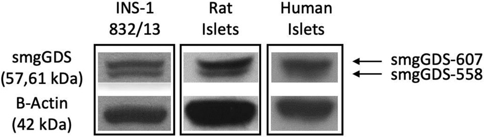
Emerging evidence implicates novel roles for small G protein GDP dissociation stimulator (smgGDS) in G protein activation and subsequent targeting to relevant subcellular compartments for effector regulation. Given the well-established roles of small G proteins in insulin secretion, we undertook this investigation to determine the putative roles of smgGDS in insulin secretion. Immunoblotting studies revealed that both splice variants of smgGDS are expressed in human islets, rat islets and INS-1 832/13 cells. A significant inhibition (-52%) of glucose-stimulated insulin secretion (GSIS) was observed in INS-1 832/13 cells following siRNA-mediated depletion of smgGDS. In addition, insulin secretion elicited by a membrane depolarizing concentration of KCl (via increased calcium influx), forskolin (via increased cAMP generation) or IBMX (via inhibition of phosphodiesterase) was inhibited by -49%, -27%, and -28%, respectively. Subcellular distribution studies revealed no significant alterations in the abundance of smgGDS in the cytosolic and membrane fractions during the 45-min exposure of INS-1 832/13 cells to an insulinotropic concentration of glucose. Together, we present the first evidence of expression of smgGDS in human islets, rodent islets, and clonal β-cells. We also demonstrate novel regulatory roles of these proteins in insulin secretion derived from glucose metabolic events, including calcium- and cAMP-dependent signaling steps.
Keywords: Insulin secretion; Islet beta cell; RAP1GDS1; Small G proteins; smgGDS.
Published by Elsevier B.V.
Conflict of interest statement
Declaration of competing interest The authors declare that they have no known competing financial interests or personal relationships that could have appeared to influence the work reported in this paper.
Figures


Similar articles
-
Novel Roles for Geranylgeranyl Transferase-III (GGTase-III) in Insulin Secretion.Cell Physiol Biochem. 2025 Jun 30;59(3):419-426. doi: 10.33594/000000783. Cell Physiol Biochem. 2025. PMID: 40598917
-
An INS-1 832/13 𝛽-Cell Proteome Highlights the Rapid Regulation of Fatty Acid Biosynthesis in Glucose-Stimulated Insulin Secretion.Proteomics. 2025 Aug;25(15):13-26. doi: 10.1002/pmic.70005. Epub 2025 Jul 20. Proteomics. 2025. PMID: 40685699 Free PMC article.
-
METRNL represses beta-to-alpha cell trans-differentiation to maintain beta cell function under diabetic metabolic stress in mice.Diabetologia. 2025 Aug;68(8):1769-1788. doi: 10.1007/s00125-025-06459-7. Epub 2025 Jun 10. Diabetologia. 2025. PMID: 40495021
-
Evaluating glucose-dependent insulinotropic polypeptide and glucagon as key regulators of insulin secretion in the pancreatic islet.Am J Physiol Endocrinol Metab. 2024 Jul 1;327(1):E103-E110. doi: 10.1152/ajpendo.00360.2023. Epub 2024 May 22. Am J Physiol Endocrinol Metab. 2024. PMID: 38775725 Free PMC article. Review.
-
Exploring pancreatic beta-cell subgroups and their connectivity.Nat Metab. 2024 Nov;6(11):2039-2053. doi: 10.1038/s42255-024-01097-6. Epub 2024 Aug 8. Nat Metab. 2024. PMID: 39117960 Review.
Cited by
-
Hyperglycemic Stress Induces Expression, Degradation, and Nuclear Association of Rho GDP Dissociation Inhibitor 2 (RhoGDIβ) in Pancreatic β-Cells.Cells. 2024 Feb 1;13(3):272. doi: 10.3390/cells13030272. Cells. 2024. PMID: 38334664 Free PMC article.
-
Protein prenylation in islet β-cell function in health and metabolic stress.Biochem Pharmacol. 2025 Aug;238:116994. doi: 10.1016/j.bcp.2025.116994. Epub 2025 May 21. Biochem Pharmacol. 2025. PMID: 40409598 Review.
References
-
- Asahara S, Shibutani Y, Teruyama K, Inoue HY, Kawada Y, Etoh H, Matsuda T, Kimura-Koyanagi M, Hashimoto N, Sakahara M, Fujimoto W, Takahashi H, Ueda S, Hosooka T, Satoh T, Inoue H, Matsumoto M, Aiba A, Kasuga M and Kido Y (2013). "Ras-related C3 botulinum toxin substrate 1 (RAC1) regulates glucose-stimulated insulin secretion via modulation of F-actin." Diabetologia 56(5): 1088–1097. - PMC - PubMed
-
- Bergom C, Hauser AD, Rymaszewski A, Gonyo P, Prokop JW, Jennings BC, Lawton AJ, Frei A, Lorimer EL, Aguilera-Barrantes I, Mackinnon AC Jr., Noon K, Fierke CA and Williams CL (2016). "The tumor-suppressive small GTPase DiRas1 binds the noncanonical guanine nucleotide exchange factor SmgGDS and antagonizes SmgGDS interactions with oncogenic small GTPases." J Biol Chem 291(20): 10948. - PMC - PubMed
MeSH terms
Substances
Grants and funding
LinkOut - more resources
Full Text Sources
Molecular Biology Databases
