Comment
. 2024 Feb;14(2):276-289.
doi: 10.1002/2211-5463.13742. Epub 2023 Dec 19.

Dysfunction of Drosophila mitochondrial carrier homolog (Mtch) alters apoptosis and disturbs development

Affiliations
Comment

Dysfunction of Drosophila mitochondrial carrier homolog (Mtch) alters apoptosis and disturbs development

Cristina González et al. FEBS Open Bio. 2024 Feb.

Abstract

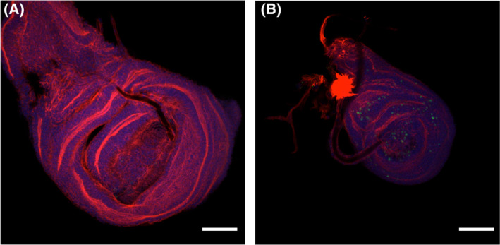
Mitochondrial carrier homologs 1 (MTCH1) and 2 (MTCH2) are orphan members of the mitochondrial transporter family SLC25. Human MTCH1 is also known as presenilin 1-associated protein, PSAP. MTCH2 is a receptor for tBid and is related to lipid metabolism. Both proteins have been recently described as protein insertases of the outer mitochondrial membrane. We have depleted Mtch in Drosophila and show here that mutant flies are unable to complete development, showing an excess of apoptosis during pupation; this observation was confirmed by RNAi in Schneider cells. These findings are contrary to what has been described in humans. We discuss the implications in view of recent reports concerning the function of these proteins.

Keywords: Drosophila; apoptosis; development; mitochondria; mitochondrial carrier homolog (MTCH).


Conflict of interest statement

The authors declare no conflict of interest.

Figures

Fig. 1
Fig. 1
Alignment of human and Drosophila proteins. Human Mtch1 (short spliced isoform starting at the third ATG, HsMTCH1S3ATG), human Mtch2 (HsMTCH2), Drosophila Mtch (DmMtch) and Drosophila CG10920 (DmCG10920) were aligned using clustalw2 with default settings. An asterisk indicates fully conserved residues; colon indicates strongly similar properties; period indicates weakly similar properties. Amino acids in bold indicate a peptide in human Mtch2 (140–161) that binds tightest to tBid [53]. Equivalent amino acids conserved in DmMtch are also marked in bold letters. Underlined amino acids in the DmMtch sequence refer to those that have been reported by Robinson et al. [17] to be conserved in mitochondrial carrier proteins, specifically referring to the Bos taurus ATP/ADP carrier shown in their Fig. 1. The proline (dark gray) that is part of the Px[D/E]xx[K/R] motif present in the SLC25 repeats, as well as polar and charged residues (light gray) highlighted by Guna et al. [30] are also marked in the human MTCH2 sequence.
Fig. 2
Fig. 2
Phylogenetic analysis of Mtch orthologs. (A) Radial phylogenetic tree of Mtch orthologs in vertebrates and invertebrates (including 12 sequenced drosophilids). Most vertebrate species contain two copies of Mtch homologs, one of which related to human MTCH1 and the other to human MTCH2. Exceptions (gray font) are cartilaginous fishes (such as sharks) that contain one MTCH1‐like gene, and bony fishes (teleosts) and amphibians that contain one MTCH2‐like gene. The invertebrate species generally contain one Mtch homolog. However, in drosophilids there are 2‐3 Mtch homologs which we call CG6851‐like and CG10920‐like groups. (B) Detailed phylogram and the syntenic relationships (Muller elements, colored dots) among the Mtch orthologs of the 12 sequenced fly genomes. Because karyotypes vary in different Drosophila species, a six‐element Muller element designation (A–F) is used as a standardized notation for the syntenic relationship, i.e. conserved chromosome regions among species. Additionally, a view of the phylogenetic relationships of the species in different groups is shown (colored bars). All drosophilid species shown contain two Mtch orthologs, except Dgri and Dwil which each have three orthologs. The Mtch orthologs can be divided clearly in two phylogenetic/syntenic groups, one being “CG6851‐like” (Muller D) and another “CG10920‐like” (Muller A). The third Mtch orthologs in Dgri (GH13246) and Dwil (Gk13597) are phylogenetically CG10920‐like but located in different Muller elements suggesting more recent gene duplication in these species.
Fig. 3
Fig. 3
Phenotypes of wild type, mutant and revertant larva. (A) Representative images of each phenotype. (B) Graph indicating the lengths, in mm, of larva (L3) from each type averaged in each case from 6 to 12 individuals (average length is indicated inside each bar). Error bars represent standard deviation. Yw, wild type; BL‐27981 (mutant 1), homozygote larva of this mutant; 27981 Jo, revertant of this mutant; BL‐28432 (mutant 2), homozygote larva of this mutant; 28432 Jo, revertant of this mutant. Statistical analysis was carried out using a two‐tailed test, with N = 3, where each N represents the average of 6 to 12 larva analyzed on different days. **P < 0.0005. Scale bar in A is 0.5 mm.
Fig. 4
Fig. 4
Increase in apoptosis in imaginal discs. Wing imaginal discs were isolated from wild type pupa (A) and from BL‐28432 homozygotes (B), stained for Actin (red) and activated caspase 3 (green) and photographed with a Nikon 90i microscope at 400× magnification. Representative images of at least 12 analyzed imaginal discs are shown. The scale bar is 100 μm.
Fig. 5
Fig. 5
Effect of RNA interference in cultured cells. (A) Mtch mRNA levels under standard and interference situations. The amount of the mRNA was determined by qPCR in exponentially growing Schneider SL2 cells in presence of dsRNA internal to LacZ, as a negative control, or Mtch dsRNA as indicated in Materials and methods. n = 6 independent experiments, mean ± SD are shown. ***P value < 0.001. (B) Quantification of viable cells and cells in early apoptosis in a Mtch interference situation and control either in presence and absence of staurosporine. Data are shown as a mean ± SD for n = 6 independent experiments. A statistical two‐tailed t‐student test was carried out. **P value < 0.01. ns, not significant.

Comment on

Similar articles

References

    1. Tait SW and Green DR (2013) Mitochondrial regulation of cell death. Cold Spring Harb Perspect Biol 5, a008706. - PMC - PubMed
    1. Elmore S (2007) Apoptosis: a review of programmed cell death. Toxicol Pathol 35, 495–516. - PMC - PubMed
    1. Luo X, Budihardjo I, Zou H, Slaughter C and Wang X (1998) Bid, a Bcl2 interacting protein, mediates cytochrome c release from mitochondria in response to activation of cell surface death receptors. Cell 94, 481–490. - PubMed
    1. Wang Y and Tjandra N (2013) Structural insights of tBid, the caspase‐8‐activated bid, and its BH3 domain. J Biol Chem 288, 35840–35851. - PMC - PubMed
    1. Zaltsman Y, Shachnai L, Yivgi‐Ohana N, Schwarz M, Maryanovich M, Houtkooper RH, Vaz FM, De Leonardis F, Fiermonte G, Palmieri F et al. (2010) MTCH2/MIMP is a major facilitator of tBID recruitment to mitochondria. Nat Cell Biol 12, 553–562. - PMC - PubMed

Publication types

Substances

Associated data

LinkOut - more resources