[Preprint]. 2023 Nov 17:2023.11.16.567274.
doi: 10.1101/2023.11.16.567274.

Macrophage Depletion Protects Against Cisplatin-Induced Ototoxicity and Nephrotoxicity

Affiliations

Macrophage Depletion Protects Against Cisplatin-Induced Ototoxicity and Nephrotoxicity

Cathy Yea Won Sung et al. bioRxiv. .

Update in

Abstract

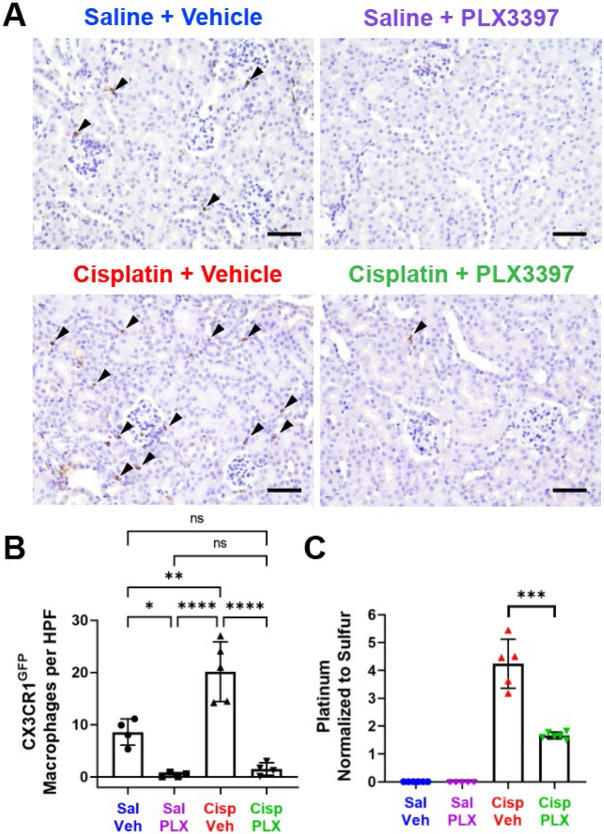
Cisplatin is a widely used and highly effective anti-cancer drug with significant side effects including ototoxicity and nephrotoxicity. Macrophages, the major resident immune cells in the cochlea and kidney, are important drivers of both inflammatory and tissue repair responses. To investigate the roles of macrophages in cisplatin-induced ototoxicity and nephrotoxicity, we used PLX3397, an FDA-approved inhibitor of the colony-stimulating factor 1 receptor (CSF1R), to eliminate tissue-resident macrophages during the course of cisplatin administration. Mice treated with cisplatin alone (cisplatin/vehicle) had significant hearing loss (ototoxicity) as well as kidney injury (nephrotoxicity). Macrophage ablation using PLX3397 resulted in significantly reduced hearing loss measured by auditory brainstem responses (ABR) and distortion-product otoacoustic emissions (DPOAE). Sensory hair cells in the cochlea were protected against cisplatin-induced death in mice treated with PLX3397. Macrophage ablation also protected against cisplatin-induced nephrotoxicity, as evidenced by markedly reduced tubular injury and fibrosis as well as reduced plasma blood urea nitrogen (BUN) and neutrophil gelatinase-associated lipocalin (NGAL) levels. Mechanistically, our data suggest that the protective effect of macrophage ablation against cisplatin-induced ototoxicity and nephrotoxicity is mediated by reduced platinum accumulation in both the inner ear and the kidney. Together our data indicate that ablation of tissue-resident macrophages represents a novel strategy for mitigating cisplatin-induced ototoxicity and nephrotoxicity.

Keywords: Ablation; Auditory Brainstem Response (ABR); CSF1R; Cisplatin; Hair cells; Kidney; Macrophages; Nephrotoxicity; Ototoxicity; PLX3397.


Figures

Figure 1.
Figure 1.. Initial protocol in Experiment 1 resulted in macrophage ablation via PLX3397 with partial repopulation.
(A) Experimental design showing auditory tests, PLX3397 treatment, and three cycles of cisplatin administration. Mice received PLX3397-formulated chow for seven days to facilitate macrophage depletion. During cisplatin administration, mice received PLX3397 via oral gavage once every three days. Upon observation of macrophage repopulation on day 14 (see Supplemental Figure 1), daily treatment of PLX3397 via oral gavage was initiated. The days on which mice received PLX3397 via oral gavage are denoted with blue circles. (B) Mice treated with the combination of cisplatin and PLX3397 exhibited significantly less weight loss compared to mice treated with cisplatin and vehicle. Mean±SEM, n=10-16 mice per experimental group. Statistical comparisons (asterisks or n.s.) are color-coded as described in Methods section. (C) Cochleae harvested after endpoint auditory tests were immunolabeled for Kir4.1 (yellow) to visualize cochlear structures and GFP to visualize CX3CR1GFP-positive macrophages (green). (C’) Representative confocal images (cochlear modiolus; from red box in panel C) and (D) quantification of macrophages per cochlear section. PLX3397 resulted in >93% depletion of all macrophages. (C”) Schematic diagram of macrophage distribution in the middle cochlear turn (from green box in panel C). Scale bar, 200 μm (cochlear section) and 50 μm (mid-modiolar section). (D) Quantification of macrophages per cochlear section. Mean±SD, n=6 cochleae per experimental group. One-way ANOVA with Tukey’s multiple comparisons test.
Figure 2.
Figure 2.. Partial macrophage ablation protected against cisplatin-induced hearing loss and OHC dysfunction (Experiment 1).
(A-C) Auditory sensitivity was measured via ABRs at baseline and endpoint. Threshold shifts are reported as the difference between baseline and endpoint ABR thresholds. (A) Cisplatin/vehicle-treated mice (red line) demonstrated significant hearing loss (threshold shifts) at all frequencies compared to saline/vehicle-treated mice (blue line). PLX3397 alone (purple line) did not cause hearing loss. PLX3397 provided significant protection against cisplatin-induced hearing loss (green line). (B-C) PLX3397 protected against cisplatin-induced hearing loss in both (B) female and (C) male mice, with better protection in males than in female (*p=0.0277). P values were calculated using one-way ANOVA with Tukey’s multiple comparisons test. Mean±SEM, n=10-16 mice per experimental group. Statistical comparisons (asterisks or n.s.) are color-coded as described in Methods. (D-I) OHC function was measured via DPOAEs. An emission at 2f1-f2 was considered present when its amplitude was greater than −5dB (dotted line). Mice co-treated with saline and PLX3397 maintained normal DPOAE amplitudes across an f2 range of 8-22.4kHz, with a small but significant decrease at 32 and 40kHz compared to control mice (saline/vehicle-treated mice). Mice treated with cisplatin/vehicle showed a significant reduction in DPOAE amplitudes. Macrophage ablation via PLX3397 treatment significantly protected against cisplatin-induced loss of DPOAE amplitudes. Statistical analyses were performed using two way-ANOVA with Tukey’s multiple comparisons test (main column effect). Mean±SEM, n=10-16 mice per experimental group.
Figure 3.
Figure 3.. Partial macrophage depletion via PLX3397 increased OHC survival and decreased cisplatin accumulation in the cochleae of cisplatin-treated mice (Experiment 1).
(A-B) PLX3397 alone did not affect the number of OHCs. Cisplatin/vehicle-treated mice show a significant reduction in the number of OHCs in the higher frequency regions (22.4 kHz and above). PLX3397 protected against cisplatin-induced OHC death. The number of IHCs was not different in any of the four treatment groups. (A) Scale bar, 20 μm. (B) Quantification of OHC and IHC numbers. The solid lines (blue, purple, red, green) represent OHC counts, and dotted lines represent IHC counts. Mean±SEM, n=6 cochleae per experimental group. Statistical analysis was performed using two way-ANOVA with Tukey’s multiple comparisons test (main column effect). (C) Platinum concentrations in microdissected inner ear tissues were measured using ICP-MS. Cochleae of control (no cisplatin) mice do not contain measurable platinum. Cochleae from mice treated with cisplatin showed significant increases in platinum in all cochlear regions. Mice treated with cisplatin and PLX3397 exhibited significantly lower platinum concentrations in all cochlear tissues, with stria vascularis showing the largest difference. Mean±SD, n=4-7 inner ears per experimental group. Statistical analysis was performed using one way-ANOVA with Tukey’s multiple comparisons test.
Figure 4.
Figure 4.. PLX3397 in rodent chow followed by daily oral gavage resulted in sustained macrophage ablation and a significant reduction in weight loss induced by cisplatin (Experiment 2).
(A) Experimental design of Experiment 2. Mice received either control chow or PLX3397-formulated chow for seven days to facilitate macrophage depletion. Following initiation of cisplatin administration, mice received daily PLX3397 treatment via oral gavage, which continued until euthanasia, ensuring sustained macrophage ablation. The days on which mice received PLX3397 through oral gavage are indicated with blue circles. (B) Mice co-administered cisplatin and vehicle exhibited significant weight loss, whereas those co-administered cisplatin and PLX3397 demonstrated significantly reduced cisplatin-induced weight loss. Mean±SEM, n=8-9 mice per experimental group. Statistical analysis was performed using one-way ANOVA with Tukey’s multiple comparisons test. Statistical comparisons (asterisks or n.s.) are color-coded as described in Methods. (C-D) Following sustained macrophage ablation via PLX3397, more than 96% of all macrophages were ablated in cochleae collected after endpoint auditory testing when compared to saline/vehicle-treated mice. (C) Scale bars in 200 μm (cochlear section) and 50 μm (modiolus, magnified inset). (D) Quantification of macrophages in whole cochlear sections. Mean±SD, n=5-6 cochleae per experimental group. Statistical analysis was performed using one-way ANOVA with Tukey’s multiple comparisons test.
Figure 5.
Figure 5.. Sustained depletion of macrophages via PLX3397 provided complete protection against cisplatin-induced hearing loss and OHC dysfunction (Experiment 2).
Hearing loss was assessed by ABRs, and OHC function was evaluated using DPOAEs. (A-C) Auditory tests were performed before (baseline) PLX3397 treatment and after (endpoint) completion of the cisplatin administration protocol. Hearing loss is reported as threshold shifts (the difference between baseline and endpoint ABR thresholds). PLX3397 in saline-treated mice (purple line) did not cause hearing loss when compared to saline/vehicle-treated mice (blue line). Cisplatin/vehicle-treated mice (red line) exhibited significant hearing loss (threshold shifts) at all frequencies . Cisplatin-treated mice receiving PLX3397 showed comparable ABR threshold shifts to saline-treated mice, indicating that PLX3397 resulted in complete protection against cisplatin-induced hearing loss. This protection was observed in both (B) female and (C) male mice. (D-I) PLX3397 in saline-treated mice did not elicit significant changes in OHC function (DPOAE amplitudes). Cisplatin/vehicle-treated mice (red line) exhibited significantly reduced DPOAE amplitudes compared to saline-treated mice. PLX3397 resulted in complete protection against cisplatin-induced OHC dysfunction. An emission at 2f1-f2 was considered present when its amplitude exceeded the threshold of −5dB (dotted line). The grey line represents the biological noise floor. For both ABRs and DPOAEs, data are shown as Mean±SEM, n=8-9 mice per experimental group. Statistical analysis was performed using two way-ANOVA with Tukey’s multiple comparisons test (main column effect).
Figure 6.
Figure 6.. Sustained macrophage ablation protected against cisplatin-induced OHC death and resulted in reduced cisplatin accumulation in the cochlea (Experiment 2).
(A-B) Cochlear wholemounts were stained for myosin 7a (cyan) to visualize hair cells. (A) Representative images and (B) quantitative analysis revealed that cisplatin administration resulted in death of OHCs at the 22.4 kHz region and above. PLX3397 provided near-complete protection against cisplatin-induced OHC loss, resulting in OHC numbers that were comparable to those in saline-treated mice. IHC counts did not differ in any of the experimental groups. (A) Scale bar, 100 μm. (B) Data are shown as Mean±SEM, n=6 cochleae per experimental group. Statistical analysis was performed using two way-ANOVA with Tukey’s multiple comparisons test (main column effect). (C) Platinum levels were measured using ICP-MS in microdissected inner ear tissues. Platinum was not detected in cochleae of mice that received saline/vehicle or saline/PLX3397 treatments. A significant increase in platinum was observed in all tissue samples from mice treated with cisplatin/vehicle, with the highest levels observed in the stria vascularis. Mice co-administered with cisplatin and PLX3397 exhibited significantly reduced platinum levels in the organ of Corti and spiral ligament, with the stria vascularis showing the largest reduction. No significant differences in platinum levels were observed in the SGNs or vestibular tissues when compared with mice that received cisplatin/vehicle treatment. Data are shown as Mean±SD, n=4-6 cochleae per experimental group. Statistical analysis was performed using one-way ANOVA with Tukey’s multiple comparisons test.
Figure 7.
Figure 7.. Sustained depletion of macrophages protects against cisplatin-induced kidney damage (Experiment 2).
Kidney function, tubular injury, and fibrosis were evaluated after endpoint auditory testing. Macrophage ablation using PLX3397 protected against cisplatin-induced increases in (A) plasma blood urea nitrogen (BUN) levels and (B) Neutrophil gelatinase-associated lipocalin (NGAL) levels. (C) Tubular injury was assessed semi quantitatively using a tubular injury score (see Methods). Cisplatin resulted in increased tubular injury scores, while PLX3397 protected against cisplatin-induced injury. (D) The percentage of fibrotic area in the entire surface area of the section was calculated. PLX3397 protected against cisplatin-induced fibrosis. (E-F) Representative images of cortico-medullary junction are presented, demonstrating (E) tubular injury using periodic acid-Schiff staining and (F) fibrosis using Masson-Trichrome staining. (A-D) Data are expressed as mean±SD, n=8–9 blood samples kidneys per experimental group. P-values were calculated using one-way ANOVA with Tukey’s multiple comparisons test. (E-F) The scale bar indicates 50 μm.
Figure 8.
Figure 8.. PLX3397 results in ablation of CX3CR1GFP-positive cells and significant reduction in cisplatin accumulation in the kidney (Experiment 2).
(A) Representative images of renal CX3CR1GFP-positive cells (arrowheads), identified through immunohistochemical staining. Scale bar indicates 50 μm. (B) The number of CX3CR1GFP-positive cells in each field was quantified (n = 4-5 mice per experimental group). Compared to control mice (saline/vehicle-treated mice), PLX3397 treatment in saline-treated mice and cisplatin-treated mice ablated 95.63% and 82.52% of CX3CR1GFP-positive cells, respectively. (C) Platinum levels in the kidney tissues were analyzed by ICP-MS (n = 5–6 mice per experimental group). Cisplatin resulted in increased platinum in the kidney, while PLX3397 significantly reduced cisplatin accumulation. Platinum levels were normalized to sulfur contained in the kidney. Data are expressed as mean±SE. P-values were calculated using one-way ANOVA with Tukey’s multiple comparisons test.
Figure 9.
Figure 9.. Working model of the mechanisms underlying the protective effects of macrophage ablation against cisplatin-induced hearing loss and kidney injury.
(A) In the absence of PLX3397, macrophages are present, and the cochlear BLB is permeable enough to permit cisplatin to cross the BLB and enter the cochlea, where it results in the death of OHCs, loss of ribbon synapses and SGNs, and hearing loss. In the presence of PLX3397, perivascular macrophages are ablated, and cisplatin entry into the cochlea is reduced, resulting in protection against cisplatin-induced hearing loss. (B) In the absence of PLX3397, renal macrophages are present and may promote cisplatin entry into proximal tubule cells causing renal fibrosis and kidney dysfunction. In the presence of PLX3397, macrophages are ablated in the kidney and cisplatin accumulation in the kidney is reduced, thus providing protection against cisplatin-induced tubular injury and interstitial fibrosis.

References

    1. Dasari S., Tchounwou P. B., Cisplatin in cancer therapy: molecular mechanisms of action. Eur J Pharmacol 740, 364–378 (2014). - PMC - PubMed
    1. Wozniak K., Blasiak J., Recognition and repair of DNA-cisplatin adducts. Acta Biochim Pol 49, 583–596 (2002). - PubMed
    1. Yao X., Panichpisal K., Kurtzman N., Nugent K., Cisplatin nephrotoxicity: a review. Am J Med Sci 334, 115–124 (2007). - PubMed
    1. Zhang J., Ye Z. W., Tew K. D., Townsend D. M., Cisplatin chemotherapy and renal function. Adv Cancer Res 152, 305–327 (2021). - PMC - PubMed
    1. Rybak L. P., Mukherjea D., Ramkumar V., Mechanisms of Cisplatin-Induced Ototoxicity and Prevention. Semin Hear 40, 197–204 (2019). - PMC - PubMed

Publication types