[Preprint]. 2023 Nov 13:2023.11.13.566911.
doi: 10.1101/2023.11.13.566911.

The Dmc1 recombinase physically interacts with and promotes the meiotic crossover functions of the Mlh1-Mlh3 endonuclease

Affiliations

The Dmc1 recombinase physically interacts with and promotes the meiotic crossover functions of the Mlh1-Mlh3 endonuclease

Gianno Pannafino et al. bioRxiv. .

Update in

Abstract

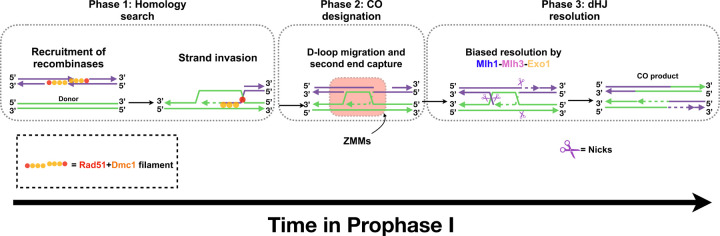
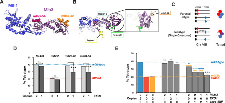
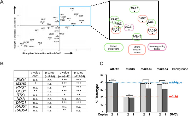
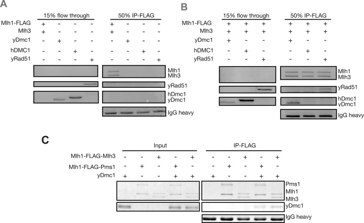
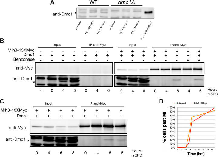
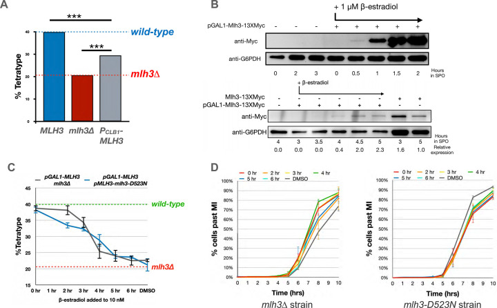
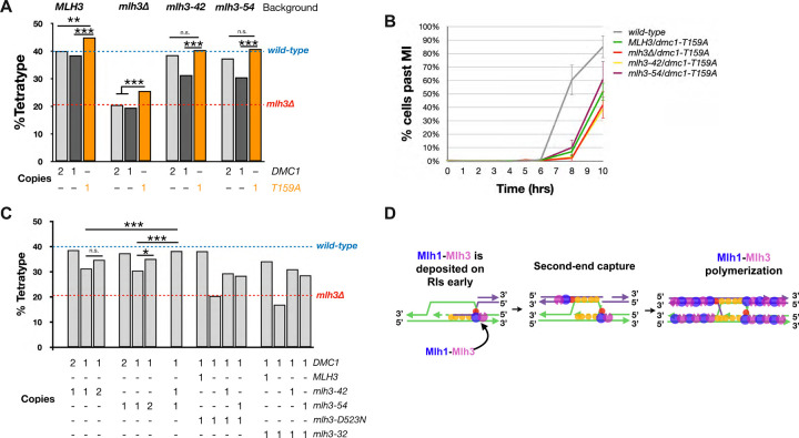
The accurate segregation of homologous chromosomes during the Meiosis I reductional division in most sexually reproducing eukaryotes requires crossing over between homologs. In baker's yeast approximately 80 percent of meiotic crossovers result from Mlh1-Mlh3 and Exo1 acting to resolve double-Holliday junction (dHJ) intermediates in a biased manner. Little is known about how Mlh1-Mlh3 is recruited to recombination intermediates and whether it interacts with other meiotic factors prior to its role in crossover resolution. We performed a haploinsufficiency screen in baker's yeast to identify novel genetic interactors with Mlh1-Mlh3 using sensitized mlh3 alleles that disrupt the stability of the Mlh1-Mlh3 complex and confer defects in mismatch repair but do not disrupt meiotic crossing over. We identified several genetic interactions between MLH3 and DMC1, the recombinase responsible for recombination between homologous chromosomes during meiosis. We then showed that Mlh3 physically interacts with Dmc1 in vitro and at times in meiotic prophase when Dmc1 acts as a recombinase. Interestingly, restricting MLH3 expression to roughly the time of crossover resolution resulted in a mlh3 null-like phenotype for crossing over. Our data are consistent with a model in which Dmc1 nucleates a polymer of Mlh1-Mlh3 to promote crossing over.

Keywords: Dmc1; Holliday junction resolution; Mlh1; Mlh3; crossing over; meiotic recombination.


Conflict of interest statement

Conflicts of interest. None declared.

Figures

Fig. 1.
Fig. 1.. Model of canonical resolvase role for Mlh1-Mlh3 in meiotic recombination.
A model for the Type I CO pathway in yeast meiotic prophase. Phase I, homology search: Following Spo11-mediated DSB formation and 5’ to 3’ resection of DSBs, a recombinase filament (Dmc1 filament initiates at the end of the Rad51 filament) forms at the 3’ resected ends. This filament initiates a search for the allelic locus on the homologous donor chromosome. Phase 2, CO designation: ZMM-facilitated stabilization of SEIs, D-loop migration, and second-end capture commit recombination intermediates to the Type I crossover pathway. Phase 3, dHJ resolution: Mlh1-Mlh3 and Exo1 participate in biased resolution of dHJs to produce crossover products.
Fig. 2.
Fig. 2.. Rationale for using mlh3-42 and mlh3-54 as baits (MMR− CO+) in gene dosage suppression analysis.
A. Crystal structure of the Mlh1-Mlh3 C-terminus (PDB 6RMN, Dai et al. 2020) with mutated residues highlighted by space filling (R552A, D553A, K555A, D556A for mlh3-42 and R682A, E684A for mlh3-54). B. Three regions within the Mlh3 C-terminus predicted to mediate the formation of an Mlh1-Mlh3 polymer (Dai et al. 2020) with amino acids changed in mlh3-42 highlighted as sticks for resolution purposes. C. Spore-autonomous fluorescence assay was used to measure tetratypes (single crossover events) in the CEN8-THR1 interval on chromosome VIII (Thacker et al. 2011). D. Crossover analysis in the CEN8-THR1 interval on chromosome VIII expressed as percent tetratype for indicated EXO1 gene dosage in wild-type, mlh3Δ, mlh3-32, and mlh3-54 strains. Dashed lines indicate wild-type, mlh3Δ, and exo1Δ levels of crossing over. E. Crossing over in the CEN8-THR1 interval on chromosome VIII expressed as percent tetratype for indicated gene dosages of MLH3, EXO1, and exo1-MIP. Significance was assessed according to χ2 p-values where p>0.05 = n.s. (not significant), p<0.05 = *, p<0.01 = **, and p<0.001 = ***. Blue asterisks indicate comparisons to wild-type and orange asterisks indicate comparisons to mlh3Δ. To account for multiple comparisons, we applied a Benjamini-Hochberg correction at a 5% false discovery rate (Benjamini and Hochberg, 1995).
Fig. 3.
Fig. 3.. EXO1, MSH5, PMS1, RTK1, and DMC1 are allele-specific interactors of MLH3.
A. Analysis of the strength of genetic interactions with mlh3-42 (x-axis) and mlh3-54 (y-axis) for 34 meiotic genes from various stages of meiotic prophase. See Materials and methods for rationale and Supplementary File 4 underlying data for raw tetrad counts and significance measurements. B. Statistical confirmation of allele-specific interactions (see Materials and methods). C. DMC1 displays dosage-specific interaction with mlh3-42 and mlh3-54 strains. Dashed lines indicate wild-type and mlh3Δ levels of crossing over. Significance was assessed as described in the Materials and methods.
Fig. 4.
Fig. 4.. In vitro co-immunoprecipitation reactions performed with yMlh1-FLAG-Mlh3, yMlh1-FLAG-Pms1, yDmc1, yRad51, and hDmc1.
A. Unbound protein flow throughs showing that yDmc1, yRad51 and human Dmc1 do not bind non-specifically to Dynabeads Protein G conjugated to anti-FLAG M2 antibody. B, C. Immunoprecipitation reactions containing the indicated purified proteins and Dynabeads Protein G conjugated to anti-FLAG M2 antibody. In all panels proteins were electrophoresed in 10% SDS-PAGE. See Materials and methods for details.
Fig. 5.
Fig. 5.. Mlh3 interacts with Dmc1 in vivo.
A. Western blot showing specificity of yDmc1 antibody. 20 μg of whole cell extracts prepared in the indicated NaCl concentrations were loaded per lane. 5 ng of purified yDmc1 was loaded in the yDmc1 lane. B. Time course pulldown of C-terminally tagged Mlh3-13xMyc at 0, 4, 6 hr following transfer to sporulation media. Controls include dmc1Δ input (lane 1, left most lane) and inputs/pulldowns for untagged, wild-type strains (lanes 2–7). Inputs/pulldowns for Mlh3-13xMyc are in lanes 8–13. Lanes 14–15 show Mlh3-13xMyc pulldowns at 4 and 6 hr when Benzonase was added to lysis buffer (125 Units/mL). C. Time course pulldown showing inputs/pulldowns for Mlh3-13xMyc at 0, 4, 6, 8 hr. D. Meiotic progression, as measured by completion of Meiosis I, is presented for the Mlh3-13X Myc tagged strain compared to an untagged strain. The amount of sample loaded in panels B and C were normalized based on cell mass.
Fig. 6.
Fig. 6.. Modulating expression of MLH3 in meiotic prophase.
A. MLH3 expression was placed under the control of the cyclin CLB1 promoter to drive late meiotic prophase expression. Crossing over in the CEN8-THR1 interval is presented. B. Western blot showing temporal lag between addition of β-estradiol (1μM, added at T= 0 hr, top, and 10 nM, added at T = 3 hrs, bottom) and subsequent Mlh3 protein expression. C. Comparison of crossover rates for strains expressing MLH3 from the GAL1 promoter upon addition of β-estradiol to sporulation media to a final concentration of 10 nM from 0 to 6 hr post transfer to sporulation media. Error bars represent standard error for three independent transformants analyzed on different days. D. Meiotic progression, as measured by completion of Meiosis I (MI), of sporulated cultures presented in panel C. Error bars represent standard error for the same three independent experiments shown in Panel C. Underlying data can be found in Supplementary File 4 and Supplementary File 5.
Fig. 7.
Fig. 7.. DMC1 haploinsufficiency phenotype is rescued by dmc1-T159A and functional MLH3 supplementation.
A. Crossover analysis at CEN8-THR1 for the indicated wild-type and dmc1-T159A strains. B. Meiotic progression of strains shown in panel A. Error bars represent standard error. C. CEN8-THR1 interval crossover data for strains containing 1 or 2 copies of DMC1 in mlh3-42, −54, -D523N (endonuclease defective), and −32 (crossover defective strains. D. A model proposing that retention of Dmc1 in meiotic recombination intermediates nucleates the formation of stable Mlh1-Mlh3 polymers required for dHJ resolution. Asterisks indicate significance (p>0.05 = n.s., p<0.05 = *, p<0.01 = **, and p<0.001 = ***).

Similar articles

References

    1. Abdullah MF, Hoffmann ER, Cotton VE, Borts RH. 2004. A role for the MutL homologue MLH2 in controlling heteroduplex formation and in regulating between two different crossover pathways in budding yeast. Cytogenet Genome Res. 107:180–190. - PubMed
    1. Ahuja JS, Harvey CS, Wheeler DL, Lichten M. 2021. Repeated strand invasion and extensive branch migration are hallmarks of meiotic recombination. Mol Cell. 81:4258–4270. - PMC - PubMed
    1. Allers T, Lichten M. 2001. Differential timing and control of noncrossover and crossover recombination during meiosis. Cell. 106:47–57. - PubMed
    1. Altmannova V, Firlej M, Müller F, Janning P, Rauleder R, Rousova D, Schäffler A, Bange T, Weir JR. 2023. Biochemical characterisation of Mer3 helicase interactions and the protection of meiotic recombination intermediates. Nucleic Acids Res. 51:4363–4384. doi: 10.1093/nar/gkad175. - DOI - PMC - PubMed
    1. Al-Sweel N, Raghavan V, Datta A, Ajith VP, Di Vietro L, Khondakar N, et al. 2017. mlh3 separation of function and endonuclease defective mutants display an unexpected effect on meiotic recombination outcomes. PLoS Genet. 13: e1006974. - PMC - PubMed

Publication types