[Preprint]. 2023 Nov 15:2023.11.15.565967.
doi: 10.1101/2023.11.15.565967.

Creation of de novo cryptic splicing for ALS/FTD precision medicine

Affiliations

Creation of de novo cryptic splicing for ALS/FTD precision medicine

Oscar G Wilkins et al. bioRxiv. .

Update in

  • Creation of de novo cryptic splicing for ALS and FTD precision medicine.
    Wilkins OG, Chien MZYJ, Wlaschin JJ, Barattucci S, Harley P, Mattedi F, Mehta PR, Pisliakova M, Ryadnov E, Keuss MJ, Thompson D, Digby H, Knez L, Simkin RL, Diaz JA, Zanovello M, Brown AL, Darbey A, Karda R, Fisher EMC, Cunningham TJ, Le Pichon CE, Ule J, Fratta P. Wilkins OG, et al. Science. 2024 Oct 4;386(6717):61-69. doi: 10.1126/science.adk2539. Epub 2024 Oct 3. Science. 2024. PMID: 39361759 Free PMC article.

Abstract

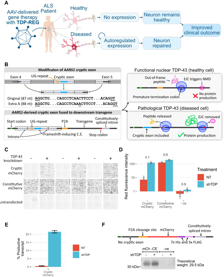
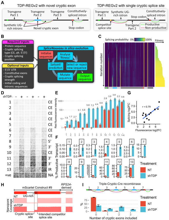
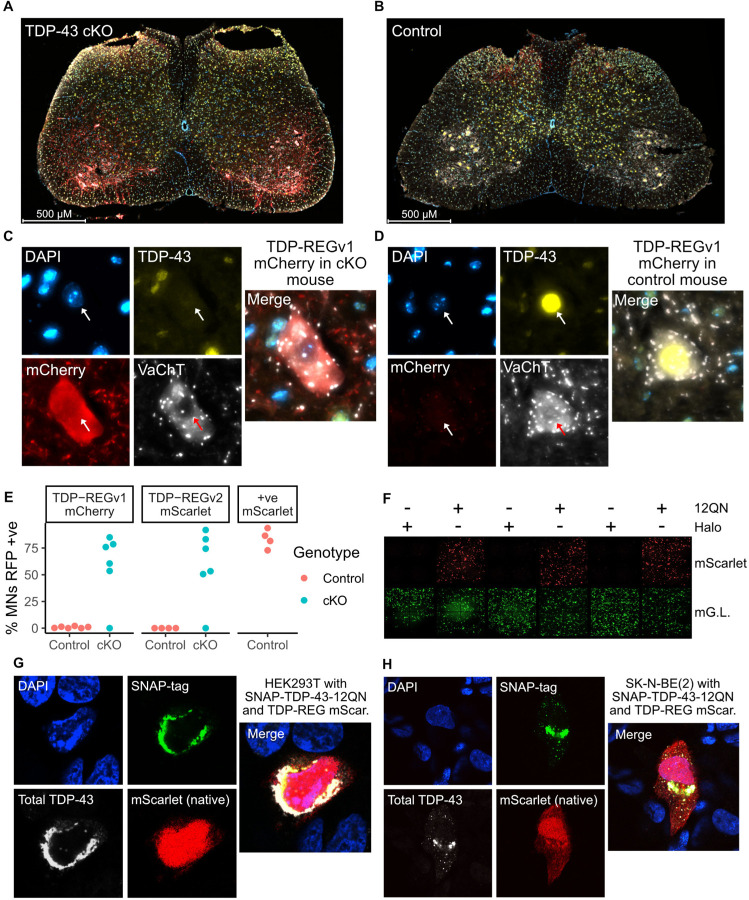
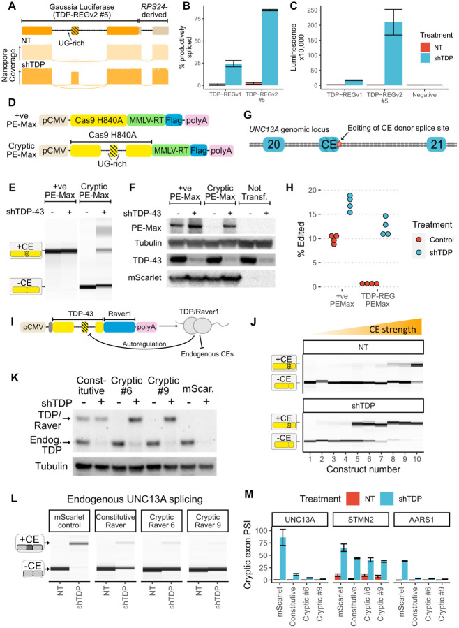
A system enabling the expression of therapeutic proteins specifically in diseased cells would be transformative, providing greatly increased safety and the possibility of pre-emptive treatment. Here we describe "TDP-REG", a precision medicine approach primarily for amyotrophic lateral sclerosis (ALS) and frontotemporal dementia (FTD), which exploits the cryptic splicing events that occur in cells with TDP-43 loss-of-function (TDP-LOF) in order to drive expression specifically in diseased cells. In addition to modifying existing cryptic exons for this purpose, we develop a deep-learning-powered algorithm for generating customisable cryptic splicing events, which can be embedded within virtually any coding sequence. By placing part of a coding sequence within a novel cryptic exon, we tightly couple protein expression to TDP-LOF. Protein expression is activated by TDP-LOF in vitro and in vivo, including TDP-LOF induced by cytoplasmic TDP-43 aggregation. In addition to generating a variety of fluorescent and luminescent reporters, we use this system to perform TDP-LOF-dependent genomic prime editing to ablate the UNC13A cryptic donor splice site. Furthermore, we design a panel of tightly gated, autoregulating vectors encoding a TDP-43/Raver1 fusion protein, which rescue key pathological cryptic splicing events. In summary, we combine deep-learning and rational design to create sophisticated splicing sensors, resulting in a platform that provides far safer therapeutics for neurodegeneration, potentially even enabling preemptive treatment of at-risk individuals.


Conflict of interest statement

Competing interests: PF and OGW have filed a patent application relating to TDP-REG technology. PF, ALB, MJK and OGW have filed a patent application relating to the use of ASO therapies for correcting cryptic splicing in UNC13A.

Figures

Fig. 1.
Fig. 1.. Regulatory upstream CE controls downstream transgene expression (TDP-REGv1).
A: Schematic showing the intended function and purpose of this work. B: Schematic of TDP-REGv1. Top left: the genomic locus surrounding the human AARS1 CE region was modified, with the middle parts of the introns removed to reduce size, and an extra adenosine added to the CE sequence to enable frame-shifting. The AG and GT splice sites of the CE are underlined. Bottom left: the modified sequence from part A was incorporated into a “minigene”. Top right: when the CE is excluded, the start codon is out-of-frame with the transgene, triggering nonsense-mediated decay (NMD) due to the downstream exon junction complex (EJC). Bottom right: when the CE is included, the transgene is in-frame with the start codon, resulting in protein expression. C: Fluorescence microscopy images (red channel) showing SK-N-BE(2) cells with (shTDP) or without (NT = “not treated”) TDP-43 knockdown, transfected with a vector fusing the upstream AARS1-derived sequence to mCherry (“cryptic mCherry”) or a constitutive vector. D: Quantification of the images in Part C; numbers show log2-fold-change in TDP-43 knockdown cells; each dot shows the average of one well (three wells per condition), with error bars showing the standard deviation within each well (four per well). E: Summary of Nanopore sequencing results for the cells in Part C; error bars show standard deviation across three replicates. F: Top: schematic of mCh -CE vector, without the CE but with a downstream His/Tri-FLAG dual tag to enable sensitive detection. Bottom: Western blot of cells transduced with the above vector (“mCh -CE”) or a completely different vector (“-ve” - a Prime Editing vector). Samples were enriched with a His-tag pulldown, then blotted with an anti-FLAG antibody.
Fig. 2.
Fig. 2.. AI-guided design of novel cryptic splicing events (TDP-REGv2):
A: Diagrams of internal cryptic exon (left) and single intron (right) designs; for the single intron design, an alternative 5’ splicing design is shown. B: Schematic of the SpliceNouveau algorithm for designing new cryptic splicing-dependent expression vectors. C: Heatmap showing the in silico evolution trajectory for an example internal cryptic exon design (below), with associated “fitness” of each (right), which was initialised with a constitutively spliced intron from RPS24 at the 3’ end. As the iteration number increases, splice sites are “evolved” at the desired positions, and off-target splice sites are depleted. The splice sites flanking the cryptic exon are weaker than the constitutive splice sites, as specified by the user. D: Fluorescence microscopy images showing mScarlet expression for 13 constructs generated by SpliceNouveau and a positive control. Each replicate consists of four images taken from different parts of the well. E: Quantification of D; each dot represents the average of the four images for each replicate; error bars show the standard deviation of images in each well. F: Percentage of productive transcripts (i.e. transcripts which are predicted to produce full-length mScarlet), as determined by Nanopore sequencing; error bars show standard deviation across three replicates. G: Correlation of fold changes in fluorescence and productive isoform fraction; Pearson correlation shown. H: Diagram of mScarlet construct 9 (top); representative Nanopore pileup with and without TDP-43 knockdown. I: Schematic of construct encoding Cre recombinase, split across seven exons; exons 2, 4 and 6 are flanked by UG-rich regions (top); number of cryptic exons included in each transcript without and with shTDP, assessed by Nanopore sequencing (bottom); error bars show standard deviation across three replicates.
Fig. 3.
Fig. 3.. Functionality of TDP-REG in different biological contexts:
A: Spinal cord of a TDP-43 cKO mouse injected with TDP-REGv1 mCherry AAV (blue = DAPI, yellow = TDP-43, red = mCherry, white = VAChT). B: Equivalent to part A, but with control mouse. C and D: Magnified representative motor neurons from figures in parts A and B respectively. E: Manual quantification of the percentage of motor neurons (MNs) with clear mCherry (TDP-REGv1 mCherry), mScarlet (TDP-REGv2 mScarlet #7 - see fig. S4A) or positive control mScarlet for cKO and control mice (only control mice for positive control AAV); N = 4–6 per condition. F: Fluorescence microscopy imaging of HEK293T cells transduced with an mScarlet TDP-REG reporter, a constitutive mGreenLantern vector, and either SNAP-TDP-43–12QN or Halo-tag expression vector. G and H: Confocal microscopy of HEK293T and SK-N-BE(2) cells, respectively, co-transfected with a TDP-REG mScarlet reporter (#7) and SNAP-TDP-43–12QN.
Fig. 4.
Fig. 4.. Biosensors, genome editing and splicing rescue:
A: Schematic of TDP-REGv2 Gluc vector #5 (top); representative Nanopore pileups with (shTDP) and without (NT=”not treated” with doxycycline) TDP-43 knockdown. B: Quantification of Nanopore data for Gluc TDP-REGv1 and TDP-REGv2 #5 for SK-N-BE(2) cells with and without TDP-43 knockdown; error bars show standard deviation across four replicates. C: Quantification of luminescence from supernatant of cells used for Part B; error bars show standard deviation across four replicates. D: Schematic of constitutive and cryptic (TDP-REGv2) Prime Editing vector (based on Addgene PE-Max vector). E: Capillary electrophoresis results from RT-PCR of cryptic or constitutive PE-Max vector transfected into SK-N-BE(2) cells with or without TDP-43 knockdown. F: Western blot of FLAG-tagged PE-Max vectors (co-transfected with FLAG-tagged mScarlet) in SK-N-BE(2) cells with or without TDP-43 knockdown. G: Diagram of UNC13A genomic locus surrounding the UNC13A CE between exons 20 and 21; the position of genome editing is shown. H: Quantification of intended editing of UNC13A in SK-N-BE(2) cells with either vector, +/− TDP-43 knockdown, assessed by targeted Nanopore sequencing. I: Schematic of TDP-43/Raver1 with an internal cryptic exon. The design features an additional dual N-terminal NLS; the constitutive vector encodes the same amino acid sequence, but with the introns removed. J: RT-PCR analysis of ten constructs designed (as in part A), using the 2FL mutation to block auto-repression of the cryptic exon; constructs were transfected into SK-N-BE(2) cells without (NT) or with (shTDP) doxycycline-inducible TDP-43 knockdown. K: Western blot of SK-N-BE(2) cells stably expressing constitutive or cryptic (vectors 6 and 9) TDP-43/Raver1, or mScarlet, with or without endogenous TDP-43 knockdown. Endogenous TDP-43 and TDP-43/Raver1 are labelled in the anti-TDP-43 blot; tubulin loading control is shown below. All lines are polyclonal piggyBac lines; three polyclonal lines were made per construct with consistent results. L: RT-PCR analysis of endogenous UNC13A cryptic splicing for the same lines as Part K, showing results from a single replicate. M: Quantification of UNC13A, STMN2 and AARS1 cryptic splicing, using all three polyclonal replicates; error bars show standard deviation across three replicates (one replicate for mScarlet control “NT” was excluded because the RNA failed QC).

References

    1. Scotter E. L., Chen H.-J., Shaw C. E., TDP-43 Proteinopathy and ALS: Insights into Disease Mechanisms and Therapeutic Targets. Neurotherapeutics. 12, 352–363 (2015). - PMC - PubMed
    1. McAleese K. E., Walker L., Erskine D., Thomas A. J., McKeith I. G., Attems J., TDP-43 pathology in Alzheimer’s disease, dementia with Lewy bodies and ageing. Brain Pathol. 27, 472–479 (2017). - PMC - PubMed
    1. Nelson P. T., Dickson D. W., Trojanowski J. Q., Jack C. R., Boyle P. A., Arfanakis K., Rademakers R., Alafuzoff I., Attems J., Brayne C., Coyle-Gilchrist I. T. S., Chui H. C., Fardo D. W., Flanagan M. E., Halliday G., Hokkanen S. R. K., Hunter S., Jicha G. A., Katsumata Y., Kawas C. H., Keene C. D., Kovacs G. G., Kukull W. A., Levey A. I., Makkinejad N., Montine T. J., Murayama S., Murray M. E., Nag S., Rissman R. A., Seeley W. W., Sperling R. A., White C. L. 3rd, Yu L., Schneider J. A., Limbic-predominant age-related TDP-43 encephalopathy (LATE): consensus working group report. Brain. 142, 1503–1527 (2019). - PMC - PubMed
    1. Ling J. P., Pletnikova O., Troncoso J. C., Wong P. C., TDP-43 repression of nonconserved cryptic exons is compromised in ALS-FTD. Science. 349, 650–655 (2015). - PMC - PubMed
    1. Klim J. R., Williams L. A., Limone F., Guerra San Juan I., Davis-Dusenbery B. N., Mordes D. A., Burberry A., Steinbaugh M. J., Gamage K. K., Kirchner R., Moccia R., Cassel S. H., Chen K., Wainger B. J., Woolf C. J., Eggan K., ALS-implicated protein TDP-43 sustains levels of STMN2, a mediator of motor neuron growth and repair. Nat. Neurosci. 22, 167–179 (2019). - PMC - PubMed

Publication types