

This is a preprint.
Hyper-physiologic mechanical cues, as an osteoarthritis disease relevant environmental perturbation, cause a critical shift in set-points of methylation at transcriptionally active CpG sites in neo-cartilage organoids
- PMID: 38014245
- PMCID: PMC10680909
- DOI: 10.21203/rs.3.rs-3568544/v1
Hyper-physiologic mechanical cues, as an osteoarthritis disease relevant environmental perturbation, cause a critical shift in set-points of methylation at transcriptionally active CpG sites in neo-cartilage organoids
Update in
-
Hyper-physiologic mechanical cues, as an osteoarthritis disease-relevant environmental perturbation, cause a critical shift in set points of methylation at transcriptionally active CpG sites in neo-cartilage organoids.Clin Epigenetics. 2024 May 10;16(1):64. doi: 10.1186/s13148-024-01676-0. Clin Epigenetics. 2024. PMID: 38730337 Free PMC article.
Abstract
Background: Osteoarthritis (OA) is a complex, age-related multifactorial degenerative disease of diarthrodial joints marked by impaired mobility, joint stiffness, pain, and a significant decrease in quality of life. Among other risk factors, such as genetics and age, hyper-physiological mechanical cues are known to play a critical role in the onset and progression of the disease (1). It has been shown that post-mitotic cells, such as articular chondrocytes, heavily rely on methylation at CpG sites to adapt to environmental cues and maintain phenotypic plasticity. However, these long-lasting adaptations may eventually have a negative impact on cellular performance. We hypothesize that hyper-physiologic mechanical loading leads to the accumulation of altered epigenetic markers in articular chondrocytes, resulting in a loss of the tightly regulated balance of gene expression that leads to a dysregulated state characteristic of the OA disease state.
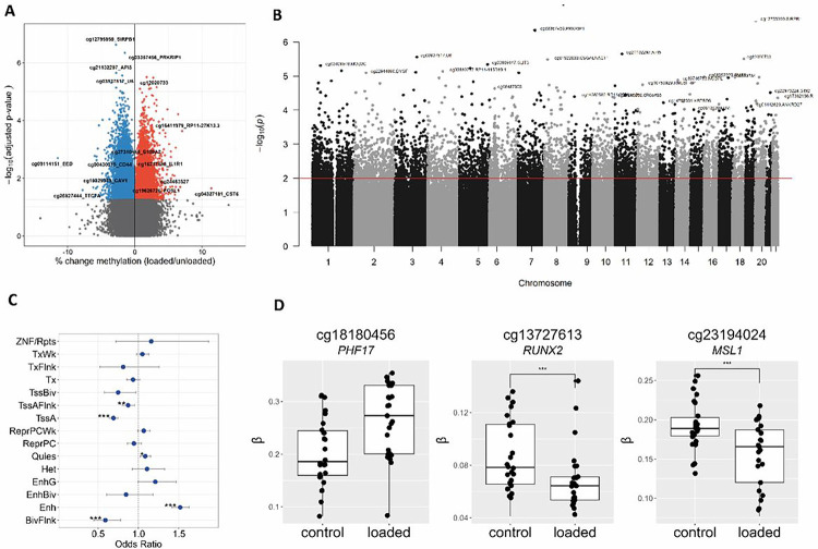
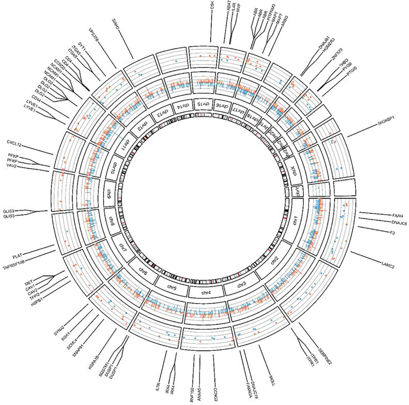
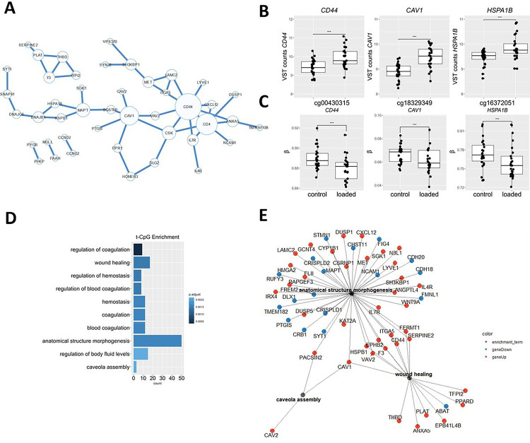
Results: We showed that hyper-physiological loading evokes consistent changes in ML-tCpGs associated with expression changes in ITGA5, CAV1, and CD44, among other genes, which together act in pathways such as anatomical structure morphogenesis (GO:0009653) and response to wound healing (GO:0042060). Moreover, by comparing the ML-tCpGs and their associated pathways to tCpGs in OA pathophysiology, we observed a modest but particular interconnected overlap with notable genes such as CD44 and ITGA5. These genes could indeed represent lasting detrimental changes to the phenotypic state of chondrocytes due to mechanical perturbations that occurred earlier in life. The latter is further suggested by the association between methylation levels of ML-tCpGs mapped to CD44 and OA severity.
Conclusion: Our findings confirm that hyper-physiological mechanical cues evoke changes to the methylome-wide landscape of chondrocytes, concomitant with detrimental changes in positional gene expression levels (ML-tCpGs). Since CAV1, ITGA5, and CD44 are subject to such changes and are central and overlapping with OA-tCPGs of primary chondrocytes, we propose that accumulation of hyper-physiological mechanical cues can evoke long-lasting, detrimental changes in set points of gene expression that influence the phenotypic healthy state of chondrocytes. Future studies are necessary to confirm this hypothesis.
Keywords: Chondrocytes; DNA Methylation; Environmental stressors; Mechanical Loading; Osteoarthritis.
Conflict of interest statement
Competing interests None
Figures
Similar articles
-
Hyper-physiologic mechanical cues, as an osteoarthritis disease-relevant environmental perturbation, cause a critical shift in set points of methylation at transcriptionally active CpG sites in neo-cartilage organoids.Clin Epigenetics. 2024 May 10;16(1):64. doi: 10.1186/s13148-024-01676-0. Clin Epigenetics. 2024. PMID: 38730337 Free PMC article.
-
DNA Methylation in Osteoarthritis.Curr Genomics. 2015 Dec;16(6):419-26. doi: 10.2174/1389202916666150817212711. Curr Genomics. 2015. PMID: 27019616 Free PMC article.
-
STAT3 promotes a youthful epigenetic state in articular chondrocytes.Aging Cell. 2023 Feb;22(2):e13773. doi: 10.1111/acel.13773. Epub 2023 Jan 13. Aging Cell. 2023. PMID: 36638270 Free PMC article.
-
Effects of shear stress on articular chondrocyte metabolism.Biorheology. 2000;37(1-2):95-107. Biorheology. 2000. PMID: 10912182 Review.
-
The interplay between biochemical mediators and mechanotransduction in chondrocytes: Unravelling the differential responses in primary knee osteoarthritis.Phys Life Rev. 2024 Mar;48:205-221. doi: 10.1016/j.plrev.2024.02.003. Epub 2024 Feb 12. Phys Life Rev. 2024. PMID: 38377727 Review.
References
-
- Chen CT, et al. (2001) Chondrocyte necrosis and apoptosis in impact damaged articular cartilage. J Orthop Res 19(4):703–711. - PubMed
-
- Lee JH, Fitzgerald JB, Dimicco MA, & Grodzinsky AJ (2005) Mechanical injury of cartilage explants causes specific time-dependent changes in chondrocyte gene expression. Arthritis Rheum 52(8):2386–2395. - PubMed
-
- Kurz B, et al. (2001) Biosynthetic response and mechanical properties of articular cartilage after injurious compression. J Orthop Res 19(6):1140–1146. - PubMed
Publication types
Grants and funding
LinkOut - more resources
Full Text Sources
Miscellaneous
