[Preprint]. 2023 Nov 21:2023.11.18.567607.
doi: 10.1101/2023.11.18.567607.

Control of Biofilm Formation by an Agrobacterium tumefaciens Pterin-Binding Periplasmic Protein Conserved Among Pathogenic Bacteria

Affiliations

Control of Biofilm Formation by an Agrobacterium tumefaciens Pterin-Binding Periplasmic Protein Conserved Among Pathogenic Bacteria

Jennifer L Greenwich et al. bioRxiv. .

Update in

Abstract

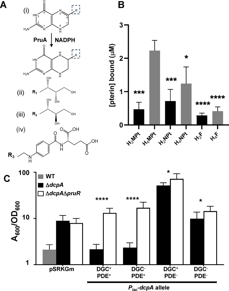
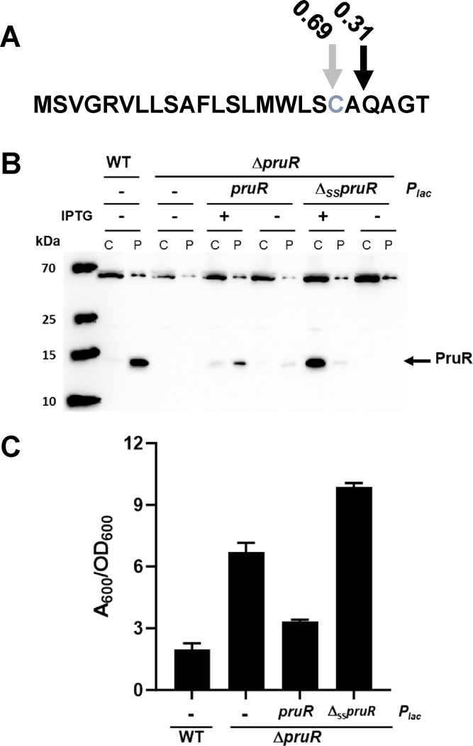
Biofilm formation and surface attachment in multiple Alphaproteobacteria is driven by unipolar polysaccharide (UPP) adhesins. The pathogen Agrobacterium tumefaciens produces a UPP adhesin, which is regulated by the intracellular second messenger cyclic diguanylate monophosphate (cdGMP). Prior studies revealed that DcpA, a diguanylate cyclase-phosphodiesterase (DGC-PDE), is crucial in control of UPP production and surface attachment. DcpA is regulated by PruR, a protein with distant similarity to enzymatic domains known to coordinate the molybdopterin cofactor (MoCo). Pterins are bicyclic nitrogen-rich compounds, several of which are formed via a non-essential branch of the folate biosynthesis pathway, distinct from MoCo. The pterin-binding protein PruR controls DcpA activity, fostering cdGMP breakdown and dampening its synthesis. Pterins are excreted and we report here that PruR associates with these metabolites in the periplasm, promoting interaction with the DcpA periplasmic domain. The pteridine reductase PruA, which reduces specific dihydro-pterin molecules to their tetrahydro forms, imparts control over DcpA activity through PruR. Tetrahydromonapterin preferentially associates with PruR relative to other related pterins, and the PruR-DcpA interaction is decreased in a pruA mutant. PruR and DcpA are encoded in an operon that is conserved amongst multiple Proteobacteria including mammalian pathogens. Crystal structures reveal that PruR and several orthologs adopt a conserved fold, with a pterin-specific binding cleft that coordinates the bicyclic pterin ring. These findings define a new pterin-responsive regulatory mechanism that controls biofilm formation and related cdGMP-dependent phenotypes in A. tumefaciens and is found in multiple additional bacterial pathogens.

Keywords: Biofilm; Biological Sciences; Microbiology; cyclic diguanylate monophosphate; protein structure; pterin; regulation.


Conflict of interest statement

Competing Interest Statement: K.J.F.S. has a significant interest in Situ Biosciences, a contract research organization that conducts research unrelated to this study. All other authors declare no conflicts of interest. The funders had no role in study design, data collection, and analysis, decision to publish, or preparation of the manuscript.

Figures

Figure 1.
Figure 1.. PruR binds pterins and is required to control both the DGC and PDE activity of DcpA.
(A) PruA reaction and relevant pterin molecule structures. (i): PruA uses NADPH as a cofactor and catalyzes the reduction of a 7,8-dihydropterin substrate to a 5,6,7.8-tetrahydropterin. Atoms are numbered in the dihydropterin. R indicates side groups as shown in (ii)-(iv); (ii): R1- monapterin; (iii): R2- neopterin; (iv): R3 – pABA-glutamate (folate). (B) In vitro pterin binding assays were performed as described in the supplemental methods with purified ΔSS-PruR (50 μM), NADPH, and with or without His6-PruA. HPLC fractioned reactions were examined by fluorescence for the oxidized pterins (excitation: 356 nm, emission: 450 nm). UV absorbance at 283 nm was used to measure folate relative to standards. PruR was incubated with the following pterin or folate species: H2MPt, dihydromonapterin; H4MPt, PruA-generated-tetrahydromonapterin; H2NPt, dihydroneopterin; H4NPt, PruA-generated tetrahydroneopterin; H2F, dihydrofolate; and H4F, tetrahydrofolate. Bars are averages of triplicate assays with error bars as standard deviations. Analyzed by standard one-way ANOVA and post-hoc Tukey analysis (P values relative to H4MPt, *, <0.05, *** <0.001, ****, <0.0001). (C) Biofilm assays of. A. tumefaciens C58 WT (gray bar), a ΔdcpA mutant (black bars) or a ΔdcpAΔpruR double mutant (white bars) containing the vector control or a plasmid-borne Plac-dcpA fusion expressing either the wild type dcpA or catalytic site mutants (DGC, E308A; PDE, E431A; DGCPDE, both). Ratio of acetic acid-solubilized CV absorbance (A600) from 48 h biofilm assays normalized to the OD600 planktonic turbidity from the same culture. Assays performed in triplicate and error bars are standard deviation; P values calculated comparing complementation of dcpA in the ΔdcpA strain compared to the ΔdcpAΔpruR strain by standard two-tailed t-test. (P values, * <0.05, **** <0.0001.)
Figure 2.
Figure 2.. PruR is a periplasmic protein.
(A) Signal P prediction of the PruR N-terminal signal sequence. Black arrow, predicted signal peptidase cleavage site; Grey arrow, putative cleavage and lipidation site; Grey text, predicted lipidated Cys19. (B) Western blot of SDS-PAGE using α-PruR polyclonal antisera to probe extracts of wild type A. tumefaciens C58 WT and the ΔpruR mutant on its own or expressing either the Plac-pruR plasmid or the PlacSSpruR plasmid (both plasmids also expressing dcpA). Cultures were grown to similar densities with or without induction with 400 μM IPTG and fractionated to separate the cytoplasmic/membrane fraction (C lanes) from the periplasmic fraction (P lanes). (C) Biofilm assays of WT or a ΔpruR mutant derivative with the empty vector plasmid (−) or harboring a plasmid-borne Plac fusion expressing either pruR or the ΔSSpruR (both plasmids also express dcpA). Ratio of acetic acid-solubilized CV A600 from 48 h biofilm assays normalized to the OD600 planktonic turbidity of the same culture. Assays performed in triplicate and error bars are standard deviation; P values calculated by standard two-tailed t-test (P values, *** <0.01).
Figure 3.
Figure 3.. PruR forms a complex with the periplasmic region of DcpA.
(A) Western blot probing for PruR with α–PruR polyclonal antisera . Whole cell suspensions were either untreated or incubated with DSS crosslinker (0.75 mM) for wild type A. tumefaciens C58 and the ΔdcpA mutant. Wild type A. tumefaciens harboring a plasmid ectopically expressing either pruR-dcpA or pruA from Plac (400 μM IPTG) was also subjected to same analysis. Black arrows; PruR-DcpA complex, 87 kDa; DcpA, 71 kDa; PruR; 16 kDa. (B) Similar western blot to panel A probing for PruR following DSS crosslinking of whole cell suspensions of either A. tumefaciens wild type strain or the ΔdcpA mutant. Either the cytoplasmic (DcpAcyt) or periplasmic (DcpAperi) domains of DcpA are ectopically expressed from Plac (400 μM IPTG). Red triangles, PruR-DcpA, 87 kDa; Blue triangles, expected size of PruR-DcpAcyt, ~68 kDa; Black triangles, PruR-DcpAperi,38 kDa. α-PruR used at 1:40,000 and antibody binding detected with GAR-HRP secondary antibody and chemiluminescent substrate on a BioRad ChemiDoc. Non-specific bands serve as protein loading controls.
Figure 4.
Figure 4.. Deletion of pruA diminishes the interaction between DcpA and PruR.
Western blot probing for the PruR-DcpAperi complex following in vivo DSS crosslinking in A. tumefaciens wild type or a ΔpruA mutant expressing the Plac-pruR-dcpAcyt plasmid (400 μM IPTG). Probed with polyclonal antibody preparations (A) α-PruR, 1:40,000; (B) α-DcpAperi, 1:20,000. Antibody binding was detected with GAR-HRP secondary antibody and a chemiluminescent substrate on a BioRad ChemiDoc. Non-specific bands serve as protein loading controls.
Figure 5.
Figure 5.. The pruR-dcpA operon is conserved across Proteobacteria in animal and human pathogens.
(A) Domain structure for PruR and DcpA; transmembrane domains and signal sequence are black lines; DGC domain is green; PDE domain is lime green; Pt indicates the pterin-binding activity of the protein. (B) Presumptive operon structure for homologs of the pruR-dcpA operon from multiple pathogens. A subset of DcpA homologs are truncated relative to DcpA, and contain only the DGC domain. Ochrobactrum anthropi ATCC 49188, Klebsiella pneumoniae pneumoniae ATCC 700721; Vibrio cholerae, O1 biovar El Tor str. N16961; Vibrio vulnificus CMCP6; Pseudomonas aeruginosa PA01. Color coding as in panel A – gene and domain sizes are proportional.
Figure 6.
Figure 6.. The structure of PruR is a degenerate SUOX fold conserved in multiple Alpha- and Gammaproteobacteria
(A) The overall structure of PruR from A. tumefaciens is depicted. The secondary structure elements are labeled and shown in red (α helices), yellow (β-strands) and green (loops). (B) Superposition of PruR from A. tumefaciens (orange, 7kos; violet, 7kou), K. pneumoniae (pink, 7rkb) V. cholerae (wheat, 7kp2), V. vulnificus (light green, 7kom). The peptide main chains of all structures are depicted as ribbons. (C) Multiple sequence alignment of PruR proteins with secondary structure elements from A. tumefaciens (PDB code 7kos) mapped above. Lime green circles mark pterin binding site residues conserved across all proteins.
Figure 7.
Figure 7.. Comparison of PruR structure to conserved MoCo-binding domains.
(A) Superposition of A. tumefaciens PruR (orange, 7kou) with the MoCo binding domain of chicken liver SUOX (violet, PDB 1sox) and E. coli YedY (salmon, PDB 1xdq). The MoCo and conserved C185 of SUOX are shown as balls-and-sticks (carbon, violet; oxygen, red; nitrogen, dark blue; sulfur, yellow), the molybdenum is shown as a green sphere, water molecules as small cyan spheres and hydrogen bonds as black, dashed lines. W70, conserved in PruR, is also shown as sticks. (B) Multiple sequence alignment of A. tumefaciens PruR with the MoCo binding domain of chicken liver sulfite oxidase and E. coli YedY. The magenta oval marks PruR W70 that aligns with the conserved C185 in canonical MoCo-binding domains. The green oval marks an active site tyrosine conserved in pterin-binding proteins. (C) A zoomed in view of superposition of A. tumefaciens (orange, 7kou) and K. pneumoniae (pink, 7rkb) PruR. The neopterin and conserved residues of the pterin binding pocket are shown as sticks. Residue sidechains are numbered according to A. tumefaciens. (D) Superposition of all observed neopterin binding modes from crystal structures of K. pneumoniae (7RKB) and V. cholerae (7KP2). The binding pocket is represented as an electrostatic surface potential (blue-to-red, positive-to-negative charge) and neopterin as stick models. Carbons are in yellow (alternative conformation A, V. cholerae), grey (alternative conformation B, V. cholerae, 7kp2) and green (K. pnuemoniae, 7rkb), with oxygen in red and nitrogen in blue.

References

    1. Ciofu O., Moser C., Jensen P. O., Hoiby N., Tolerance and resistance of microbial biofilms. Nat Rev Microbiol 20, 621–635 (2022). - PubMed
    1. Sauer K. et al., The biofilm life cycle: expanding the conceptual model of biofilm formation. Nat Rev Microbiol 20, 608–620 (2022). - PMC - PubMed
    1. Berne C., Ellison C. K., Ducret A., Brun Y. V., Bacterial adhesion at the single-cell level. Nat Rev Microbiol 16, 616–627 (2018). - PubMed
    1. Jenal U., Reinders A., Lori C., Cyclic di-GMP: second messenger extraordinaire. Nat Rev Microbiol 15, 271–284 (2017). - PubMed
    1. Randall T. E. et al., Sensory perception in bacterial cyclic diguanylate signal transduction. J Bacteriol 204, e0043321 (2022). - PMC - PubMed

Publication types