[Preprint]. 2023 Nov 17:2023.11.17.567549.
doi: 10.1101/2023.11.17.567549.

Reversible histone deacetylase activity catalyzes lysine acylation

Affiliations

Reversible histone deacetylase activity catalyzes lysine acylation

Takeshi Tsusaka et al. bioRxiv. .

Update in

  • Reversible histone deacetylase activity catalyzes lysine acylation.
    Tsusaka T, Najar MA, Schwarz B, Bohrnsen E, Oses-Prieto JA, Neudorf H, Lee C, Little JP, Burlingame AL, Bosio CM, Burslem GM, Goldberg EL. Tsusaka T, et al. Nat Chem Biol. 2025 Mar 26. doi: 10.1038/s41589-025-01869-5. Online ahead of print. Nat Chem Biol. 2025. PMID: 40140626

Abstract

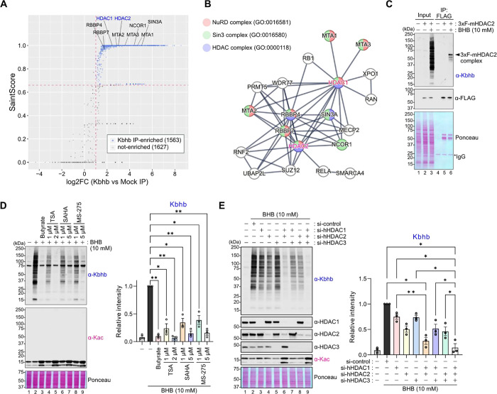
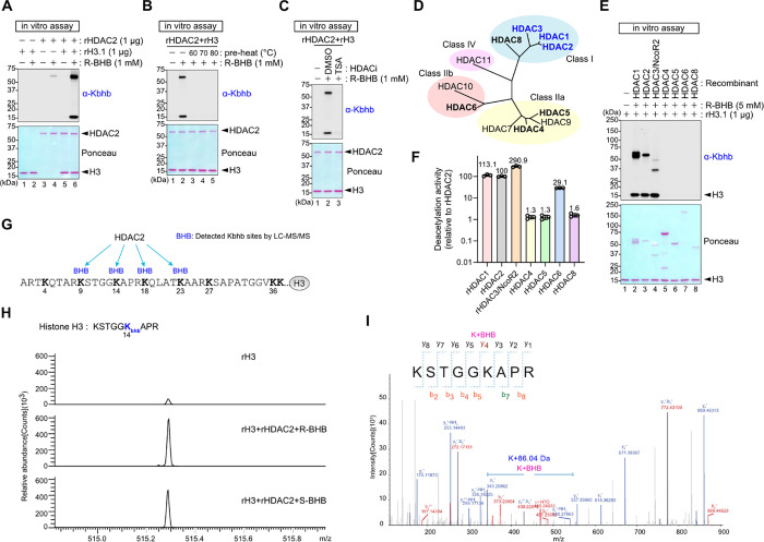
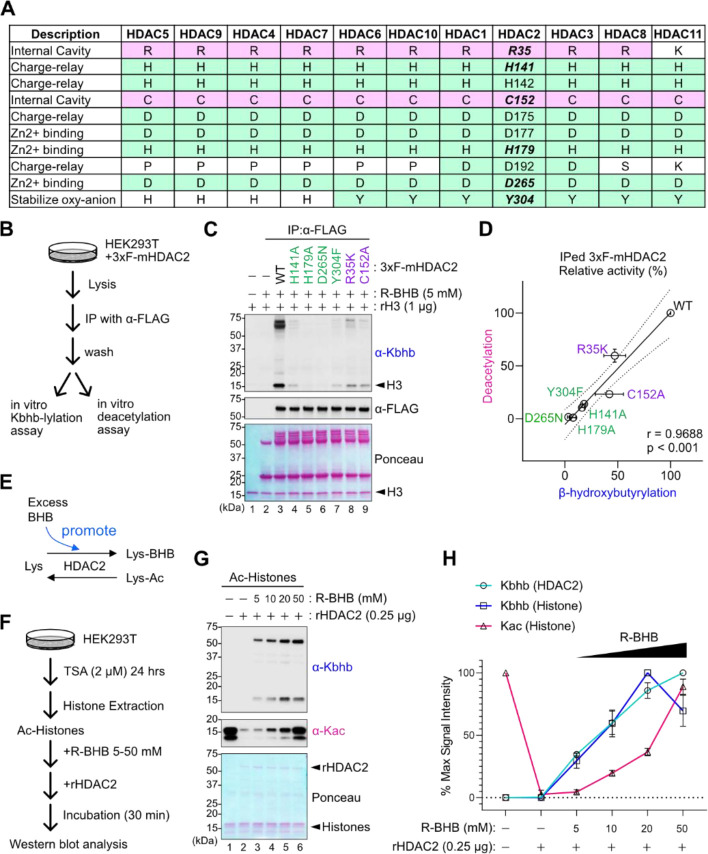
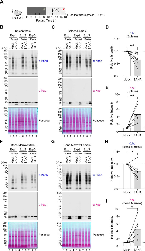
Starvation and low carbohydrate diets lead to the accumulation of the ketone body, β-hydroxybutyrate (BHB), whose blood concentrations increase more than 10-fold into the millimolar range. In addition to providing a carbon source, BHB accumulation triggers lysine β-hydroxybutyrylation (Kbhb) of proteins via unknown mechanisms. As with other lysine acylation events, Kbhb marks can be removed by histone deacetylases (HDACs). Here, we report that class I HDACs unexpectedly catalyze protein lysine modification with β-hydroxybutyrate (BHB). Mutational analyses of the HDAC2 active site reveal a shared reliance on key amino acids for classical deacetylation and non-canonical HDAC-catalyzed β-hydroxybutyrylation. Also consistent with reverse HDAC activity, Kbhb formation is driven by mass action and substrate availability. This reverse HDAC activity is not limited to BHB but also extends to multiple short-chain fatty acids. The reversible activity of class I HDACs described here represents a novel mechanism of PTM deposition relevant to metabolically-sensitive proteome modifications.


Conflict of interest statement

Declaration of Interests The authors declare no competing interests.

Figures

Fig. 1.
Fig. 1.. Class I HDACs are enriched in the Kbhb proteome and are required for Kbhb formation.
(A) Scatter plot of Saint Score and log2(fold-change; FC, Kbhb vs Mock IP). Kbhb IP-enriched proteins were determined based on criteria as indicated in the Materials and Methods and are indicated in blue. Dots for HDAC-related proteins are indicated with their name. (B) The network of HDAC-related proteins in the Kbhb proteome are visualized by STRING analysis. Proteins that are in known HDAC complexes are highlighted in the corresponding color. (C) Validation of Kbhb modification on the HDAC2 complex. HDAC2 KO HEK293T cells expressing 3xFLAG-mHDAC2 was treated with or without 10 mM BHB for 24 hours and used for immunoprecipitation with anti-FLAG beads. Input and IPed samples were analyzed by western blot for Kbhb modification. (D) Left: Representative western blots of HEK293T cells treated with 10 mM BHB in combination with each HDAC inhibitor at the indicated concentrations for 24 hours. Representative of N=3 independent experiments. Right: Relative intensities of anti-Kbhb signals to the lane 2 (10 mM BHB treatment). Signals were normalized to ponceau S staining. Data are represented as mean +/− SEM of independently performed experiments and each symbol represents an individual experiment. Statistical differences were calculated by 1-way ANOVA followed by Dunnett’s correction for multiple comparisons. (E) Left: Representative western blots of HEK293T cells transfected with siRNA(s). Cells were treated with 10 mM BHB for 4 hours. Representative of N=3 indpendent experiments. Right: Relative intensities of anti-Kbhb signals to the lane 2 (10 mM BHB treatment). Signals were normalized by ponceau S staining. Data are represented as mean +/− SEM and each symbol represents an independently performed experiment. Statistical differences were calculated by 1-way ANOVA followed by Tukey’s test for multiple comparisons. *p<0.05, **p<0.01.
Fig. 2.
Fig. 2.. HDAC1, 2, and 3 catalyze lysine β-hydroxybutyrylation vitro.
(A-C) Western blots of an in vitro lysine β-hydroxybutyrylation assay with recombinant HDAC2 (rHDAC2) and histone H3 (rH3) and R-BHB. Reaction was performed at 37°C for 1 hour. Protein loading was visualized by ponceau S staining. Kbhb was detected by anti-Kbhb antibody. rHDAC2 was inactivated by heating for 1 hour at the indicated temperatures in (B). TSA at 5 μM was added in the reaction mixture in (C). (D) Phylogenic tree of human HDAC1–11. Recombinant proteins used in (E) and (F) are indicated in bold. HDACs that are capable of lysine β-hydroxybutyrylation in (E) are highlighted in blue. (E) Representative western blots of an in vitro lysine β-hydroxybutyrylation assay with rHDAC1–8 (except for HDAC7) and rH3 and R-BHB. Approximately 1 μg HDACs were used for each reaction. (F) Relative deactylation activity of each rHDAC as compared to rHDAC2 was measured by fluorogenic peptide deacetylation assay. The same amounts of rHDACs as in (E) were used. (G-I) LC-MS/MS analysis on H3 after in vitro lysine β-hydroxybutyrylation assay. Similar results were obtained from N=2 independent experiments. Detected Kbhb modified sites on H3 are summarized in (G). MS-spectrum of the indicated peptide is shown in (H). Both R- and S-BHB induced Kbhb on lysine 14. MSMS-spectra for the indicated peptide with detected y and b ions are shown in (I).
Fig. 3.
Fig. 3.. Lysine β-hydroxybutyrylation requires HDAC deacetylation residues.
(A) Table of residues important for HDAC deacetylation activity, color-coded according to their proposed functions. The numbers in the HDAC2 column indicate the amino-acid numbers for HDAC2. The amino-acid residues with mutations are shown in italic and bold. (B) Schematic of experimental workflow. HDAC2 KO HEK293T cells expressing 3xFLAG-mHDAC2 were used for immunoprecipitation with α-FLAG antibody. The immunoprecipitants were used for in vitro lysine β-hydroxybutyrylation assay and in vitro deacetylation assay. (C) Representative western blots of in vitro lysine β-hydroxybutyrylation with IPed 3xF-mHDAC2 WT or indicated mutants. (D) Scatter plot of deacetylation and lysine β-hydroxybutyrylation activity for each mutant. Both activities are relative activity to WT 3xF-mHDAC2. Data are represented as mean +/− SEM of independently performed experiments. The line with 95% confidence bands (dotted line) was generated by simple linear regression. N=4 experiments for β-hydroxybutyrylation and N=3 experiments for deacetylation. Pearson correlation coefficient (r) with two-tailed p-value is indicated. (E) Schematic of the hypothesis that excess BHB may promote the lysine β-hydroxybutyrylation reaction. (F-H) Schematic of experimental workflow (F). Highly acetylated histone proteins (Ac-Histones) were isolated from TSA-treated HEK293T cells, and Ac-Histones (2.5 μg) and rHDAC2 (0.25 μg) with R-BHB at the indicated concentrations were incubated for 30 mins. The same isolated histones were pooled and used for all the three experiments. Kbhb and Kac were evaluated by western blot in N=3 independent experiments, with one representative experiment shown in (G). Relative intensities of anti-Kbhb and anti-Kac signals as compared to each maximum intensity are graphed in (H). Data are represented as mean +/− SD of N=3 independently performed experiments.
Fig. 4.
Fig. 4.. HDAC2 can acylate target proteins with other short-chain fatty acids in vitro.
(A) Chemical structures of the indicated short-chain fatty acids are shown. (CX) next to each name indicates the number of carbons in each fatty acid. (B) Comparison of the concentration ranges of short-chain fatty acids in blood based on reported literature values. (C-F) Representative western blots of in vitro acyations by rHDAC2 with the short-chain fatty acids, Butyrate (C), Lactate (D), Propionate (E), and Acetate (F), at the indicated concentrations.
Fig. 5.
Fig. 5.. HDACs contribute to ketosis-induced Kbhb formation in mice
(A) Schematic of experimental design. C57BL/6 mice were fasted starting from night time and injected with SAHA twice at 12 and 15 hr after fasting was initiated. WB: western blot analysis. (B-E) Western blots of splenocytes are shown in (B-C). Exp1–3: N=3 independent experiments are shown on each blot, each containing a fed control, a mock-treated, and a SAHA-treated mouse as indicated. The signals were normalized by ponceau S staining and intensities relative to Mock sample for Kbhb and Kac are plotted in (D) and (E), respectively. Circles indicate males and triangles indicate females. Unpaired t-test with two-tailed P values and lines connect the control and SAHA-treated mice from the same independent experiment. (F-I) Western blots of bone marrow cells are shown in (F-G). Exp1–3: N=3 independent experiments are shown on each blot. The signals were normalized by ponceau S staining and intensities relative to Mock sample for Kbhb and Kac are plotted in (H) and (I), respectively. Circles indicate males and triangles indicate females. Unpaired t-test with two-tailed P values and lines connect the control and SAHA-treated mice from the same independent experiment. *p<0.05, **p<0.01

Similar articles

Cited by

References

    1. Cahill G. F. Jr., Veech R. L., Ketoacids? Good medicine? Trans Am Clin Climatol Assoc 114, 149–161; discussion 162–143 (2003). - PMC - PubMed
    1. Puchalska P., Crawford P. A., Metabolic and Signaling Roles of Ketone Bodies in Health and Disease. Annu Rev Nutr 41, 49–77 (2021). - PMC - PubMed
    1. Youm Y. H. et al., The ketone metabolite beta-hydroxybutyrate blocks NLRP3 inflammasome-mediated inflammatory disease. Nat Med 21, 263–269 (2015). - PMC - PubMed
    1. Shimazu T. et al., Suppression of oxidative stress by beta-hydroxybutyrate, an endogenous histone deacetylase inhibitor. Science 339, 211–214 (2013). - PMC - PubMed
    1. Taggart A. K. et al., (D)-beta-Hydroxybutyrate inhibits adipocyte lipolysis via the nicotinic acid receptor PUMA-G. J Biol Chem 280, 26649–26652 (2005). - PubMed

Publication types