. 2023 Dec;16(6):e004200.
doi: 10.1161/CIRCGEN.123.004200. Epub 2023 Nov 28.

Genotype-Phenotype Taxonomy of Hypertrophic Cardiomyopathy

Affiliations

Genotype-Phenotype Taxonomy of Hypertrophic Cardiomyopathy

Lara Curran et al. Circ Genom Precis Med. 2023 Dec.

Abstract

Background: Hypertrophic cardiomyopathy (HCM) is an important cause of sudden cardiac death associated with heterogeneous phenotypes, but there is no systematic framework for classifying morphology or assessing associated risks. Here, we quantitatively survey genotype-phenotype associations in HCM to derive a data-driven taxonomy of disease expression.

Methods: We enrolled 436 patients with HCM (median age, 60 years; 28.8% women) with clinical, genetic, and imaging data. An independent cohort of 60 patients with HCM from Singapore (median age, 59 years; 11% women) and a reference population from the UK Biobank (n=16 691; mean age, 55 years; 52.5% women) were also recruited. We used machine learning to analyze the 3-dimensional structure of the left ventricle from cardiac magnetic resonance imaging and build a tree-based classification of HCM phenotypes. Genotype and mortality risk distributions were projected on the tree.

Results: Carriers of pathogenic or likely pathogenic variants for HCM had lower left ventricular mass, but greater basal septal hypertrophy, with reduced life span (mean follow-up, 9.9 years) compared with genotype negative individuals (hazard ratio, 2.66 [95% CI, 1.42-4.96]; P<0.002). Four main phenotypic branches were identified using unsupervised learning of 3-dimensional shape: (1) nonsarcomeric hypertrophy with coexisting hypertension; (2) diffuse and basal asymmetrical hypertrophy associated with outflow tract obstruction; (3) isolated basal hypertrophy; and (4) milder nonobstructive hypertrophy enriched for familial sarcomeric HCM (odds ratio for pathogenic or likely pathogenic variants, 2.18 [95% CI, 1.93-2.28]; P=0.0001). Polygenic risk for HCM was also associated with different patterns and degrees of disease expression. The model was generalizable to an independent cohort (trustworthiness, M1: 0.86-0.88).

Conclusions: We report a data-driven taxonomy of HCM for identifying groups of patients with similar morphology while preserving a continuum of disease severity, genetic risk, and outcomes. This approach will be of value in understanding the causes and consequences of disease diversity.

Keywords: genotype; hypertension; hypertrophy; magnetic resonance imaging; phenotype.


Conflict of interest statement

Disclosures Dr Ware has consulted for MyoKardia, Inc, Foresite Labs, and Pfizer and receives research support from Bristol Myers Squibb outside the submitted work. Dr O’Regan has consulted for Bayer AG and Bristol Myers Squibb and also receives research support from Bayer AG outside the submitted work. Dr Halliday is on an advisory board for AstraZeneca. The other authors report no conflicts.

Figures

Figure 1.
Figure 1.
Study flowchart. A, Details of the analysis pipeline using segmentations of cardiac magnetic resonance cine imaging to build a 3-dimensional (3D) model of phenotypic variation in UK Biobank and hypertrophic cardiomyopathy (HCM) participants. Association models and clustering analysis is then performed on the data. B, Details of patients with HCM recruited to the study and reasons for exclusion. CMR indicates cardiac magnetic resonance; DDRtree, discriminative dimensionality reduction via learning a tree; ED, end diastole; ES, end systole; SARC-NEG, genotype negative; SARC-P/LP, pathogenic/likely pathogenic sarcomeric variants; SARC-VUS, variants of uncertain significance; T, last cardiac phase; UMAP, uniform manifold approximation and projection; UKB, UK Biobank; and PRS, polygenic risk score.
Figure 2.
Figure 2.
Unsupervised clustering of patients with hypertrophic cardiomyopathy (HCM) using demographic and anthropometric data, clinical characteristics, and conventional cardiac imaging parameters. A, Participant clinical features segmented in 3 clusters with a K-means algorithm, optimized with a silhouette score, projected in the 2-dimensional (2D) space of the first 2 uniform manifold approximation and projection (UMAP) components. B, Genotype status of participants in the 2D UMAP space with genotype prevalence by cluster. SARC-NEG indicates genotype negative; SARC-P/LP, pathogenic/likely pathogenic sarcomeric variants; and SARC-VUS, variants of uncertain significance. *P≤0.05, **P≤0.01, ***P≤0.001,****P≤0.0001; n=436.
Figure 3.
Figure 3.
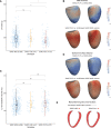
Genotype-phenotype associations in hypertrophic cardiomyopathy (HCM). A and C, Dot and boxplots of left ventricular (LV) mass and end-diastolic volume in patients with HCM stratified by genotype. SARC-NEG indicates genotype negative; SARC-P/LP, pathogenic/likely pathogenic sarcomeric variants; and SARC-VUS, variants of uncertain significance. B and D, Three-dimensional modeling of LV geometry with vertex-wise standardized β-coefficients projected on the epicardial surface. These show the extent of association between genotype and wall thickness or surface-to-surface distance (comparing regional shape change) for different comparisons, adjusting for the covariates of age, sex, and race. Yellow contour lines show significant regions (P<0.05) after multiple testing correction. LV projections are septal (left) and anterior (right). E, Long-axis cross sections showing the myocardial outline in red for each genotype compared with control participants in UK Biobank (dashed outline). *P≤0.05, **P≤0.01.
Figure 4.
Figure 4.
Phenotypic tree of hypertrophic cardiomyopathy (HCM) morphology. A, Three-dimensional models of the left ventricle in patients with HCM were reduced to a 2-dimensional tree structure where each point represents 1 individual. The tree maps undifferentiated states in the center to more characteristic morphologies in the distal branches while preserving a continuous stratification. For each branch, we show a wall thickness shape model at end systole where the colors represent β-coefficients for the comparison between branches. Corresponding features at end diastole are shown in Figure S1. Branch 5 showed an undifferentiated mixed phenotype. B, Each participant in the tree labeled by probability of pathogenic/likely pathogenic sarcomeric variant (SARC-P/LP) genotype, polygenic score (PGS) for HCM, and predicted survival probability at median age. C, Continuous and discrete phenotypic variables found to be significantly associated to at least 1 branch. For late gadolinium enhancement, labels are as follows: 1, none; 2, minimal; 3, moderate, and 4, severe. The significance for the enrichment of discrete variables is reported within the bars. ACE indicates angiotensin-converting enzyme inhibitor; ARB, angiotensin receptor blocker; BP, blood pressure; LGE, late gadolinium enhancement; LV, left ventricular; LVH, left ventricular hypertrophy; LVOTO, left ventricular outflow tract obstruction; SARC-NEG, genotype negative; and SV, stroke volume. Only the significant pairs are reported with the symbols: *P≤0.05, **P≤0.01, ***P≤0.001, ****P≤0.0001; n=436.
Figure 5.
Figure 5.
Cumulative hazard plot. All-cause mortality in individuals with hypertrophic cardiomyopathy (HCM) carrying pathogenic/likely pathogenic sarcomeric variants (SARC-P/LP) compared with genotype negative (SARC-NEG; hazard ratio, 2.66 [95% CI, 1.42–4.96]; P=0.002).

References

    1. Maron BJ, Desai MY, Nishimura RA, Spirito P, Rakowski H, Towbin JA, Rowin EJ, Maron MS, Sherrid MV. Diagnosis and evaluation of hypertrophic cardiomyopathy: JACC state-of-the-art review. J Am Coll Cardiol. 2022;79:372–389. doi: 10.1016/j.jacc.2021.12.002 - PubMed
    1. Tadros R, Francis C, Xu X, Vermeer AMC, Harper AR, Huurman R, Kelu Bisabu K, Walsh R, Hoorntje ET, Te Rijdt WP, et al. . Shared genetic pathways contribute to risk of hypertrophic and dilated cardiomyopathies with opposite directions of effect. Nat Genet. 2021;53:128–134. doi: 10.1038/s41588-020-00762-2 - PMC - PubMed
    1. Harper AR, Goel A, Grace C, Thomson KL, Petersen SE, Xu X, Waring A, Ormondroyd E, Kramer CM, Ho CY, et al. ; HCMR Investigators. Common genetic variants and modifiable risk factors underpin hypertrophic cardiomyopathy susceptibility and expressivity. Nat Genet. 2021;53:135–142. doi: 10.1038/s41588-020-00764-0 - PMC - PubMed
    1. Butters A, Lakdawala NK, Ingles J. Sex differences in hypertrophic cardiomyopathy: interaction with genetics and environment. Curr Heart Fail Rep. 2021;18:264–273. doi: 10.1007/s11897-021-00526-x - PMC - PubMed
    1. de Marvao A, McGurk KA, Zheng SL, Thanaj M, Bai W, Duan J, Biffi C, Mazzarotto F, Statton B, Dawes TJW, et al. . Phenotypic expression and outcomes in individuals with rare genetic variants of hypertrophic cardiomyopathy. J Am Coll Cardiol. 2021;78:1097–1110. doi: 10.1016/j.jacc.2021.07.017 - PMC - PubMed

Publication types