

Severe bronchiectasis resulting from chronic bacterial bronchitis and bronchopneumonia in a jungle cat
- PMID: 38014741
- PMCID: PMC10734597
- DOI: 10.1177/10406387231216181
Severe bronchiectasis resulting from chronic bacterial bronchitis and bronchopneumonia in a jungle cat
Abstract
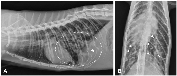
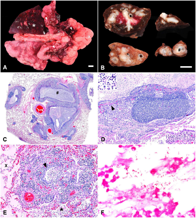
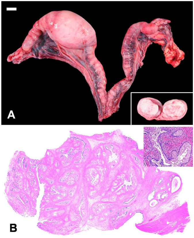
Bronchiectasis is irreversible bronchial dilation that can be congenital or acquired secondary to chronic airway obstruction. Feline bronchiectasis is rare and, to our knowledge, has not been reported previously in a non-domestic felid. An ~10-y-old female jungle cat (Felis chaus) was presented for evaluation of an abdominal mass and suspected pulmonary metastasis. The animal died during exploratory laparotomy and was submitted for postmortem examination. Gross examination revealed consolidation of the left caudal lung lobe and hila of the cranial lung lobes. Elsewhere in the lungs were several pale-yellow pleural foci of endogenous lipid pneumonia. On cut section, there was severe distension of bronchi with abundant white mucoid fluid. The remaining lung lobes were multifocally expanded by marginal emphysema. Histologically, ectatic bronchi, bronchioles, and fewer alveoli contained degenerate neutrophils, fibrin, and mucin (suppurative bronchopneumonia) with rare gram-negative bacteria. Aerobic culture yielded low growth of Proteus mirabilis and Escherichia coli. There was chronic bronchitis, marked by moderate bronchial gland hyperplasia, lymphoplasmacytic inflammation, and lymphoid hyperplasia. The palpated abdominal mass was a uterine endometrial polyp, which was considered an incidental, but novel, finding. Chronic bronchitis and bronchopneumonia should be considered as a cause of bronchiectasis and a differential diagnosis for respiratory disease in non-domestic felids.
Keywords: Escherichia coli; Felidae; Proteus; exotic animal; lung; respiratory disease; zoo animal.
Conflict of interest statement
Declaration of conflicting interestsThe authors declared no conflicts of interest regarding the research, authorship, and/or publication of this article.
Figures
Similar articles
-
A novel Filobacterium sp can cause chronic bronchitis in cats.PLoS One. 2021 Jun 9;16(6):e0251968. doi: 10.1371/journal.pone.0251968. eCollection 2021. PLoS One. 2021. PMID: 34106938 Free PMC article.
-
Bronchiectasis, Chronic Suppurative Lung Disease and Protracted Bacterial Bronchitis.Curr Probl Pediatr Adolesc Health Care. 2018 Apr;48(4):119-123. doi: 10.1016/j.cppeds.2018.03.003. Epub 2018 Mar 27. Curr Probl Pediatr Adolesc Health Care. 2018. PMID: 29602647 Review.
-
Clinical, radiographic, and pathologic features of bronchiectasis in cats: 12 cases (1987-1999).J Am Vet Med Assoc. 2000 Feb 15;216(4):530-4. doi: 10.2460/javma.2000.216.530. J Am Vet Med Assoc. 2000. PMID: 10687008
-
[Bronchopneumonia, obstructive bronchitis and bronchiectasis in children with adenovirus infections].Lijec Vjesn. 1997 Jan;119(1):11-5. Lijec Vjesn. 1997. PMID: 9213724 Croatian.
-
[Feline asthma and chronic bronchitis - an overview of diagnostics and therapy].Tierarztl Prax Ausg K Kleintiere Heimtiere. 2019 Jun;47(3):175-187. doi: 10.1055/a-0917-6245. Epub 2019 Jun 18. Tierarztl Prax Ausg K Kleintiere Heimtiere. 2019. PMID: 31212350 Review. German.
References
-
- Big Cat Rescue. State laws exotic cats. 2022. [cited 2023 Mar 30]. https://bigcatrescue.org/state-laws-exotic-cats/
-
- Cannon MS, et al.. Quantitative and qualitative computed tomographic characteristics of bronchiectasis in 12 dogs. Vet Radiol Ultrasound 2013;54:351–357. - PubMed
-
- Furman AC, et al.. Lung abscess in patients with AIDS. Clin Infect Dis 1996;22:81–85. - PubMed
-
- Gelberg HB, McEntee K. Hyperplastic endometrial polyps in the dog and cat. Vet Pathol 1984;21:570–573. - PubMed
-
- Hawkins EC, et al.. Demographic, clinical, and radiographic features of bronchiectasis in dogs: 316 cases (1988–2000). J Am Vet Med Assoc 2003;223:1628–1635. - PubMed
MeSH terms
LinkOut - more resources
Full Text Sources
Medical
Miscellaneous
